|
Exhibition Hall 11:45 - 12:45 |
|
|
|
Computer # |
|
2792.
 |
73 |
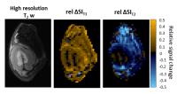
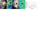
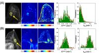
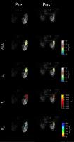
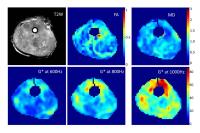
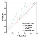
Effects of Temporal Resolution on Quantitative DCE-MRI
Prediction of Breast Cancer Therapy Response 
Wei Huang1, Aneela Afzal1, Alina
Tudorica1, Yiyi Chen1, Stephen Y-C
Chui1, Arpana Naik1, Megan Troxell1,
Kathleen Kemmer1, Karen Y Oh1, Nicole
Roy1, Megan L Holtorf1, and Xin Li1
1Oregon Health & Science University, Portland,
OR, United States
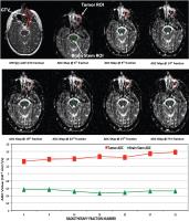
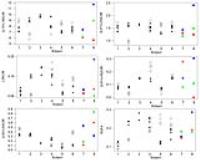
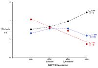
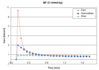
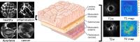
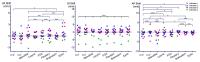
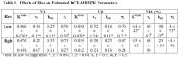
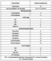
15 breast cancer patients undergoing neoadjuvant
chemotherapy (NACT) consented to two DCE-MRI studies at the
same time points before, during, and after NACT: one with
high temporal resolution (tRes) and the other with low tRes.
There were systematic errors in estimated pharmacokinetic (PK)
parameters from the low tRes data compared to the high tRes
data. However, the abilities of PK parameters for early
prediction of pathologic response to NACT were not affected
by poorer tRes.
|
|
2793.
 |
74 |
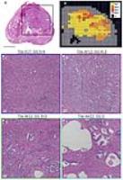
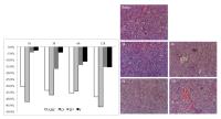
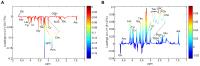
Effect of Neoadjuvant Chemotherapy on in-vivo MRS determined
tCho and Membranous and Cytoplasmic b-catenin Expression in
Breast Cancer Patients
Naranamangalam R Jagannathan1, Khushbu Agarwal1,
Uma Sharma1, Sandeep Mathur2,
Vurthaluru Seenu3, and Rajinder Parshad3
1Department of NMR and MRI Facility, All India
Institute of Medical Sciences, New Delhi, India, 2Department
of Pathology, All India Institute of Medical Sciences, New
Delhi, India, 3Department
of Surgical Disciplines, All India Institute of Medical
Sciences, New Delhi, India
We evaluated the changes in tCho levels and β-catenin
expression (membrane and cytoplasm) after III neoadjuvant
chemotherapy in breast cancer patients. Significant
reduction in β-catenin expression (membranous and
cytoplasmic) was observed after therapy. Post-therapy, tCho
reduced significantly in tumors with Grades 1 and 2
membranous β-catenin expression and also in tumors with IRS
0 and Grade 1 cytoplasmic β-catenin. Prior to therapy, tCho
was positively associated with cytoplasmic β-catenin while
negatively with membranous protein. However post-therapy
tCho was negatively associated with both cytoplasmic and
membranous β-catenin. This signifies antiproliferative and
apoptosis induction effects of chemotherapy drugs on breast
cancer patients.
|
|
2794.
 |
75 |
Correlation of diffusion weighted MR imaging with the prognosis
of locally advanced gastric carcinoma to neoadjuvant
chemotherapy
Lei Tang1, Ying-Shi Sun1, Zi-Yu Li2,
Xiao-Ting Li 1,
Fei Shan2, Zi-Ran Li2, and Jia-Fu Ji2
1Radiology, Peking University Cancer Hospital &
Institute, Beijing, China, People's Republic of, 2GI
surgery, Peking University Cancer Hospital & Institute,
Beijing, China, People's Republic of
The percentage changes of ADC after neoadjuvant chemotherapy
of gastric carcinoma have correlation with long-term
prognosis. The significantly increased ADC after
chemotherapy is more prone to signify long-term survival,
and has potential to be a surrogate imaging biomarker for
the prediction of the prognosis. ADCentire for the whole
lesion is better than ADCmin for high signal area in the
prognosis prediction.
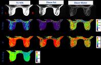
|
 |
2795.
 |
76 |
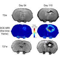
T2*-weighted imaging and DCE-MRI as complementary tools to
characterize the continuous process of radionecrosis and
neovascularization
Jérémie P. Fouquet1, Julie Constanzo1,
Laurence Masson-Côté1,2, Luc Tremblay1,
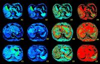
Philippe Sarret3, Sameh Geha4, Kevin
Whittingstall5, Benoit Paquette1, and
Martin Lepage1
1Department of Nuclear Medicine and Radiobiology,
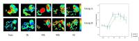
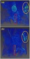
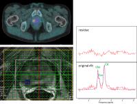
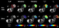
Université de Sherbrooke, Sherbrooke, QC, Canada, 2Service
of Radiation Oncology, Centre Hospitalier Universitaire de
Sherbrooke, Sherbrooke, QC, Canada, 3Department
of Pharmacology-Physiology, Université de Sherbrooke,
Sherbrooke, QC, Canada, 4Department
of Pathology, Université de Sherbrooke, Sherbrooke, QC,
Canada, 5Department
of Diagnostic Radiology, Université de Sherbrooke,
Sherbrooke, QC, Canada
Radiation dose delivered to healthy tissues during brain
tumors radiosurgery can cause important side effects. We
imaged an animal model of brain irradiation with DCE-MRI and
T2*-weighted imaging at different time points after
treatment. DCE-MRI allowed the discrimination of areas with
high vessel permeability and necrotic regions. T2*-weighted
imaging enabled the visualization of a necrotic core and
micro-lesions at its periphery. Micro-lesions were initially
co-localized with permeable vessels and later evolved into
necrosis. Together, DCE-MRI and T2*-weighted images provided
a coherent picture on the phenomena involved in
radionecrosis progression, which could help in the
management of associated problems.
|
|
2796.
 |
77 |
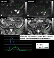
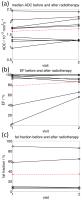
DIFFUSION-WEIGHTED IMAGING (DWI) AS A TREATMENT RESPONSE
BIOMARKER IN PROSTATE CANCER BONE METASTASES
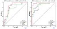
Raquel Perez-Lopez1,2, Matthew D. Blackledge1,2,
Joaquin Mateo1,2, David J. Collins1,2,
Veronica A. Morgan1,2, Alison MacDonald1,2,
Diletta Bianchini1,2, Zafeiris Zafeiriou1,2,
Pasquale Rescigno1,2, Michael Kolinsky1,2,
Daniel Nava Rodrigues1,2, Helen Mossop1,
Nuria Porta1, Emma Hall1, Martin O.
Leach1,2, Johann S. de Bono1,2, Dow-Mu
Koh1,2, and Nina Tunariu1,2
1The Institute of Cancer Research, Sutton, United
Kingdom, 2The
Royal Marsden NHS Foundation Trust, Sutton, United Kingdom
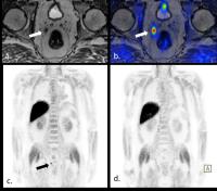
We hypothesized that changes in the median apparent
diffusion coefficient (mADC) and volume of bone metastases
(BM), quantified by whole body (WB) diffusion-weighted
imaging (DWI), are response biomarkers in metastatic
castration-resistant prostate cancer (mCRPC). 21 patients
completed WB-DWI at baseline and after 12 weeks of treatment
in a sub-study within a clinical trial of olaparib in mCRPC,
performed on a 1.5-T Siemens Avanto scanner. Four different
segmentation techniques were explored including axial
skeleton analyses and simpler methods including 5 target
lesions. Changes in mADC and volume of BM associated with
response to therapy. The simplified approach also showed
promising results, warranting further evaluation.
|
|
2797.
 |
78 |
Response Assessment to Tumor Treating Fields in Patients with
Glioblastoma using Physiologic and Metabolic MR Imaging
Sanjeev Chawla1, Sumei Wang1, Gaurav
Verma1, Aaron Skolnik1, Sulaiman
Sheriff2, Katelyn M Reilly1, Lisa
Desiderio1, Andrew Maudsley2, Steven
Brem3, Katherine Peters4, Harish
Poptani5, and Suyash Mohan1
1Radiology, Perelman School of Medicine at the
University of Pennsylvania, Philadelphia, PA, United States, 2Radiology,
University of Miami, Miami, FL, United States, 3Neurosurgery,
Perelman School of Medicine at the University of
Pennsylvania, Philadelphia, PA, United States, 4Neurology,
Duke University Medical Center, Durham, NC, United States, 5Department
of Cellular and Molecular Physiology, University of
Liverpool, Liverpool, United Kingdom
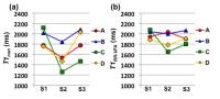
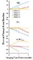
Tumor treating fields (TTFields) are a novel antimitotic
treatment modality for treatment of patients with
glioblastoma (GBM). To assess response to TTFields, 4 GBM
patients underwent diffusion, perfusion and 3D-echo-planar
spectroscopic imaging prior to initiation of TTFields and at
one and two month follow-up periods. A trend towards
increased MD and a decrease in FA and rCBVmax was
noted in most patients at 2-month relative to baseline
indicating inhibited tumor growth and vascularity. Cho/Cr
values did not exhibit any trend probably due to
heterogeneity in response. These preliminary data indicate
the potential of advanced MR imaging in assessing response
to TTFields.
|
|
2798.
|
79 |
The value of functional MRI on predicting therapeutic outcome of
TACE on hepatocellular carcinoma
Ma Xiaohong1, Zhao Xinming1, Ouyang
Han1, and Zhou Chunwu1
1Diagnostic Radiology, Cancer Hospital, Chinese
Academy of Medical Sciences, Peking Union Medical College,
Beijing, China, People's Republic of
The purpose of this study was to explore the efficacy of
functional MRI (diffusion-weighted imaging (DWI), IntraVoxel
incoherent motion (IVIM) and perfusion-weighted imaging
(PWI)) quantitative analysis in predicting therapeutic
outcome of TACE on HCC. The Dfast, Ktrans,
ΔDfast and
ΔKtrans of
HCC acquired before and after TACE obviously correlated with
PFS and was valuable in the prediction of the clinical
outcome of HCC treated with TACE.
|
|
2799.
 |
80 |
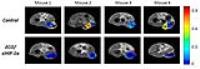
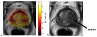
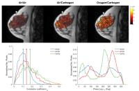
Early Assessment of Antiangiogenic Effects of Sorafenib using
IVIM in Mouse Model with Hepatocellular Carcinoma
Yong Zhang1, Bing Wu1, Xin Chen2,
and Zaiyi Liu2
1GE Healthcare MR Research China, Beijing, China,
People's Republic of, 2Radiology,
Guangdong General Hospital, Guangzhou, China, People's
Republic of
Antiangiogenic therapy is efficient to treat hypervascular
tumor such as hepatocelluar carcinoma (HCC). Unlike
chemotherapy and radiation therapy, tumor dimension doesn’t
change in its early phase. Hence traditional reponse
criteria based on morphological change fails to early assess
the therapeutic response of antiangiogenic treatment. This
study used intravoxel incoherent motion (IVIM) theory to
separate perfusion and diffusion characteristics in HCC over
sereval time points after antiangiogenic Sorafenib
administration. It was found that IVIM-derived pure
diffusivity and pseudo-diffusivity were able to detect the
microvascular collapse and cellular edema in HCC at early
phase of antiangiogenic medication.
|
|
2800.
 |
81 |
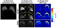
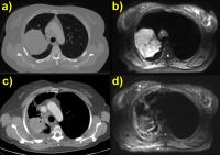
Lung tumour radiotherapy treatment response assessment using
Active Breathing Coordinated (ABC) Diffusion-Weighted Magnetic
Resonance Imaging
Evangelia Kaza1, Matthew Blackledge1,
David John Collins1, Erica Scurr2,
Helen McNair3, Richard Symonds-Tayler1,
Fiona McDonald2, Martin Osmund Leach1,
and Dow-Mu Koh2
1The Institute of Cancer Research and Royal
Marsden Hospital, London, United Kingdom, 2The
Royal Marsden NHS Foundation Trust, London, United Kingdom, 3Department
of Radiotherapy, Royal Marsden NHS Foundation Trust and
Institute of Cancer Research, London, United Kingdom
Imaging with an Active Breathing Coordinator (ABC) modified
for MR use was performed on lung cancer patients to acquire
spatially matching diffusion-weighted images (DWI) before,
during and after Radiotherapy. DWI spatially matched the CT
and depicted mediastinal nodal involvement as well as
internal tumour heterogeneity. ADC maps provided information
about changes in solid and fluid components throughout
therapy. Treatment response was evaluated by applying
multi-parametric tumour heterogeneity characterisation using
Gaussian Mixture Modelling. Differences in ADC and volume
behavior of separate cancerous tissue components at various
treatment time points may indicate tumour sub-volumes and
provide detailed cancer characterisation.
|
|
2801.
 |
82 |
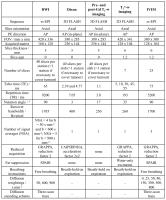
Defining the baseline functional imaging characteristics of
retroperitoneal sarcomas
Jessica M Winfield1,2, Aisha Miah3,
Dirk Strauss4, Khin Thway5, Andrew
Hayes4, Daniel Henderson3, David J
Collins1,2, Nandita M deSouza1,2,
Martin O Leach1,2, Sharon L Giles1,2,
Veronica A Morgan1,2, and Christina Messiou1,2
1MRI, Royal Marsden Hospital, Sutton, United
Kingdom, 2Division
of Radiotherapy and Imaging, Cancer Research UK Cancer
Imaging Centre, Institute of Cancer Research, London, United
Kingdom,3Department of Radiotherapy, Royal
Marsden Hospital, London, United Kingdom, 4Department
of Surgery, Royal Marsden Hospital, London, United Kingdom, 5Department
of Histopathology, Royal Marsden Hospital, London, United
Kingdom
Soft tissue sarcomas are often highly heterogeneous tumours
and post-treatment changes cannot be described by standard
size criteria. Functional imaging may provide a non-invasive
method of assessing response to treatment. Knowledge of
baseline functional imaging characteristics and the
repeatability of estimated parameters is essential in
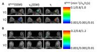
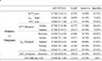
development of future studies. In this study, 22 patients
with retroperitoneal sarcoma were imaged before treatment.
Whole-tumour assessments of apparent diffusion coefficient
(ADC), parameters of the intra-voxel incoherent motion model
(IVIM: diffusion coefficient D, fraction f,
fast exponential component D*), transverse relaxation rate
(R2*), fat fraction and enhancing fraction (EF)
showed large ranges of median estimates, indicating wide
inter-tumour heterogeneity. The large standard deviation of
parameters within tumours reflects the intra-tumour
heterogeneity. In 21 patients, a second examination was
carried out to assess repeatability of ADC, D, f,
D* and R2*. Excellent repeatability of fitted
parameters, particularly ADC, indicates high sensitivity to
treatment-induced changes.
|
|
2802.
 |
83 |
Gaussian mixture modelling of combined functional imaging
parameters provides new insight into tumour heterogeneity
Jessica M Winfield1,2, Matthew D Blackledge2,
Aisha Miah3, Dirk Strauss4, Khin Thway5,
David J Collins1,2, Martin O Leach1,2,
Sharon L Giles1,2, Daniel Henderson3,
and Christina Messiou1,2
1MRI, Royal Marsden Hospital, Sutton, United
Kingdom, 2Division
of Radiotherapy and Imaging, Cancer Research UK Cancer
Imaging Centre, Institute of Cancer Research, London, United
Kingdom,3Department of Radiotherapy, Royal
Marsden Hospital, London, United Kingdom, 4Department
of Surgery, Royal Marsden Hospital, London, United Kingdom, 5Department
of Histopathology, Royal Marsden Hospital, London, United
Kingdom
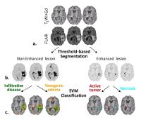
Multi-parametric functional imaging may enable non-invasive
assessment of response to treatment in soft tissue sarcomas.
Image analysis is complicated, however, by the highly
heterogeneous nature of these tumours, which can include
regions of cellular tumour, fat, necrosis and cystic change
that may respond differently to treatment. In this study,
patients with retroperitoneal sarcoma were imaged before and
after radiotherapy using DW-MRI, Dixon and
pre-/post-contrast T1-w imaging for evaluation of
enhancing fraction (EF). Gaussian mixture modelling was
applied to classify pixels in the tumour volume according to
their functional imaging behaviour, combining ADC, fat
fraction and EF to characterise tumour components. This
method enabled segmentation of highly heterogeneous tumours
and estimation of mean ADC and volume of each tumour
component. Heterogeneous changes post-radiotherapy were
summarised in tissue classification maps, which combine
multiple functional imaging parameters. Combined analysis of
functional imaging parameters may provide greater insight
into tumour behaviour, for example identification of viable
tumour.
|
|
2803.
 |
84 |
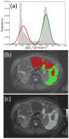
Threshold functional imaging maps depict intra-tumour
heterogeneity of response to radiotherapy in retroperitoneal
sarcomas
Jessica M Winfield1,2, Aisha Miah3,
Dirk Strauss4, Khin Thway5, David J
Collins1,2, Martin O Leach1,2, Sharon
L Giles1,2, Daniel Henderson3, Shane
Zaidi6, and Christina Messiou1,2
1MRI, Royal Marsden Hospital, Sutton, United
Kingdom, 2Division
of Radiotherapy and Imaging, Cancer Research UK Cancer
Imaging Centre, Institute of Cancer Research, London, United
Kingdom,3Department of Radiotherapy, Royal
Marsden Hospital, London, United Kingdom, 4Department
of Surgery, Royal Marsden Hospital, London, United Kingdom, 5Department
of Histopathology, Royal Marsden Hospital, London, United
Kingdom, 6Department
of Clinical Oncology, Royal Marsden Hospital, London, United
Kingdom
Functional imaging provides scope for non-invasive
assessment of response to radiotherapy and/or systemic
agents in retroperitoneal sarcomas and investigation of
heterogeneity of response in this highly heterogeneous
tumour type. In this study 9 patients with retroperitoneal
sarcoma were imaged before treatment and 2-4 weeks after
radiotherapy. Whilst some tumours exhibited large increases
in median ADC and enhancing fraction after radiotherapy, the
overall changes for the cohort were not significant and
there were no clear changes in fat fraction. Thresholded ADC
maps and enhancement maps, however, reveal localised
post-radiotherapy changes in ADC and enhancement that are
not fully characterised by whole-tumour metrics.
|
|
2804.
 |
85 |
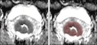
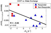
Chemical exchange saturation transfer (CEST) and relaxometry as
biomarkers for assessing response of brain metastases to
stereotactic radiosurgery
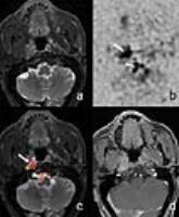
Hatef Mehrabian1,2, Kimberly L Desmond3,
Anne L Martel1,2, Arjun Sahgal1,4,
Hany Soliman1,4, and Greg J Stanisz1,2
1Physical Sciences, Sunnybrook Research
Institute, Toronto, ON, Canada, 2Medical
Biophysics, University of Toronto, Toronto, ON, Canada, 3Medical
Physics and Applied Radiation Sciences, McMaster University,
Hamilton, ON, Canada, 4Radiation
Oncology, Odette Cancer Centre, Toronto, ON, Canada
Quantitative MRI techniques that probe the metabolic and
micro-structural changes in the tumor have the potential to
assess response of brain metastases to stereotactic
radiosurgery early after treatment. Two techniques were
investigated here: a) Chemical
Exchange Saturation Transfer (CEST), b) Relaxometry.
Among all model parameters, early changes in the
intracellular-extracellular water exchange rate in
relaxometry, and peak amplitude of nuclear overhauser effect
at the ipsilateral normal appearing white matter in CEST
provided the strongest correlation with tumor volume change
one-month post-treatment. We also demonstrated that these
two parameters were highly correlated suggesting they could
provide complementary information about treatment effects.
|
|
2805.
 |
86 |
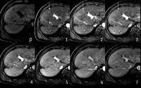
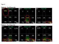
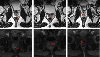
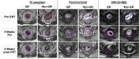
MRI in Assessing Response to Neoadjuvant Chemo-radiation in
Locally Advanced Rectal Cancer Using DCE-MR and DWI Data Sets:
Before, During and After the Treatment
Ke Nie1, Liming Shi2, Ning Yue1,
Jabbour Salma1, Xi Hu2, Liwen Qian2,
Tingyu Mao2, Qin Chen2, Xiaonan Sun2,
and Tianye Niu2,3,4
1Radiation Oncology, Rutgers-Cancer Institute of
New Jersey, Rutgers-Robert Wood Johnson Medical School, New
Brunswick, NJ, United States, 2Radiation
Oncology, Sir Run Run Shaw Hospital, Zhejiang University of
Medicine, Hangzhou, China, People's Republic of, 3Institute
of Translational Medicine, Hangzhou, China, People's
Republic of, 4Radiology,
Sir Run Run Shaw Hospital, Zhejiang University of Medicine,
Hangzhou, China, People's Republic of
We are one of the first to investigate the predictive value
of combined anatomical, DCE-MRI and DWI for good
pathological response at different time points during the
pre-operative chemo-radiation treatment (CRT) in patients
with locally advanced rectal cancer (LARC). The
pre-treatment ADC and internal heterogeneity enhancement
measured by texture features from DCE-MRI and the relative
change of the ADC values during the treatment showed good
prognostic value with pathological response. Overall, this
study provides new information of the optimal use of MRI in
predicting response to the pre-operative CRT, which may
further help tailor the treatment into the era of
personalized medicine.
|
|
2806.
 |
87 |
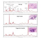
Altered lipid metabolism on 1H NMR as response biomarkers in
prostate cancer cells and tumors following radiotherapy
Gigin Lin1, Yu-Chun Lin1, Hsi-Mu Chen 1,
and Chiun-Chieh Wang2
1Medical Imaging and Intervention, Chang Gung
Memorial Hospital, Taoyuan, Taiwan, 2Radiation
Oncology, Chang Gung Memorial Hospital, Taoyuan, Taiwan
The intracellular storage and utilization of lipids are
critical for cancer cells to maintain energy homeostasis. In
this study, we investigated the changes of lipid metabolites
in murine TRAMP-C prostate cancer cells and tumors following
radiotherapy. The lipid profile following radiotherapy
demonstrated increased levels of fatty acids and
triacylglycerols, before the change of tumor size. The
increase of lipids signals can potentially serve as early
response biomakers in clinical setting for prostate cancer
patients following radiotherapy.
|
|
2807.
 |
88 |
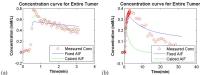
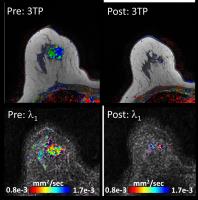
Impact of T1 and B1 correction on quantitative DCE-MRI for
assessing longitudinal therapy response in breast cancer - Permission Withheld
Dattesh D Shanbhag1, Parita Sanghani1,
Reem Bedair 2,
Venkata Veerendranadh Chebrolu1, Sandeep N Gupta3,
Scott Reid 4,
Fiona Gilbert 2,
Andrew Patterson 2,
Rakesh Mullick1, and Martin Graves2
1GE Global Research, Bangalore, India, 2University
of Cambridge, Cambridge, United Kingdom, 3GE
Global Research, Niskayuna, NY, United States, 4GE
Healthcare, Leeds, United Kingdom
In this work, we investigated the impact of incorporating T1 and/or
B1 maps
on PK parameters in breast cancer patients and impact of
these PK maps on assessing therapy response in longitudinal
data. DCE-MRI PK parameters in six breast cancer patients
was investigated with four different processing schemes
comprising combinations of T1 and
B1 map
with DCE and its trend assessed in longitudinal data . We
demonstrate that in breast tumor imaging, a DCE protocol
incorporating T1 and
B1 mapping
can be more reliable in reflecting tumor heterogeneity and
predicting therapy response longitudinally.
|
|
2808.
 |
89 |
Monitoring Breast Cancer Response to Neoadjuvant Chemotherapy by
Diffusion Tensor Imaging - Permission Withheld
Edna Furman-Haran1, Noam Nissan2,
Hadassa Degani2, and Julia Camps Herrero3
1Department of Biological Services, The Weizmann
Institute of Science, Rehovot, Israel, 2Department
of Biological Regulation, The Weizmann Institute of Science,
Rehovot, Israel, 3Radiology,
Hospital de la Ribera, Alzira, Spain
We have evaluated the ability of diffusion tensor imaging
(DTI) to assess breast cancer response to neoadjuvant
chemotherapy. Changes in lesion size and diffusion
parameters in response to therapy were determined. Diameter
and volume measurement derived from DTI were compared to
those derived from dynamic contrast enhanced (DCE) MRI and
to post surgery pathological reports. A high congruence was
found between DTI and DCE-MRI for tumor size and response
evaluation, with both methods showing a good agreement with
pathology results.
|
|
2809.
 |
90 |
Evaluation of neoadjuvant chemotherapy combined with bevacizumab
in breast cancer using MR metabolomics
Leslie R. Euceda1, Tonje H. Haukaas1,2,
Guro F. Giskeødegård1, Riyas Vettukattil1,
Geert Postma3, Laxmi Silwal-Pandit2,4,
Jasper Engel5, Lutgarde M.C. Buydens3,
Anne-Lise Børresen-Dale2,4, Olav Engebraaten6,
and Tone F. Bathen1,2
1Department of Circulation and Medical Imaging,
The Norwegian University of Science and Technology,
Trondheim, Norway, 2K.G.
Jebsen Center for Breast Cancer Research, Institute of
Clinical Medicine, University of Oslo, Oslo, Norway, 3Institute
for Molecules and Materials, Radboud University Nijmegen,
Nijmegen, Netherlands, 4Department
of Genetics, Institute for Cancer Research, Oslo University
Hospital, The Norwegian Radium Hospital, Oslo, Norway, 5NERC
Biomolecular Analysis Facility Metabolomics Node (NBAF-B),
School of Biosciences, University of Birmingham, Birmingham,
United Kingdom, 6Department
of Oncology, Department of Tumor Biology, Oslo University
Hospital, Oslo, Norway
This study used HR MAS magnetic resonance based metabolic
profiles from breast tumor tissue to explore the metabolic
changes occurring as an effect of overall neoadjuvant
therapy, discriminate therapy responders from nonresponders,
and determine metabolic differences between patients
receiving or not receiving the antiangiogenic drug
bevacizumab. Changes as an effect of chemotherapy were
detected and responders were successfully discriminated from
nonresponders after treatment, showing potential for
assessment of patient benefit to treatment and the
understanding of underlying mechanisms affecting response.
Although metabolic differences based on bevacizumab
administration were not prominent, glutathione was
identified to be possibly affected by the drug.
|
|
2810.
 |
91 |
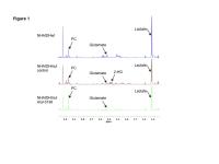
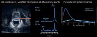
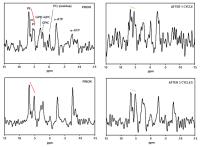
Early detection of changes in phospholipid metabolism during
neoadjuvant chemotherapy using phosphorus magnetic resonance
spectroscopy at 7 tesla
Erwin Krikken1, Wybe J.M. van der Kemp1,
Hanneke W.M. van Laarhoven2, Dennis W.J. Klomp1,
and Jannie P. Wijnen1
1Radiology, University Medical Center Utrecht,
Utrecht, Netherlands, 2Medical
Oncology, Academic Medical Center Amsterdam, Amsterdam,
Netherlands
Neoadjuvant chemotherapy plays an important role in the
treatment of breast cancer patients. During chemotherapy,
the phospholipid metabolism changes which can be measured by 31P-MRS
at 7 tesla. Eight patients were examined, using the AMESING
sequence to receive metabolic signals in the tumor. The 31P-MRS
data were analyzed on group level, which enables the
detection of changes the levels of phospholipid metabolites
in an early stage of the treatment, directly after the first
cycle of chemotherapy.
|
|
2811.
 |
92 |
The Role of Heterogeneity Analysis for Differential Diagnosis in
Diffusion-Weighted Images of Meningioma Brain Tumors
Mojtaba Safari1, Anahita Fathi Kazerooni1,2,
Maryam Babaie3, Mahnaz Nabil4, Mahsa
Rostamie1, Parvin Ghavami1, Morteza
Saneie Taheri3, and Hamidreza Saligheh Rad1,2
1Quantitative MR Imaging and Spectroscopy Group
(QMISG), Research Center for Molecular and Cellular Imaging
(RCMCI), Tehran University of Medical Sciences, Tehran,
Iran, 2Department
of Medical Physics and Biomedical Engineering, School of
Medicine, Tehran University of Medical Sciences, Tehran,
Iran, 3Radiology
Department, School of Medicine, Shahid Beheshti University
of Medical Sciences, Tehran, Iran, 4Department
of Mathematics, Islamic Azad University, Qazvin Branch,
Qazvin, Iran
Meningioma brain tumors constitute the majority of adult
primary brain tumors, in which the role of apparent
diffusion coefficient (ADC) is controversial. We hypothesize
that analysis of the heterogeneity within a tumorous
ecological region can reveal biological tissue properties,
which could further assist decision making about the optimum
patient-specific treatment strategy. In the present work, we
propose an automated computer-aided diagnosis method for
phenotyping meningioma brain tumors, based on features
representing spatial heterogeneity in ADC-maps, with
classification accuracy of 85.1%. In conclusion, it is
demonstrated that heterogeneity of meningioma brain tumors
can be a potential discriminating biomarker of tumor
malignancy.
|
|
2812.
 |
93 |
Microvascular Heterogeneity Assessed Using DCE-MRI Predicts
Disease-Free Survival in Cancers of the Cervix, Bladder, and
Head and Neck
Ben R Dickie1,2, Lucy E Kershaw1,2,
Bernadette M Carrington3, Suzanne Bonington3,
Susan E Davidson3, Catharine ML West1,
and Chris J Rose4
1Institute of Cancer Sciences, The University of
Manchester, Manchester, United Kingdom, 2Christie
Medical Physics and Engineering, Christie NHS Foundation
Trust, Manchester, United Kingdom,3Diagnostic
Radiology, Christie NHS Foundation Trust, Manchester, United
Kingdom, 4Centre
for Imaging Sciences, The University of Manchester,
Manchester, United Kingdom
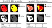
There is a clinical need for non-invasive imaging biomarkers
capable of accurately predicting outcomes in locally
advanced cancers. Microvascular heterogeneity measurements
obtained from dynamic contrast enhanced-MRI have shown
prognostic utility however no attempt has been made to
compare the prognostic value of the available methods across
disease and identify which type of heterogeneity
(statistical or spatial) is important for survival. In this
study we identify heterogeneity biomarkers that are
universally prognostic across cancers of the cervix,
bladder, and head and neck and compare their
prognostic value to standard clinicopathologic factors such
as disease stage.
|
|
2813.
 |
94 |
Oscillatory shear strain impacts metastatic cancer cell spread
Marlies Christina Hoelzl1, Marco Fiorito2,
Ondrej Holub3, Gilbert Fruhwirth4, and
Ralph Sinkus1
1Biomedical Engineering, King's College London,
London, United Kingdom, 2Imaging
Chemistry and Biology, King's College London, London, United
Kingdom, 3London,
United Kingdom, 4Imaging
Chemistry and Biology, King's College London, Lodnon, United
Kingdom
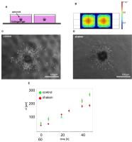
Major reasons of cancer related deaths are repercussion of
the dissemination of cancer cells from the primary tumour
site and an outgrowth at the secondary metastatic site. The
microenvironment where the cancer cells reside with various
signals, are central factors to provide cancer cell spread
throughout the body; signals can be (bio)chemical or
mechanical nature. Translation of mechanical forces,
displacements and deformations into biochemical signals
(i.e. mechanotransduction) affects their adhesion, spread
and survival. We show here, that focussed shear waves
operating at specific frequency and amplitude affects the
metastatic behaviour of cancer cells by reducing the
invasive behaviour and growth.
|
|
2814.
 |
95 |
Can Diffusion Weighted MRI Assess Early Response of
Lymphadenopathy to Induction Chemotherapy in Nasopharyngeal
Cancer: A Heterogeneity Analysis Approach
Manijeh Beigi1, Anahita Fathi Kazerooni1,
Mojtaba Safari2, Marzieh Alamolhoda3,
Ahmad Ameri4, Shiva Moghadam5, Mohsen
Shojaee Moghadam6, and Hamidreza SalighehRad2
1, Tehran University of Medical Sciences,
Quantitative MR Imaging and Spectroscopy Group, Research
Center for Cellular and Molecular Imaging, Institute for
Advanced Medical Imaging, Tehran, Iran,2Tehran
University of Medical Sciences, Quantitative MR Imaging and
Spectroscopy Group, Research Center for Cellular and
Molecular Imaging, Institute for Advanced Medical Imaging,
Tehran, Iran,3Statistics, Shiraz University of
Medical Science, Shiraz, Iran, 4Jorjani
Radiotherapy Center, Shahid Beheshti of Medical Sciense,
Tehran, Iran, 5Shahid
Beheshti University of Medical Science, Tehran, Iran,6Payambaran
MRI center, Tehran, Iran
Induction chemotherapy is an effective way to control
subclinical metastasis in locally-advanced nasopharyngeal
cancer patients. Diffusion-weighted MRI is a noninvasive
imaging technique allowing some degree of tissue
characterization by showing and quantifying molecular
diffusion. Histogram analysis on ADC map could be carried
out to reveal physiological alterations early after IC. For
this purpose, several quantitative metrics from ADC-map were
explored to obtain the most accurate feature(s) as potential
predictive biomarker for early response of the lymphnode to
IC. If the outcome can be predicted at an early stage of the
treatment, the patient could be spared from unnecessary
treatment toxicity.
|
|
2815.
 |
96 |
Monitoring Changes of the Tumor Microenvironment Following
Administration of a Novel Vascular Disrupting Agent OXi6197
Using Multi-parametric MRI
Heling Zhou1, James Campbell1, Zhang
Zhang2, Debabrata Saha2, Rebecca
Denney1, Mary Lynn Trawick3, Kevin G
Pinney3, and Ralph P Mason1
1Radiology, UT Southwestern Medical Center,
Dallas, TX, United States, 2Radiation
Oncology, UT Southwestern Medical Center, Dallas, TX, United
States, 3Chemistry
and Biochemistry, Baylor University, Waco, TX, United States
Vascular disrupting agents (VDAs), selectively damage the
endothelial cells of tumor blood vessels, inducing ischemia
and consequent hypoxia and cell death. We investigated the
impact of a novel indole-based VDA (OXi6197) to tumor
perfusion and oxygenation using multi-parametric MRI on a
lung tumor animal model. DCE MRI showed decreased blood flow
after administration of VDA. Oxygen sensitive MRI, BOLD and
TOLD, showed progression of hypoxia at 24 hours.
Multimodality imaging provides useful information to
evaluate the efficacy of VDA. The findings in this study
will be important for dose optimization and potential
combination therapy in the future.
|
|