ISMRT Poster Presentations
ISMRT Clinical Posters
ISMRM & ISMRT Annual Meeting & Exhibition • 04-09 May 2024 • Singapore

| 17:00 |
5122.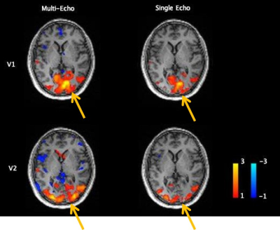 |
MULTI-ECHO FUNCTIONAL IMAGING ON AN ULTRA HIGH-PERFORMANCE
HEAD-ONLY GRADIENT SYSTEM
Gail Helene Kohls1,
Nastaren Abad2,
H Douglas Morris1,
Mauren N Hood1,
James Kevin Demarco1,
and Thomas TK Foo2
1Radiology, USU/WRNMMC, Bethesda, MD, United States, 2GE Global Research, Niskayuna, NY, United States
Motivation: This abstract focuses on educating on
how high-gradient MRI systems can utilize the multi-echo
functional MRI (ME-fMRI) techniques. Goal(s): We explain the blood-oxygen-level-dependent (BOLD) signal technique and how additional echoes can improve the signal fidelity. Approach: The ME-fMRI, using three or more echoes allow for the pixelwise T2* decay to be modeled, and as BOLD contrast is a function of T2* evolution over time, an experiment sampling the voxel-wise T2* signal decay can be used to separate BOLD from artifact signal constituents. Results: Gradient systems with ultra-rapid slew rates (> 400 T/m/s) allow ME-fMRI to reduce artefacts from flow, motion, and susceptibility effects. Impact: MRI systems with ultra-high gradient systems can reduce artefacts from flow, motion, and susceptibility effects in the BOLD contrast technique using an ME-fMRI technique with three or more echoes to improve fidelity of fMRI. |
| 17:00 |
5123.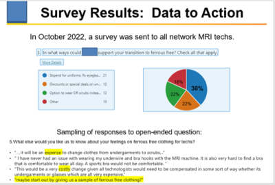 |
GOT SAFETY?? Our journey into creating, embracing and
growing a culture of MRI safety excellence!
Jean Ellis-Land1,
Katherine Sedler2,
and Monica Weiser3
1MRI, SLUHN, Harleysville, PA, United States, 2MRI, SLUHN, Lehighton, PA, United States, 3MRI, SLUHN, Coaldale, PA, United States Motivation: The ever-evolving complexities of implants, clothing and technology in MRI, combined with the increased recent reported safety incidents around the world, motivated us to reinvent our safety program. Goal(s): Our goal was to create and institute a robust, multifaceted safety program that provides awareness into how each of us contributes to a safe environment in the MRI suite. Approach: We assessed needs through surveys and observation. Support and education are provided via weekly emails, site visits, quarterly meetings, and clearly written policies. Results: Greater technologist engagement through increased incident documentation, adoption of a safer workflow, and greater participation at safety meetings.
Impact: Got Safety?? |
| 17:00 | 5124. |
Play therapy and non-sedative strategies for supplementary
MR examination in children aged 3-7 years
Yin Ting Chiu1,
Mei Yu Poon1,
Kwok Chun Wong1,
Kin Fen Kevin Fung1,
Yee Ling Elaine Kan1,
and Wing Kei Carol Ng1
1Department of Radiology, The Hong Kong Children's Hospital, Hong Kong, Hong Kong Keywords: Motivation: MRI scans in young pediatric patients are challenging due to motion sensitivity and anxiety triggers yet its high contrast resolution and radiation-free make it pivotal especially in directing an appropriate treatment plan. Goal(s): Identify key factors for successful sedation-free supplementary MRI scans in children, focusing on a child-friendly environment, trust-building, communication, tailored techniques, and parental support. Approach: The MR team of the Hong Kong Children's Hospital implemented strategies like child-friendly rooms, audio-visual systems, child-life specialists, optimized protocols, shorter scan times, and parental accompany.
Results: Achieved high success rates with
improved patient experience, shorter procedures, and
better MRI facility utilization. Impact: With the increasing demand for MRI, the efficient use of MR suits has become a global issue. Introducing non-sedating MR scanning skills for young pediatrics could save both preparation and table time and therefore boost the scanning efficiency. |
| 17:00 | 5125. |
Remote assist in a single site MRI department.
Jack Jaspers1
1LUMC Leiden, Leiden, Netherlands Keywords: Motivation: A worldwide increase of MRI staff shortage and at the same time an increasing number of MRI requests. Goal(s): Deployment of remote assist performed by properly trained MRI staff in a single site MRI department. Approach: After a test period, remote assist was evaluated based on a survey conducted among the MRI staff members. Results: Due to slow technical performance of the remote assist hardware and software and a normal patient schedule during the test period, the first experience was disappointing. A new test period will be planned where patient scheduling and technical performance will be tested and evaluated again. Impact: Technical performance of the remote assist hardware and software (direct respond required) and patient scheduling need further improvement. |
| 17:00 | 5126. |
Dynamic Contrast-Enhanced MR Lymphangiogram of the Central
Lymphatic System in Children: Overcoming the challenges in a
non-hybrid environment
Mei Yu Poon1,
Chi Yan Iris Chung1,
Kin Fen Kevin Fung1,
Wing Kei Carol Ng1,
and Yee Ling Elaine Kan1
1Radiology, Hong Kong Children’s Hospital, Hong Kong SAR, Hong Kong Keywords: Motivation: Performing dynamic contrast-enhanced MR lymphangiogram (DCMRL) in children require general anesthesia. In a non-hybrid environment, the nodal puncture for DCMRL is often performed in the angiography suite. Transporting a pediatric patient under general anesthesia with needles to an MRI environment presents unique challenges. Goal(s): Develop a safe and effective workflow for DCMRL in non-hybrid setting. Approach: Meticulous planning and collaborations between radiology and anesthesiology teams ensure safe execution of DCMRL. Using contrast-enhanced ultrasound (CEUS) also improve the logistic of intranodal DCMRL by minimizing patient transfer. Results: Careful coordination between Radiology and Anesthetic team results in successful and safe intranodal and intrahepatic DCMRL. . Impact: The ability to perform DCMRL in non-hybrid environment has significant implications for accurate diagnosis of lymphatic diseases, enabling improved treatment planning and better patient outcomes. |
| 17:00 | 5127. |
To evaluate the role that MR Spectroscopy and MR Perfusion
play in characterizing breast masses on MR Mammograms with
HPE correlation.
Abhishek Sehrawat1
1Radiodiagnosis & Imaging, All India Institute of Medical Sciences, Bhopal, India Motivation: To evaluate the role that MR Spectroscopy and MR Perfusion play in characterizing breast masses on MR Mammograms with HPE correlation. Goal(s):
Approach: 160 patients were included in
prospective research design for eight months.
Results: MRI diagnosis and histopathological
results correlated in 86.1% patients. Dynamic Contrast
Enhanced MR perfusion kinetics have been highly
sensitive in distinguishing benign and malignant
lesions. Impact: MR mammography has been demonstrated that MR Perfusion is extremely sensitive in identifying benign from malignant breast lesions. In order to provide sufficient information to differentiate between breast tumors, MR Spectroscopy has proven useful adjunct to MR Perfusion. |
| 17:00 | 5128. |
How to deal with MRI artefacts
Catherine Wangui muchuki1
1Radiology, Kenyatta National Hospital, Nairobi, Kenya Motivation: Artefacts are features that appear in an image not present in the original object.The study was informed by the various artefacts encountered during scanning.
Goal(s): Different artefacts occured during
scanning affecting the diagnostic quality ,classified
as patient related, signal processing and hardware
related. Approach: Information was collected on patients undergoing scanning on 3T Philip ingenia machine recording all artefacts. The consent form was used as a measure of accuracy of patient information. Results: Artefacts noted were motion artefacts, metallic and orthopaedic. Various methods were applied of elimination including field of view, spin echo sequences, short echo time and use of oversampling techniques. Impact: knowledge of different types of artefacts and their origin was necessary to eliminate them to reduce their negative influence on MR images by adjusting acquisition parameters. |
| 17:00 |
5129.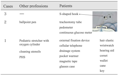 |
Incident reports of bringing ferromagnetic objects into the
magnetic resonance imaging room in the past 10 years
Miho Uemura1,
Yoshihiro Akatsuka1,
Mitsuhiro Nakanishi1,
Keishi Ogura1,
and Osamu Asanuma1
1Division of Radiology and Nuclear Medicine, Sapporo Medical University Hospital, Sapporo, Japan Motivation: The fatal accidents in the MRI room are caused by the bringing of ferromagnetic objects. It is necessary to identify the trends in incidents. Goal(s): Our goal was to investigate the relationship between the frequency of bringing ferromagnetic objects and years of MRI experience. Approach: We compiled incident reports for the past 10 years, extracted reports related to the bringing of ferromagnetic objects. Results: There were 26 reports of ferromagnetic objects brought introduced, and half of the reports were from technologists with less than one year of MRI experience. Impact: In order to prevent the adsorption accidents of the ferromagnetic objects, it is important to understand the causes of such incidents and the characteristics of our own facilities. Sharing this survey will lead to the prevention of similar incidents. |
| 17:00 |
5130. |
Usefulness of Compress Sense Technique With 1.5T Magnetic
Resonance Hydrography of inner ear: Comparison of Image
Quality and Acquisition Time
cheng tang1,
jianjian huang2,
peng peng1,
shunzu lu1,
yiwu lei1,
huiting zhang3,
and chen zhao3
1THE FIRST AFFILIATED HOSPITAL OF GUANGXI MEDICAL UNIVERSITY, nanning, China, 2Guangdong Sencond Provincial General Hospital, guangzhou, China, 3MR Scientific Marketing, Siemens Healthnieers, wuhan, China Keywords: Motivation: Minimize scanning time while ensuring the quality of MRH images. Goal(s): This study investigated the feasibility of using compressed perception acceleration sequences to evaluate the quality of MRH images. Approach: We compared data from traditional sequences and CS sequences with different acceleration factors. Results: The results showed that CS4 can guarantee diagnostic requirement with 44%time reduction. Conventional images were more commonly used than CS SPACE images for radiologists. However, CS4 SPACE images still get more than 3-point. This suggests that CS4 SPACE images is still considered acceptable. Impact: This preliminary study showed that for the MRH of inner ear, T2 SPACE sequence with 4-fold CS technique provided similar image quality to conventional T2 SPACE with 44% shorter scan time. Therefore, we can save a lot of examination time. |
| 17:00 | 5131. |
Perspective on planning PET-MR protocols for research
practices and long-term goals
Shufen Zheng1
1Clinical Imaging Research Centre, National University Singapore, Singapore, Singapore Keywords: Motivation: Even though PET-MR has been around for more than a decade, it is not highly available in all imaging centers due to high cost and complexity. Thus, seeking experience in setting up PET-MR protocols is challenging and can be quite daunting for first-timers and scans involving new tracers. Goal(s): To create an informative checklist which can ease planning a PET-MR protocol based on clinical experience and from literature review. This aim to overcome pitfalls and include considerations in setting up PET-MR protocols. Approach: Systematic planning of PET-MR protocol. Results: PET-MR radiographers and researchers will be able to plan their protocols efficiently and effectively. Impact: This informative checklist can assist planning a PET-MR protocol on scanner from scratch. Using this guide, the PET-MR radiographers and researchers will become more confident and efficient in planning the PET-MR scanning protocols. This can also boost data-sharing among others. |
| 17:00 | 5132. |
Value of Compressed Sensing in Non-Contrast Enhanced
Coronary Magnetic Resonance Angiography in Elderly Patients
yue jiang1,
wenjing liu1,
yong yuan1,
guangming lu2,
tong chen3,
weibo chen3,
dongsheng jin1,
and yan e zhao1
1jiangsu province official hospital, Nan jing, China, 2general hospital of eastern theater command, nan jing, China, 3philips healthcare, shanghai, China Keywords: Motivation: Coronary artery examination in elderly patients with low renal function and poor respiratory coordination has always been a clinical pain point. Goal(s): Explore the feasibility of balanced steady-state free precession BTFE sequence for non-drug coronary imaging and find the optimal compressed sensing coefficient under this sequence Approach: Acquire cs4, 6, and 8 images under BTFE sequence, record acquisition time and analyze objective indicators and subjective scores Results: CS6 can achieve relatively high imaging quality in a short time Impact: Our study may inform efforts to more rapidly perform non-contrast enhanced coronary MRI angiography in elderly patients. |
| 17:00 |
5133. |
Native T1 Mapping in Post-COVID Vaccine-related Myocarditis
in a paediatric population in 3T: The Singapore Paediatric
Hospital’s experience.
Ker Sin Tan1,
Chee Chung Au1,
and Marielle Valerie Fortier1
1Diagnostic and Interventional Imaging, KK Women's and Children's Hospital, Singapore, Singapore Motivation: To present our experience with native T1 mapping in Post-COVID Vaccine-related myocarditis. Goal(s): To evaluate paediatric native T1 mapping values in Post-COVID Vaccine-related myocarditis Approach: Data was collated over August 2021 - August 2023 in 15 patients (13 boys and 2 girls), aged 9 to 16 years old. Patients were scanned in a 3 Tesla MRI scanner. Results: Majority of T1 values ranged 1201-1250ms. 17 segments had T1 values above 1250ms which correlated with LGE, (mostly in the basal region), ranging 1256ms – 1606ms. Thus, we perceived the normal range of T1 values to be 1100ms-1250ms and those above as increased T1 values. Impact: Our study evaluated the characteristics of post-COVID vaccine-related myocarditis, a relatively new disease, in children aged 9 to 16 years-old using native T1 Mapping at 3T MRI, filling up the current gap in data in paediatric myocardial native T1 values |
| 17:00 |
5134.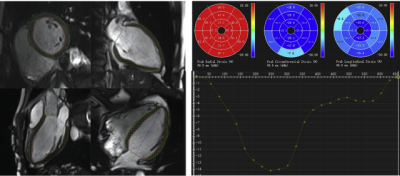 |
Feasibility of single-shot compressed sensingcine imaging in
arrhythmia patients
Nan Zhang1,
Fan Du1,
and Xiuzheng Yue2
1Zhongshan Hospital of Fudan University, Shanghai, China, 2Philips Healthcare, Beijing, China Motivation: The quality of cardiac film sequences in patients with arrhythmia is poor, which affects the clinical diagnosis. Goal(s): To observe the value of single-shot compressed sensing cine imaging inevaluating cardiac structure and function in arrhythmia patients Approach: MRI;ICC;Kappa Results: n arrhythmia group, there was statistical difference of myocardial thickness in 12 myocardial segments between the 2 sequences,as well as peak and average values of myocardial radial and circumferential strain. Impact: For arrhythmia patients, single-shot compressed sensing cine imaging cine sequence could improve image quality of cardiac MRI. |
| 17:00 |
5135.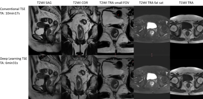 |
The Value of Deep Learning Reconstruction In Improving the
Image Quality of rectum MRI Images
Sijie Hu1,
Yueluan Jiang2,
and Nickel Marcel Dominik3
1Department of Diagnostic Radiology, National Clinical Research Center for Cancer/Cancer Hospital, Chinese Academy of Medical Sciences and Peking Union Medical College, Beijing, China, 2MR Research Collaboration, Siemens Healthineers, Beijing, China, 3MR Application Predevelopment,, Siemens Healthineers AG, Erlangen, Germany Keywords: Motivation: TSE sequences are crucial for rectum MRI, but have limitations. DL-TSE is expected to improve image quality and reduce acquisition time for rectum MRI. Goal(s): To assess the viability of employing TSE sequences with deep learning reconstruction for rectal MRI when compared to conventional TSE sequences.
Approach: This study included 16 patients with
colorectal cancer confirmed by pathology. SNR and CNR
were analyzed by SPSS 22.0 software.A P-value below 0.05
was considered statistically significant. Results: The results show that the application of deep learning can shorten the scanning time while maintaining high image resolution, and improve the diagnostic efficiency of rectal diseases. Impact: Deep learning reconstruction of TSE sequence in rectal MRI has the advantages of shortening acquisition time, improving image quality, and improving diagnostic efficiency. DL-TSE may also be extended to MRI examinations of other organs, such as the prostate and pelvis. |
| 17:00 | 5136. |
Denoising Approaches by Artificial Intelligence in PET MRI
for clinical routine application
MARCO DE SUMMA1
1Diagnostic Imaging, Oncological Radiotherapy and Hematology, Policlinico Universitario Agostino Gemelli, ROME, Italy Motivation: Two big problems encountered in hybrid MR PET exams are: long duration of the exams and the optimization of the administered activity. Goal(s): I tried to evaluate the feasibility of decreasing the time and dose using an artificial intelligence tool in reconstruction while preserving the performance of PET and MR. Approach: By analyzing the literature, it was possible to identify the optimal reconstruction strategies for PET and MR imaging that utilize artificial intelligence to save dose and time. Results: Deep learning techniques have made significant advances in data reconstruction images from examinations with low scan times or radiopharmaceutical dose Impact: Artificial intelligence in MR PET is a promising approach. The impact on the health of patients is undeniable, especially in the paediatric population. This approach reduces the dose and consequently the cost of radiopharmaceuticals and increases productivity and efficiency. |
| 17:00 |
5137.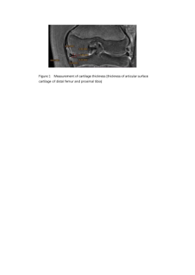 |
Measurement and analysis of objective indicators of
ossification development in MRI of knee joint
huan Wang1,
xinyu li2,
xiaoqian jia2,
Tianze Wang3,
and Jianxin Guo2
1the First Affiliated Hospital of Xi'an Jiaotong University, Xi'an,, China, 2the First Affiliated Hospital of Xi'an Jiaotong University, Xi'an, Shaanxi province, China, 3the First Affiliated Hospital of Xi'an Jiaotong University, xi'an, China Keywords:
Motivation:
Goal(s): To analyze the predictive value of MRI
ossification development of knee joint with gender and
age.
Approach: The indexes representing epiphyseal
development of distal femur and proximal tibia were
selected for measurement, and the differences of
epiphyseal development of distal femur and proximal
tibia between different gender and different age groups
were explored. Results: Males performed better on the distal femur and females on the proximal tibia for age prediction, which could be evaluated separately if necessary. Impact: The objective indicators of ossification development in MRI of knee joint have certain predictive value with gender and age, which helps to evaluate bone age objectively and accurately, and provides a new idea for epiphysis evaluation |
| 17:00 |
5138.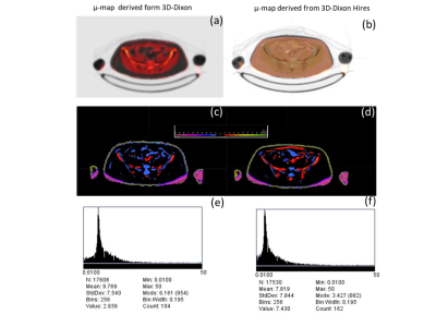 |
The Hidden Challenge: Unraveling the Reliability of Pelvic
Attenuation Maps in Simultaneous PET/MRI
Monchai Phonlakrai1,
Chidchanok Chusuwon1,
Pohnpan Kampipak1,
Paramest Wongsa1,
Phornpailin Pairodsantikul1,
and Attapon Jantarato 2
1School of Radiological Technology, Chulabhorn Royal Academy, Bangkok, Thailand, 2National Cyclotron and PET Centre, Chulabhorn Hospital, Bangkok, Thailand Motivation: PET/MRI integrate functional and anatomical information, elevating diagnostic precision. However, a key challenge lies in ensuring the reliability of attenuation (µ) map generation. Goal(s): This research assesses the accuracy of PET/MR-based µ-map from 3D-Dixon and 3D-Dixon Hires T1W in comparison to a standard µ-map from PET/CT at the voxel level. Approach: the µ-map data from 15 patients who underwent both PET/CT and PET/MRI of the pelvic region were analyzed to quantify the disparities in the generated µ-maps. Results: We found that 3D-Dixon Hires outperforms the 3D-Dixon in the creation of µ-maps, rendering it a superior choice for precise attenuation correction. Impact: Spatial resolution influences the accuracy of attenuation correction in PET/MRI, particularly when employing quantitative methods for diagnosis. Notably, the utilization of higher-resolution Dixon MRI images enhances the reliability of attenuation in tissue compartmental models for the generation of this map. |
| 17:00 |
5139.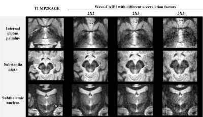 |
Evaluation of wave-CAIPI for accelerating MP2RAGE and FLAWS
in deep-brain nucleus localization
Cuiliu Liu1,
Shuyue Wang1,
Yunzhu Wu2,
Wei-Ching Lo3,
Daniel N. Splitthoff4,
Peiyu Huang1,
and Jianzhong Sun1
1Department of Radiology, Second Affiliated Hospital of Zhejiang University School of Medicine, Hangzhou, China, 2MR Research Collaboration Team, Siemens Healthineers Ltd., Shanghai, China, 3Siemens Medical Solutions, Boston, MA, USA, Boston, MA, United States, 4Siemens Healthcare, Erlangen, Germany Keywords: Motivation: We tested the acceleration method in a clinical setting demanding high-resolution and high-quality structural images. Goal(s): Deep -brain stimulation requires high-resolution 3D MRI sequences. The Wave CAIPIRINHA (Wave-CAIPI) may be expected to significantly reduce acquisition time while changing tissue contrast. Approach: We collected scanning data from 5 healthy volunteers, including conventional MP2RAGE and Wave CAIPI research applications (with acceleration factors of 2x2, 2x3, and 3x3, respectively). Results: Compared with MP2RAGE, the Wave-CAIPI (2Xx2) and Wave-CAIPI (2xX3) sequences provide good tissue contrast while shortening acquisition time. Impact: This new sequence is helpful to the localization of brain structures for pre-surgical planning. |
| 17:00 |
5140.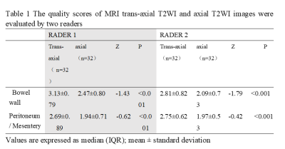 |
Optimized MRI workflow in colon cancer scan
SHA LIU1,
NAN SUN1,
ZHEN GUAN1,
XIAO TING LI1,
YU XIN YANG2,
and KE XUE2
1Peking University Cancer Hospital & Institute, BEI JING, China, 2MR Collaboration, United Imaging Research Institute of Intelligent Imaging, BEI JING, China Keywords: Motivation: Torso enhanced CT is the standard method for colon cancer staging. MRI is a complimentary method. Our purpose is to develop a MR Colon cancer scanning method with simple process and short scan time.
Goal(s): develop a new T2WI
breath-hold scanning technique that eliminates the need
for bowel preparation and reduces scan time
Approach: compared the image quality between trans-axial T2WI and axial T2WI in visualization of anatomical structure Results: Patient examination time for our colon MRI is less than 10min, compared with conventional MRI, our approach offered a simplified and expedited means of visualizing lesion details with superior clarity. Impact: Our research focused on MRI scan optimization. Without bowel preparation, using breath-hold, our creative trans-axial imaging plan becomes highly manageable and enables efficient repetition of the entire process. |
| 17:00 |
5141.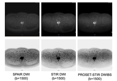 |
Enhanced Fat Suppression Effect by Integrating STIR DWI with
PROSET
Susumu Takano1,
Katsuhiro Watanabe1,
Makoto Obara2,
Masatoshi Honda2,
Yasumoto Katsumata2,
and Taro Takahara3
1Department of Radiology, Tokai University Hospital, Kanagawa, Japan, 2Medical Systems, Philips Electronics Japan, Tokyo, Japan, 3Department of Biomedical Engineering, Tokai University School of Engineering, Kanagawa, Japan Motivation: In this study, the motivation was to improve fat signal suppression in Diffusion-weighted Whole Body Imaging with Background Body Signal Suppression (DWIBS). Goal(s): The specific goals were to evaluate the fat suppression effect of the Principle of Selective Excitation Technique (PROSET) in combination with STIR DWI (PROSET-STIR DWI) compared to conventional methods. Approach: The approach involved scanning healthy volunteers using PROSET-STIR DWI, STIR DWI and SPAIR DWI. Results: The results revealed that PROSET-STIR DWI achieved superior fat suppression for Methylene signals compared to conventional methods. Overall, PROSET-STIR DWI demonstrated enhanced fat suppression, highlighting its potential in clinical applications. Impact: The PROSET-STIR DWI technique demonstrates superior fat suppression compared to conventional methods, with notable differences in Methylene fat signals. This advance can benefit medical imaging, potentially improving cancer detection and diagnostic accuracy. |
| 17:00 |
5142.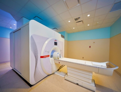 |
Operating MRI 7 Tesla Ultra High Field (UHF) in Clinical and
Research Practice
Lien M Phan1,
Krista R Runge1,
Steve H Fung2,
and Christof Karmonik1
1Translational Imaging Center, Houston Methodist Hospital Research Institute, Houston, TX, United States, 2Radiology, Houston Methodist Hospital, Houston, TX, United States Motivation: Clinical and research patients can be imaged safely at 7T while providing improved quality and resolution enabling better detection of lesions while maintaining a safe working environment for our patients and technologists. Goal(s): The goal is to guide new operator of MRI 7T on how to prepare themselves and patient for advance imaging exam. Approach: Informative approaches for equipment, safety, side effects of 7T MRI for patients and technologist. Results: Operating a 7T MRI is similar to other field strengths, but technologist should be well trained and understand the environment in UHF scanners. Impact: 7T MRI is an advance imaging technology that is very beneficial for patient care. This guidelines will provides MRI technologist some insight on the equipment, safety, and how to prepare the patient for 7T MRI studies. |
| 17:00 |
5143.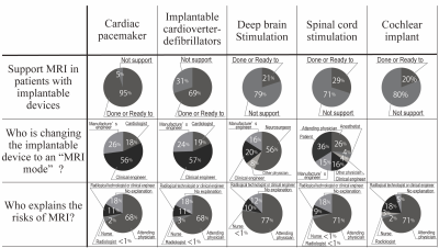 |
Investigation of Conducting for MRI for Patients with
Implantable Medical Devices and Items included in the Metal
Safety Checklist in Japan
Kousaku Saotome1,
Kunihiro Yabe2,
Kosuke Morita3,
Toshiki Tateishi4,
Tsutomu Kanazawa5,
and Tsukasa Doi6
1Department of Radiological Sciences, School of Health Sciences, Fukushima Medical University, Fukushima, Japan, 2Yamagata Prefectural Shinjo Hospital, Shinjo-shi, Japan, 3Kumamoto University Hospital, Kumamoto-shi, Japan, 4University of Fukui Hospital, Fukui-shi, Japan, 5Niigata University Medical & Dental Hospital, Niigata-shi, Japan, 6Kouseikai Takai Hospital, Tenri-shi, Japan Motivation: To improve the safety of Magnetic Resonance Imaging for Patients with Implantable Medical Devices in Japan. Goal(s): To Investigate of Conducting for Patients with Implantable Devices and Items included in the Metal Safety Checklist. Approach: A questionnaire survey was conducted at medical facilities that perform MRI examinations of patients with implantable devices. Results: Conducting MRI for Patients with Implantable Medical Devices and Items in the Metal Safety Checklist revealed variations. Impact: This study advances the discussion of Conducting for Magnetic Resonance Imaging for Patients with Implantable Medical Devices and the need to create a template for the Metal Safety Checklist in Japan. |
| 17:00 | 5144. |
MRI characterization of a giant benign adipose tissue
neoplasm of the foot
Khomotso Paulina Motiang 1
1Radiography, Sefako Makgatho Health Science University, Hartbeespoort, South Africa Motivation: MRI is the best imaging modality due to its excellent resolution and contrast differentiation of soft tissue. However, MRI is scarce and lengthy, yielding low patient throughput and delays in the diagnosis and management of health conditions. It is crucial to have an appropriate imaging protocol to minimize delays in the management of any health conditions for improved health outcomes and quality of life. Goal(s): To advocate for early diagnosis and management of health conditions for improved quality of life in developing countries. Approach: Case study Results: MRI showed a thin encapsulated lesions with a lobular pattern, which were in line with lipoma. Impact: Appropriate MRI protocols will minimize unnecessary sequences and improve patient throughput and early diagnosis for improved health outcomes in developing countries. |
| 17:00 |
5145.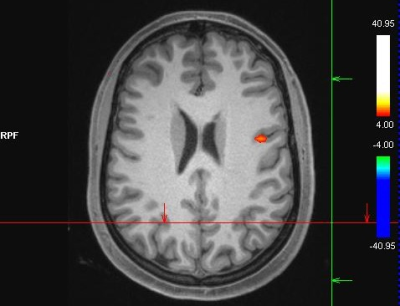 |
The Significance of fMRI in Surgical Planning for Tumour
Resection and Epilepsy Surgery: Optimal Language Paradigms
for Language Area Mapping
Shiami Luchow1
1Hunter Medical Research Institute, New Lambton Heights, Australia Motivation: The Significance of fMRI in Surgical Planning for Tumour Resection and Epilepsy Surgery: Optimal Language Paradigms for Language Area Mapping Goal(s): This paper explores the importance of fMRI in surgical planning and the most effective language paradigms for language area mapping. Approach: fMRI allows neurosurgeons to pinpoint critical brain regions responsible for language, motor function, and sensory perception. This information is pivotal in minimizing postoperative neurological deficits Results: For epilepsy surgery, it aids in delineating the epileptogenic zone as data clearly shows increase in seizure freedom with detailed imaging identifying the true extent of the lesion. Impact: Resource for radiographers on how to perform a fMRI examination, sequence parameters and corresponding language paradigms. |
The International Society for Magnetic Resonance in Medicine is accredited by the Accreditation Council for Continuing Medical Education to provide continuing medical education for physicians.