ISMRT Oral
Winning Research Poster Awards
ISMRM & ISMRT Annual Meeting & Exhibition • 04-09 May 2024 • Singapore

| 18:00 |
5181. |
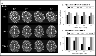
Improving Image Quality Using the Pause Function Combination
to PROPELLER Sequence in Brain MRI: a brain phantom study
Kousaku Saotome1,
Koji Matsumoto2,
Yoshiaki Kato3,
Yoshihiro Ozaki4,
Motohiro Nagai3,
Tomoyuki Hasegawa5,
Hiroki Tsuchiya6,
and Tensho Yamao1
1Department of Radiological Sciences, School of Health Sciences, Fukushima Medical University, Fukushima, Japan, 2Chiba University Hospital, Chiba, Japan, 3Kameda General Hospital, Kamogawa-shi, Japan, 4Meiwa Hospital, Nishinomiya-shi, Japan, 5Hitachinaka General Hospital, Hitachinaka-shi, Japan, 6QST Hospital, Anagawa-shi, Japan Motivation: Finding ways to use the PROPELLER sequence as an even more the robustness method to motion. Goal(s): This study investigates whether repositioning the head after pausing during PROPELLER imaging, enhances image quality. Approach: This study investigated whether image quality improved with repositioning the head after a pause during PROPELLER method with a brain phantom and driver system. Results: We found that image quality improved at all rotational angles and that pausing multiple times was effective depending on the frequency of motion. Impact: Incorporating a pause function into the PROPELLER method is expected to be clinically applicable as a practical means to further improve the robustness of the PROPELLER method to motion. |
| 18:10 |
5183. |
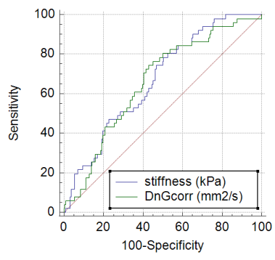
Fat-corrected non-Gaussian diffusion MRI in a non-alcoholic
fatty liver disease: diagnostic performance for liver
fibrosis
omaima said1,
Sabrina Doblas1,
Gwenaël Pagé1,
Dominique Valla1,2,
Valérie Paradis1,2,
Bernard Van Beers1,2,
and Philippe Garteiser1
1INSERM, Paris, France, 2Beaujon University Hospital, Paris, France Motivation: In nonalcoholic fatty liver disease (NAFLD), hepatic fibrosis is strongly associated with patient survival. Diffusion MRI has been proposed to assess liver fibrosis, but this evaluation is hampered in hepatic steatosis. Goal(s): Our goal was to evaluate the diagnostic performance of non-Gaussian diffusion MRI in assessing liver fibrosis in 250 patients with NAFLD. Approach: We developed a method to calculate the non-Gaussian diffusion coefficient, based on non-linear regression and fat correction. Results: With this corrected diffusion method, NAFLD patients with liver fibrosis could be differentiated from patients without it. Impact: With this corrected diffusion method, NAFLD patients with liver fibrosis could be differentiated from patients without it. |
| 18:20 |
5182. |
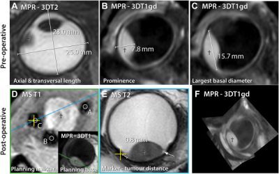
Comparison of Magnetic Resonance Imaging–Based and
Conventional Measurements for Proton Beam Therapy of Uveal
Melanoma
Guido Rene van Haren1,
Teresa Goncalves Ferreira2,
Jan Willem Beenakker3,
and Myriam Jaarsma-Coes1
1Radiology, LUMC Leiden, Leiden, Netherlands, 2Radiology, LUMC, Leiden, Netherlands, 3Radiology and ophtalmology, LUMC, Leiden, Netherlands Keywords: Motivation: Proton therapy is a relatively new option for treatment of uveal melanomas. For adequate treatment, accurate measurement of tumour dimensions and tumour-marker distance is essential. The dedicated MRI protocol for uveal melanoma patients developed in our institution could be helpful. Goal(s): In this study, we compared MRI-based measurements with conventional ophthalmic measurements. Approach: Retrospectively, MRI based measurements of tumour dimensions and marker-tumour distances in 23 patients were compared with conventional ophthalmic measurements and evaluated. Results: MRI allowed for the three-dimensional assessment of the tumour. In specific patients, it provided a more reliable measurement of tumour dimensions and marker-tumour distances. Impact: Dedicated ocular MRI proved to be a reliable tool for measurements required in the planning of proton therapy in patients with uveal melanoma. |
The International Society for Magnetic Resonance in Medicine is accredited by the Accreditation Council for Continuing Medical Education to provide continuing medical education for physicians.