|
The Richard M.
Lucas Center
for MRS/I |

|
|
|
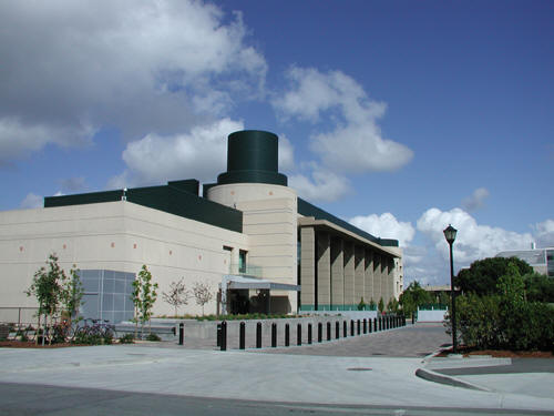
The Richard M. Lucas Center for Magnetic Resonance Spectroscopy and Imaging is located at Stanford University approximately thirty miles south of San Francisco, California, USA. We are part of the School of Medicine and Department of Radiology; and are dedicated entirely to research in MR imaging and spectroscopy, CT and X-Ray. Magnet facilities currently include three whole body systems (1.5T, 3.0T and 7.0T) and a small bore 4.7T system. Plans are underway to add a second 3.0T whole body system. The Lucas Center provides office and laboratory facilities for sixteen full-time faculty (Ph.D. and M.D.) and their complement of postdoctoral fellows and students, and the administrative and scientific staff.
|
|
|
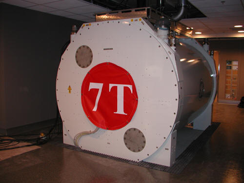 The recently installed 7.0T whole body MR system located at the Lucas Center for MRS/I. Initial studies in neurological and musculoskeletal imaging are being planned. |
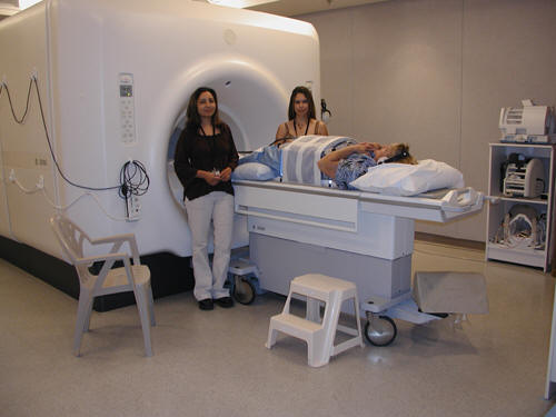 Romi Samra, R.T.(R), left, and Sandra Rodriguez, R.T.(R)(MR), right, prepare a research subject for an abdominal scan at the 3.0T MR system at the Lucas Center. |
Anne Marie Sawyer, B.S., R.T.(R)(MR), Romi Samra, R.T.(R) and Sandra Rodriguez, R.T.(R)(MR), are the MR technologists who support all of these studies. Their responsibilities, in addition to MR system operation, include teaching safety and screening procedures, MR system operation to researchers; troubleshooting MR system problems; designing new accessories, coils, and equipment; and assisting in the development of scan protocols for research studies. |