|
Combined Educational & Scientific Session
ORGANIZERS: Jenny T. Bencardino, M.D., Emily McWalter, Ph.D, Edwin H.G. Oei, M.D., Ph.D., & Philip Robinson,M.D.
Tuesday, 25 April 2017
| Room 315 |
13:45 - 15:45 |
Moderators: Christopher Burke, L. Tugan Muftuler |
Slack Channel: #s_msk
Session Number: CE04
Overview
This session will concentrate on the MRI uses in the assessment of spinal health and disease.
Target Audience
Clinical radiologists, scientists with an interest in spinal health and disease.
Educational Objectives
As a result of attending this course, participants should be able to:
-Review current applications of MRI in spinal health and disease;
-Recognize new MR techniques in the evaluation of the spine; and
-Develop research of novel MR modalities in the assessment of spinal conditions.
13:45
|
|
Update on MRI Degenerative Disc Disease: Beyond Modic Changes 
Jason Talbott
|
14:15
|
|
What's new on Spine Imaging: Beyond Degenerative Disc Disease 
Elisabeth Garwood
|
14:45
 |
0535.
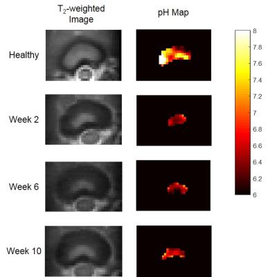 |
pH Measurement of Intervertebral Disc in the Process of Disc Degeneration Using Quantitative Chemical Exchange Saturation Transfer (qCEST) 
Zhengwei Zhou, Maxim Bez, Wafa Tawackoli, Dmitriy Sheyn, Joseph Giaconi, Zulma Gazit, Gadi Pelled, Dan Gazit, Debiao Li
Several hypotheses associate the pathogenesis of discogenic low back pain with low pH. In this study, we used qCEST MRI technique to monitor the pH changes of the intervertebral disc in the process of degeneration in porcine model. A significant pH drop was observed at 2 weeks following degeneration induction. This trend is correlated with the expression of pain markers. These results suggest that qCEST MRI has the potential to serve as a novel and non-invasive method for the diagnosis of discogenic pain.
|
14:57
 |
0536.
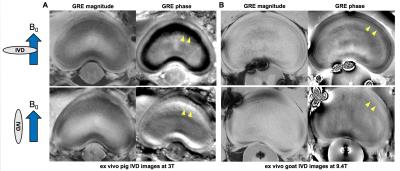 |
Feasibility study of intervertebral disc degeneration assessment using phase imaging - permission withheld
Yoonho Nam, Joonsung Lee, Eo-Jin Hwang, Joon-Yong Jung
In this study, we hypothesized that a GRE phase can be a potential indicator of annulus fibrosus integrity, because of its high sensitivity to micro-structural tissue change. With this hypothesis, we investigate the feasibility of GRE phase in the assessment of intervertebral disc degeneration in the lumbar spine. Ex vivo animal disk samples and nine human subjects were scanned and the high-pass filtered phase images were evaluated. For both ex vivo and in vivo experiments, distinctive phase contrasts were observed in the annulus fibrosus of the normal intervertebral discs.
|
15:09
|
0537.
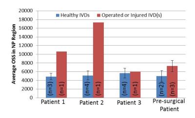 |
Detection of Internal Tissue Disruptions within the Intervertebral Disc via Magnetic Resonance Elastography: A Feasibility Study 
Benjamin Walter, Prasath Mageswaran, Elizabeth Yu, Safdar Khan, William Marras, Arunark Kolipaka
Magnetic resonance elastography (MRE) was used to non-invasively assess regions of internal tissue disruption within intervertebral discs (IVD) that were known to contain tissue damage. Internal tissue disruptions were assessed via measurements of wave continuity as they propagated throughout the IVD. Results demonstrated that injured IVD’s had an elevated octahedral shear strain (OSS) compared to non-operated control IVDs and OSS maps allowed visualization of potential location of internal tissue disruptions. Overall, results of this study suggest that MRE may provide a non-invasive way of identifying internal tissue disruptions within the IVD.
|
15:21
|
0538.
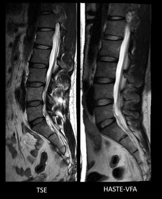 |
Clinical utility of a novel ultrafast T2 Weighted sequence for Spine Imaging 
Mahesh Bharath Keerthivasan, Ali Bilgin, Diego Martin, Jennifer Becker, Maria Altbach, Manojkumar Saranathan
T2 weighted imaging of the spine is commonly performed using Turbo Spin Echo (TSE) sequences, resulting in long scan times and vulnerability to motion artifacts. While the single-shot sequences such as HASTE could be used for rapid screening, their use is limited by poor spatial resolution and SAR limitations. We investigated the use of a variable flip angle HASTE (HASTE-VFA) sequence for ultrafast motion robust T2 weighted spine imaging and compared its performance to T2 TSE in 13 patients.
|
15:33
|
0539.
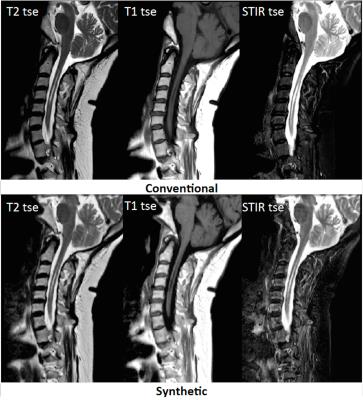 |
Feasibility of Synthetic MRI of the spine – fast and quantitative imaging 
Maria Vargas, Bénédicte Delattre, José Baiao Boto, Karl-Olof Lövblad, Sana Boudabbous
Synthetic MRI was already validated in some cerebral applications. We adapted it to evaluate the spine and showed that it produces, in general, good image quality and diagnostic confidence. Efforts will still have to be made to increase image quality in the dorso-lumbar spine. However, the non-negligible time saving and the ability to obtain quantitative measurements as well as to generate several contrasts with a single acquisition should secure an interesting and useful future for synthetic MRI in clinical routine.
|
|


