Tuesday, 25 April 2017
| Room 313A |
08:15 - 10:15 |
Moderators: Eric Achten, Masaaki Hori |
Slack Channel: #s_neuro
Session Number: O03
08:15
|
0392.
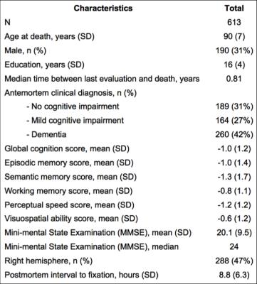 |
Age-related neuropathologies associated with white matter hyperintensities burden: a study of a community cohort of older adults. 
Nabil Alqam, Arnold Evia, Luis Campos Cardoso, Lucas Lopes, Diego Vieira Pereira, Julie Schneider, Sue Leurgans, David Bennett, Konstantinos Arfanakis
White matter hyperintensities (WMH) are lesions commonly observed in the brain of older adults, and have been associated with lower cognitive function, lower motor performance, and increased risk of dementia. The purpose of this work was to investigate the neuropathologic correlates of WMH burden by combining ex-vivo MRI and pathology on a large community cohort of older adults.
|
08:27
|
0393.
 |
Age-related changes in cerebrovascular reactivity and their relationship to cognition and vascular risk: A four-year longitudinal study 
Shin-Lei Peng, Xi Chen, Yang Li, Karen Rodrigue, Denise Park, Hanzhang Lu
Although cerebrovascular factors are the cause of cognitive impairment, the vascular decline in aging have not been characterized. In this work, we present four-year longitudinal cerebrovascular reactivity (CVR) data measured in 116 individuals. Our data revealed temporal lobe showed the fastest CVR decline and middle age manifested the fastest CVR decline. Vascular risk of hypertension results in a lower CVR when compared to normal and well-controlled subjects. Individuals with poorer general cognitive status, as indexed by a low mini-mental-state-exam (MMSE), had a lower CVR compared to participants with higher MMSE scores. These findings help elucidate age-related decline in brain hemodynamics.
|
08:39
|
0394.
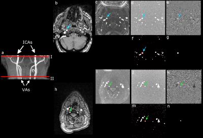 |
Cerebral Arteries Hemodynamics in Alzheimer's Disease Assessed by Phase-Contrast Velocity Mapping 
Reyes García de Eulate, Irene Goñi, Alvaro Galiano, Marta Vidorreta, Miriam Recio, Mario Riverol, Jose Luis Zubieta, María Fernández-Seara
Vascular disease increases the risk of Alzheimer's disease. The assessment of vascular dysfunction in subjects at risk for AD has the potential to contribute to the disease early diagnosis and management. In this work, the phase-contrast velocity mapping MRI technique was used to evaluate cerebral hemodynamics in patients with cognitive dysfunction and healthy controls. Results showed significant differences in hemodynamic parameters (velocity and flow) across groups with lower mean values in the AD and MCI groups compared to the CO group. PC-MRI can be used to assess hypoperfusion in an early stage of AD.
|
08:51
|
0395.
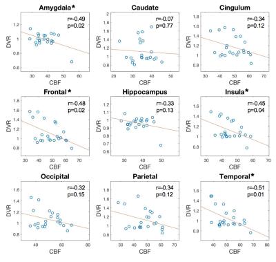 |
Association of cerebral blood flow and amyloid burden in autosomal dominant Alzheimer’s disease 
Lirong Yan, Collin Liu, Koon-Pong Wong, Sung-Cheng Huang, John Ringman, Danny Wang
The purpose of this study was to investigate the association of cerebral blood flow alternation with cerebral amyloid deposition in autosomal dominant Alzheimer’s disease. Cross-subject negative correlation between cerebral blood flow and amyloid deposition was observed, indicating brain regions with high amyloid deposition may be associated with hypoperfusion. Our finding suggests cerebral hypoperfusion may contribute to the onset and progression of AD.
|
09:03
|
0396.
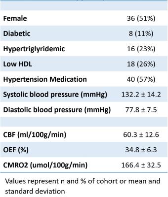 |
Association of vascular risk factors with cerebral metabolic rate 
Kevin King, Min Sheng, Peiying Liu, Christopher Maroules, Craig Rubin, Ron Peshock, Roderick McColl, Hanzhang Lu
Vascular risk factors that confer a susceptibility for dementia are thought to result in silent brain changes decades before disease onset. We hypothesized that vascular risk factors would be associated decreased Cerebral Metabolic Rate of Oxygen consumption (CMRO2). CMRO2 was derived from Arterial Spin Labelling cerebral blood flow (CBF) and oxygen extraction fraction (OEF) from TRUST MRI in this IRB approved study with informed consent on 70 participants. In stepwise linear regression higher diastolic blood pressure was correlated with decreased CMRO2 but was not associated with CBF, suggesting mechanisms other than insufficient blood flow underlie the association with metabolic rate.
|
09:15
|
0397.
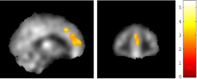 |
One-year aerobic exercise increases regional cerebral blood flow in anterior cingulate cortex: a blinded, randomized trial in patients with amnestic Mild Cognitive Impairment 
Binu Thomas, Takashi Tarumi, Min Sheng, Benjamin Tseng, Kyle Womack, Munro Cullum, Rong Zhang, Hanzhang Lu
Amnestic mild cognitive impairment (MCI) represents the early stage of Alzheimer’s disease (AD). Much research has focused on preventing the inevitable decline of MCI to AD. Aerobic exercise is considered a viable choice, and is shown to improve cognitive function in MCI. We focus on understanding the mechanisms that lead to this improvement. Pseudo-continuous-arterial-spin-labeling (PCASL) was used to assess resting cerebral blood flow (CBF) in two MCI groups. One group performed aerobic exercise, while another non-aerobic stretching. CBF was measured before and after training. CBF increase in the anterior-cingulate-cortex (ACC) was the proven mechanism that improves cognitive function in MCI.
|
09:27
|
0398.
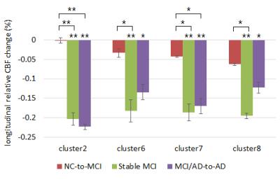 |
Cross-sectional and Longitudinal Cerebral Blood Flow Changes in the Progression from Normal Cognition to Alzheimer’s Disease Measured with Continuous Arterial Spin Labeling (CASL) 
Wenna Duan, H. Michael Gach, Arvind Balachandrasekaran, Parshant Sehrawat, Ashish Bhumkar, Paresh Boraste, James Becker, Oscar Lopez, Weiying Dai
Cross-sectional and longitudinal analysis of cerebral blood flow (CBF) versus cognitive status were performed in an elderly cohort. Voxel-based ANOVA was used to test the CBF difference between normal control (NC), mild cognitive impairment (MCI) and Alzheimer’s Disease (AD) groups at the baseline. Eight significant clusters were found between groups. The longitudinal CBF change in each cluster was compared across 4 longitudinal groups (stable NC, NC-to-MCI, stable MCI, and MCI/AD-to-AD) using a multiple linear regression model. The results indicated that CBF rises in AD-related regions of the brain during MCI and then drops dramatically in advanced MCI or early AD.
|
09:39
 |
0399.
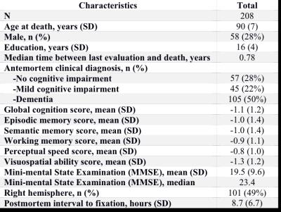 |
Effects of transactive response DNA-binding protein 43 (TDP43) pathology on amygdala volume and shape, in a community cohort of older adults 
Nazanin Makkinejad, Junxiao Yu, Aikaterini Kotrotsou, Arnold Evia, Julie Schneider, Sue Leurgans, David Bennett, Konstantinos Arfanakis
TDP43 pathology is now recognized as a common and deleterious neuropathology of the aging brain. TDP43 pathology typically originates in the amygdala, which is, however, commonly affected by other age-related neurodegenerative pathologies. The purpose of this work was to investigate the effects of TDP43 pathology on the volume and shape of the amygdala in a large community cohort of older adults.
|
09:51
|
0400.
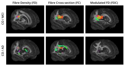 |
Fixel-based analysis of Alzheimer's Disease using multi-tissue constrained spherical deconvolution of multi-shell diffusion MRI 
Diana Giraldo, Hanne Struyfs , David Raffelt, Paul Parizel, Sebastiaan Engelborghs, Eduardo Romero, Jan Sijbers, Ben Jeurissen
In this study, we used multi-shell, multi-tissue constrained spherical deconvolution to investigate group differences in white matter between control subjects, patients with mild cognitive impairment (MCI) due to Alzheimer's Disease (AD) and patients with dementia due to AD. Using the recently proposed fixel-based analysis approach, we distinguish between different fibre populations within a single voxel and characterize them with 3 measures: fibre density, fibre cross-section and the product of these two. We found significant decreases of these metrics in MCI and AD patients compared to healthy controls.
|
10:03
|
0401.
 |
HFE Mutations Alter White Matter Diffusion and Relaxation Parametrics in Alzheimer’s Disease 
Mark Meadowcroft, Jianli Wang, Carson Purnell, Paul Eslinger, James Connor, Qing Yang
This work demonstrates that HFE mutations in cognitively normal compared to wild-type subjects lead to differences in diffusion and relaxation parametrics, such that HFE mutation carrier parametrics converge towards AD subjects. Furthermore, HFE mutations appeared to be preservative against white matter integrity loss in AD patients. Iron-loading HFE mutations appear to preserve relaxation and diffusion neuroimaging biomarkers in AD patients, but adversely affect cognitively normal subjects.
|
|


