Wednesday, 26 April 2017
| Room 312 |
16:15 - 18:15 |
Moderators: Gary Miller, Jason Talbott |
Slack Channel: #s_neuro
Session Number: O11
16:15
|
0912.
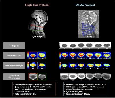 |
Regional and structural integrity of the whole cervical spinal cord using 3D-T1 MP2RAGE and multi-slice multi angle DTI and ihMT sequences at 3T: preliminary investigations on age-related changes. 
Henitsoa RASOANANDRIANINA, Guillaume DUHAMEL, Thorsten FEIWEIER, Manuel TASO, Aurélien MASSIRE, Olivier GIRARD, Maxime GUYE, Jean-Philippe RANJEVA, Virginie CALLOT
In this study, we present a 3T multi-parametric MR protocol allowing structural and diffuse evaluation of the whole cervical spinal cord (SC), within a clinically acceptable scan-time. The MRI protocol includes high-resolution anatomical T2*-weighted images allowing WM/GM atrophy evaluation, a MP2RAGE sequence allowing T1-mapping, a Multi-Slice Multi-Angle (MSMA) DTI sequence allowing evaluation of tissue structural organization and, last but not least, a MSMA inhomogeneous Magnetization transfer (ihMT) sequence allowing myelin-content evaluation in whole cervical SC. This protocol was combined with a template-based automated post-processing pipeline in a preliminary study investigating age- and region-related microstructural differences in specific regions along the cervical SC.
|
16:27
|
0913.
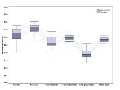 |
Quantitative measurement of myelin in distinct pathways of the spinal cord: Implications for assessing neuropathic pain after spinal cord injury 
Hanwen Liu, Emil Ljungberg, Erin MacMillan, Laura Barlow, Shannon Kolind, John Kramer, Cornelia Laule
Neuropathic pain is common in people with spinal cord injury (SCI). To better understand the type and severity of damage in the spinal cord associated with neuropathic pain, we used myelin water imaging (MWI) combined with spinal cord toolbox to study myelin content in specific pathways of spinal cord for both healthy and SCI subjects. Results show that MWI can distinguish different pathways of spinal cord and reduced myelin content in some specific pathways is found in SCI subjects. Our findings suggest that MWI with pathway-based analysis is capable of examining the correlation between damage pattern and neuropathic pain.
|
16:39
|
0914.
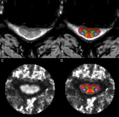 |
Multi-Parametric Spinal Cord MRI Detects Subclinical Tissue Injury in Asymptomatic Cervical Spinal Cord Compression 
Allan R. Martin, Benjamin De Leener, Julien Cohen-Adad, David W. Cadotte, Jefferson R. Wilson, Lindsay Tetreault, Aria Nouri, Adrian Crawley, David J. Mikulis, Howard Ginsberg, Michael G. Fehlings
Degenerative cervical myelopathy (DCM) involves extrinsic spinal cord (SC) compression causing tissue injury and neurological dysfunction. Asymptomatic SC compression is much more common (8-26%), but it is unknown if tissue injury occurs in this group. Our multi-parametric MRI protocol previously identified 10 measures of tissue injury in DCM. Using these techniques, we demonstrate that asymptomatic SC compression subjects show a similar pattern of tissue injury, with 8/10 measures (p=0.05) showing the same direction of changes and MTRRostral (p=0.002), MTRMCL (p=0.03), and T2*w WM/GMRostral (p=0.04) showing significant univariate differences. A logistic regression model retaining 3 MRI measures shows 86% discrimination between compressed and uncompressed subjects.
|
16:51
 |
0915.
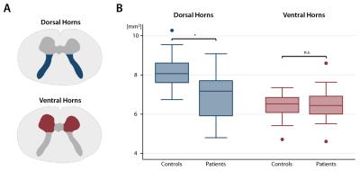 |
Ventral and dorsal horn grey matter neurodegeneration remote from stenosis in cervical spondylotic myelopathy 
Patrick Grabher, Siawoosh Mohammadi, Gergely David, Patrick Freund
Cervical spondylotic myelopathy (CSM) is the most frequent spinal cord disorder. Next to focal degeneration at the compression site, the rostral cervical white and grey matter undergo atrophic changes, the magnitude relating to clinical impairment. In this study, we assess above stenosis regional grey matter changes and demonstrate that next to bilateral dorsal horn atrophy, the normal appearing ventral horns show diffusivity changes which relate to white matter integrity loss.
|
17:03
|
0916.
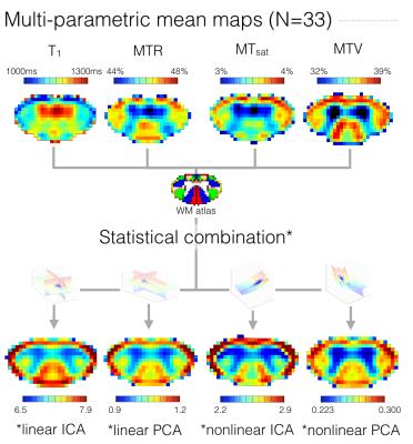 |
Statistical combinations of T1, MTR, MTsat and Macromolecular Tissue Volume to improve myelin content estimation in the human spinal cord at 3T 
Simon Lévy, Ali Khatibi, Gabriel Mangeat, Jen-I Chen, Kristina Martinu, Pierre Rainville, Nikola Stikov, Julien Cohen-Adad
Several quantitative MRI metrics have been proposed to quantify myelin in the central nervous system but each of them includes confounding factors that impair their sensitivity and specificity. Because these factors are different across metrics, data driven approaches developed for blind source separation problems to extract the common component between recordings of the same sources seem appropriate. This study compares linear and nonlinear methods to combine myelin-sensitive metrics: T1, MTR, MTsat, MTV (1 – PD). The repeatability of the resulting combined metrics as well as their sensitivity to different microstructural features are tested. A higher sensitivity is achieved with linear combinations.
|
17:15
|
0917.
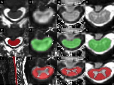 |
Multi-Parametric Cervical Spinal Cord MRI Provides An Accurate Diagnostic Tool for Detecting Clinical Myelopathy 
Allan R. Martin, Benjamin De Leener, Julien Cohen-Adad, David W. Cadotte, Jefferson R. Wilson, Lindsay Tetreault, Stefan F. Lange, Aria Nouri, Adrian Crawley, David J. Mikulis, Howard Ginsberg, Michael G. Fehlings
ddd
|
17:27
|
0918.
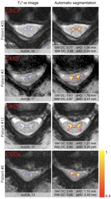 |
Fully automatic segmentation of spinal cord gray matter on patients with degenerative cervical myelopathy 
Sara Dupont, Allan Martin, Nikola Stikov, Michael Fehlings, Julien Cohen-Adad
Degenerative cervical myelopathy (DCM) occurs when arthritic changes cause extrinsic spinal cord (SC) compression, inducing motor and sensory disabilities due to gray matter (GM) and white matter (WM) injury. GM segmentation of MR images can quantify atrophy of both GM and WM and may offer biomarkers to improve diagnosis, monitoring of disease progression, and prognosis. In this study, the GM of 33 DCM patients and 8 healthy subjects was automatically segmented using the method included in the Spinal Cord Toolbox (SCT). GM segmentation results were in good accordance with the underlying anatomy, demonstrating the feasibility of automatic GM segmentation in DCM patients exhibiting severe SC compression.
|
17:39
|
0919.
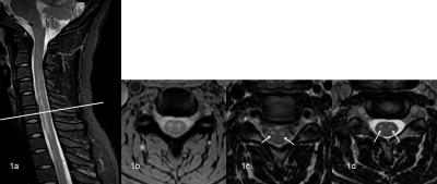 |
The use of 3D FSE in place of 2D T2-FSE, 2D T2*-GRE in the diagnosis of cervical spinal non-neoplastic lesion - permission withheld
tianyong xu, guang han, songguo liu, mingzhi pan, yang li, zhenyu zhou, bing wu
Magnetic resonance imaging has been accepted as the predominant imaging tool for diagnosis of spinal disease, such as protrusion of intervertebral disc, tumor, inflammation, trauma [1]. T2 weighted imaging is commonly used for diagonosis in many medial centers, however 2D fast spin echo (FSE) image quality is often limited by several practical factors including low fat content, high susceptibility effect and vunerability to motion and pulsation. T2* weighted imaging is another popular choice [2]. Improved contrast may be obtained between gray matter and white matter of medullar. The drawback of T2* imaging is the hyperintense gray matter signal may may cause the small cervical spinal cord lesion to be overlooked. In this work, the use of 3D FSE is assessed and compared to 2D T2 and T2* imaging, and hypothesized to overcome their shortcomings.
|
17:51
|
0920.
 |
Anthropomorphic Spinal Cord Phantom with Induced Field Inhomogeneity 
Alan Seifert, Vaishali Patel, Merin Grace, Robin Li, Mohammad Molla, Joseph Borrello, Junqian Xu
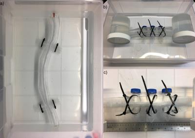
The human spinal cord exists in a particularly unfavorable magnetic field environment. Technical development of diffusion and functional MRI methods would be facilitated by a phantom to model spatially and temporally periodic field inhomogeneities. We have designed a phantom capable of simulating these specific field disturbances. The spinal canal was machined from acrylic, and the cord was cast of polyvinyl alcohol. The phantom was imaged using anatomical CT and MRI, and functional and diffusion EPI protocols. The phantom has relaxation and diffusion properties similar to the human cord, and air-filled vials create spatially periodic frequency shifts of -100 Hz.
|
18:03
|
0921.
 |
Longitudinal MRS, MRI and DTI of the spinal cord in Friedreich’s Ataxia 
Pierre-Gilles Henry, James Joers, Dinesh Deelchand, Diane Hutter, Khalaf Bushara, Gülin Öz, Christophe Lenglet
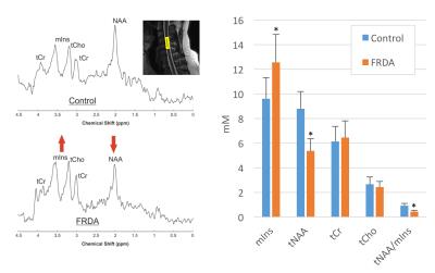
We report the first longitudinal MRS, structural MRI, and diffusion MRI data in the cervical spinal cord of subjects with Friedreich’s ataxia. We were able to detect significant changes in spinal cord area (-18%), tNAA/mIns ratio (-17%), fractional anisotropy (-11%) and mean diffusivity (+26%) in a group of 10 patients over 24 months. Our data suggest that MR of the spinal cord could be useful to assess the impact of potential treatments on neurodegeneration in upcoming clinical trials in FRDA.
|
|


