Thursday, 27 April 2017
| Room 320 |
15:30 - 17:30 |
Moderators: Alex Frydrychowicz, Susanne Schnell |
Slack Channel: #s_cv
Session Number: O23
15:30
|
1257.
 |
Simultaneous MultiSlab 4D Flow MRI for Quantification of Hemodynamics in the Carotid Bifurcation at 7 Tesla 
Sebastian Schmitter, Gregor Adriany, Steen Moeller, Edward Auerbach, Pierre-Francois Van de Moortele, Michael Markl, Kamil Ugurbil, Susanne Schnell
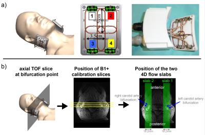
Simultaneous Multislice (SMS) imaging, also termed "Multiband" (MB), was integrated into slab-selective 4D flow MRI to quantify blood hemodynamics simultaneously in both carotid bifurcations at 7T. Therefore, sagittal oriented MB 4D flow acquisitions with 0.8mm isotropic resolution were performed in 4 volunteers using a dedicated carotid coil. The same protocol was then repeated twice, but only a single slab ("SingleBand" - SB) targeting the left or right carotid bifurcation was excited. Peak velocity and net flow quantification was performed for reconstructed MB and SB data on 3 planes each, one before and two after the bifurcation.
|
15:42
|
1258.
 |
4D Flow MRI in the Post-Myocardial Infarction Left Ventricle 
Jacob Macdonald, Jonathan Weinsaft, Christopher Francois, Oliver Wieben
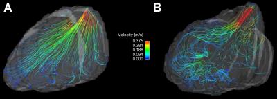
Left ventricular thrombus (LVT) formation is a serious complication of anterior ST segment elevation myocardial infarction (MI) that impacts prognosis. LVT has been linked to blood stasis in the LV apex, but conventional predictors of LVT formation are limited. This study employed 4D-flow MRI as a new means of quantifying MI induced alterations in LV flow physiology. Among a mixed cohort of post-MI subjects and controls, 4D-flow MRI demonstrated MI to be associated with increased flow stagnance in the LV apex. These findings will inform use of 4D-flow MRI in future studies to predict longitudinal risk for post-MI LVT.
|
15:54
 |
1259.
 |
Pushing the Boundaries of Low-Venc PC-MRI Acquisition Strategies with a Weighted, Regularized Optimization Reconstruction 
Michael Loecher, Daniel Ennis
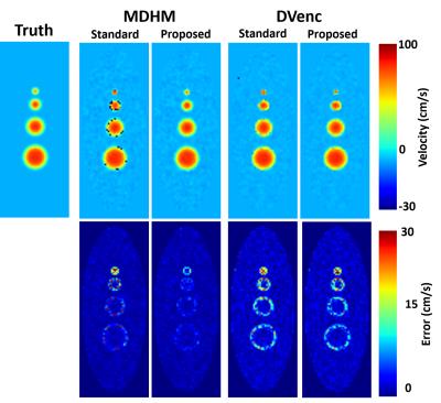
We propose a novel reconstruction method for low-Venc (high-moment) PC-MRI reconstructions to allow for significant VNR gains while being more robust to the errors associated with these methods. The reconstruction accounts for unequal signal variances due to intravoxel dephasing, and includes constraints to account for residual phase wrapping errors. The method is tested in simulations, phantoms, and volunteers.
|
16:06
|
1260.
 |
Improving left ventricular 4D Flow MRI analysis using intensity based image registration with cine-bSSFP images 
Vikas Gupta, Mariana Bustamante, Alexandru Fredriksson, Carl-Johan Carlhäll, Tino Ebbers
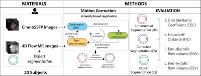
Characterization of blood flow through the left ventricle (LV) using 4D Flow MRI requires accurate segmentation of the underlying anatomy. The combination of poor resolution and low contrast in 4D flow MR images, however, makes the LV segmentation challenging and prone to observer bias. We propose an image registration based approach that allows reliable and automatic correction of LV segmentations in 4D Flow images using the high contrast in routinely acquired 2D cine-MR images. The proposed approach is shown to achieve high in plane spatial accuracy and comparable values of blood flow parameters when evaluated against manually obtained expert segmentations of 4D flow MR images.
|
16:18
 |
1261.
 |
High-Resolution 4D Real-Time Phase-Contrast Flow MRI with Sparse Sampling 
Aiqi Sun, Bo Zhao, Rui Li, Chun Yuan
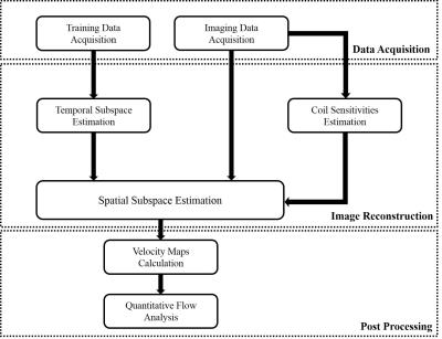
This work presents a model-based imaging method, which integrates low-rank modeling, sparse modeling with parallel imaging, to enable 4D real-time phase-contrast flow MRI without ECG gating and respiration control. The proposed method achieves real-time imaging at a spatial resolution of 2.4 mm, temporal resolution of 35 ms, with three directional flow encodings, and well resolves beat-by-beat flow variations, which cannot be achieved by the conventional cine-based method. The proposed has been evaluated by in vivo data with multiple healthy subjects and one arrhythmic patient. For the first time, we demonstrate the feasibility of real-time 4D PC flow MRI.
|
16:30
|
1262.
 |
Golden-Angle Spiral Sparse Parallel Phase-Ccontrast MR acquisition with an on-line fast GPU based reconstruction for high resolution real-time cardiovascular assessments 
Grzegorz Kowalik, Adèle Courot, Jennifer Steeden, Vivek Muthurangu
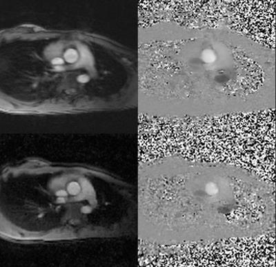
Compressed Sensing for 2D spiral PCMR with golden angle acquisition schema. The work presents a significant improvement in spatial-temporal resolution of the real-time spiral PCMR data, which are comparable with the standard high resolution cardiac gated sequences. The technique proved to be suitable for clinical use with the benefits of short acquisition times and no breathing artefacts.
|
16:42
|
1263.
 |
Compressed Sensing accelerated 4D flow MRI using a pseudo spiral Cartesian sampling technique with random undersampling in time 
Lukas Gottwald, Eva Peper, Qinwei Zhang, Valentijn Pronk, Bram Coolen, Pim van Ooij, Gustav Strijkers, Aart Nederveen
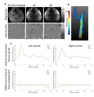
Clinical applications of three-dimensional time-resolved (4D) flow MRI are still hindered by long acquisition times. Using compressed sensing image reconstruction, undersampled 4D flow data may be recovered without a notable loss of image quality. In this study pseudo-spiral sampling on a Cartesian grid was implemented on a Philips 3T Ingenia system to facilitate random undersampling in time. The technique was tested under controlled conditions in a pulsatile phantom for different acceleration factors. An additional in vivo volunteer data set confirmed the stability of this technique.
|
16:54
|
1264.
 |
Highly accelerated Multi-Directional Velocity Encoding 4D Flow MRI: feasibility and preliminary results 
Henrik Haraldsson, Evan Kao, Yan Wang, David Saloner, Jing Liu
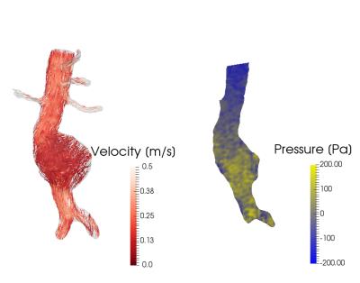
Velocity-to-noise ratio in 4D flow improves with low velocity encoding (VENC), but is usually compromised to prevent velocity aliasing. Dual-VENC and multi-directional velocity encoding schemes have been proposed to circumvent these issues, but result in an increased acquisition time. In this study, we developed a motion-robust highly accelerated 4D flow acquisition with multi-directional velocity encodings to target applications with a wide range of velocities in clinically acceptable acquisition times, for applications such as imaging of abdominal aneurysms, and simultaneous assessment of cardiac tissue phase mapping and intracardiac blood flow.
|
17:06
|
1265.
 |
Simultaneous Multi-Slice Phase Contrast Imaging for Pulse Wave Velocity Measurement in the Vessel 
Ning Jin, Jianing Pang, Shivraman Giri, Peter Speier, Dingxin Wang
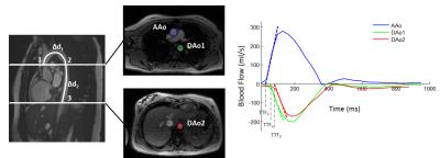
Pulse wave velocity can be derived from sequential 2D phase contrast measurements in multiple breath-holds, resulting in a long scan time and patient discomfort. Physiological conditions, may change during the measurement series, leading to flow waveform shifts and hence errors in PWV estimation. We developed a prototype SMS cine-PC sequence to measure blood flow in multiple slices simultaneously and applied it to measure PWV in the aorta.
|
17:18
|
1266.
 |
Tridimensional Axial and Circumferential WSS from 4D flow data using a finite element method and a Laplacian approach 
Julio Sotelo, Jesús Urbina, Bram Ruijsink, David Nordsletten, Joaquín Mura, Reza Razavi, Daniel Hurtado, Sergio Uribe
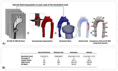
The WSS and OSI play a critical role in the progression of different vascular diseases, the multidirectional nature of WSS, can alter the balance of the endothelial cells. But the multidirectional nature of WSS only has been analyzed in 2D section. In this work, we propose a new method based on 3D finite-element and a Laplacian approach to decompose the WSS vector in an axial (WSSA) and circumferential (WSSC) component in a 3D domain. The 3D method provides an excellent agreement of the quantification of WSSA and WSSC in comparison with the actual 2D method.
|
|


