Oral
Body: Breast, Chest, Abdomen, Pelvis
Wednesday, 26 April 2017
| Room 311 |
16:15 - 18:15 |
Moderators: Shintaro Ichikawa, Riccardo Lattanzi |
Slack Channel: #s_body
Session Number: O31
16:15
 |
0902.
 |
Comprehensive T1-weighted dynamic liver MRI during free-breathing using fat/water separation, radial sampling, compressed sensing, parallel imaging, and motion-weighted reconstruction 
Thomas Benkert, Li Feng, Luke Gerges, Krishna Shanbhogue, Chenchan Huang, Daniel Sodickson, Hersh Chandarana, Kai Block
Conventional clinical liver MRI consists of several exams, including pre-contrast in-phase, opposed-phase, and fat-saturated scans as well as multiple scans with contrast-enhancement. For each of these acquisitions, accurate breath-holding is required to ensure diagnostic image quality. Here, we demonstrate how this entire protocol can be replaced by using a single comprehensive exam, where only one dataset has to be acquired during free-breathing. All relevant images can be retrospectively generated with model-based fat/water separation, which incorporates compressed sensing and parallel imaging. This approach has the potential to improve clinical workflow and eliminate the risk for failed exams caused by imperfect breath-holding.
|
16:27
|
0903.
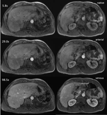 |
Image quality assessment for free-breathing dynamic liver examination using a self-navigated Cartesian acquisition with iterative reconstruction 
Benjamin Kaltenbach, Dominik Nickel, Ralph Strecker, Andreas Bucher, Thomas J. Vogl, Boris Bodelle
Free-breathing DCE-MRI of the liver is feasible using a Cartesian acquisition with self-navigation and hard-gated reconstruction in oncological patients. Compared to a standard BH-VIBE, image quality was rated marginally lower but with useful robustness regarding breathing artifacts. Therefore, the proposed sequence is a promising alternative in patients who cannot comply with breathing commands, like children or elderly patients.
|
16:39
|
0904.
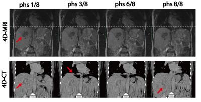 |
Respiratory resolved, self-gated, 4-dimensional MRI using Rotating Cartesian K-Space (ROCK): technical validation and initial clinical experience on an MRI-guided radiation therapy system 
Fei Han, Ziwu Zhou, Yu Gao, Percy Lee, Minsong Cao, Daniel Low, Ke Sheng, Yingli Yang, Peng Hu
Respiratory revolved 4D-MRI is used to quantify patient-specific respiratory motion to ensure optimal dose delivery in the radiation therapy of abdominal tumors. In this work, we developed a 4D-MRI technique using 3D k-space encoding, respiratory motion self-gating, and compressive sensing image reconstruction. The proposed 4D-MRI technique could provide high resolution, high quality, respiratory motion resolved 4D images with good soft-tissue contrast and are free of the “stitching” artifacts usually seen on 4D-CT, which is the current clinical standard. Results from motion phantom, healthy volunteers in a 1.5T diagnostic scanner and liver cancer patient in a 0.35T MRI-guided radiation therapy system demonstrated the feasibility of using the proposed 4D-MRI in radiation treatment planning.
|
16:51
|
0905.
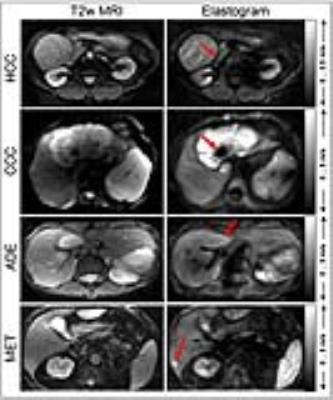 |
High resolution multifrequency MR elastography of hepatic tumors for measurement of stiffness and mechanical heterogeneity 
Mehrgan Shahryari, Jing Guo, Florian Dittmann, Heiko Tzschätzsch, Sebastian Hirsch, Eric Barnhill, Georg Böning, Uli Fehrenbach, Timm Denecke, Jürgen Braun, Ingolf Sack
High-resolution MRE of the liver based on multifrequency wave excitation and tomoelastography reconstruction is proposed for the mechanical characterization of hepatic tumors including stiffness and heterogeneity of the mechanical properties. We investigated 62 tumors in 43 patients and observed that high-grade tumors are stiffer than surrounding parenchyma, while low-grade hepatic adenoma had no altered stiffness. The intra-tumor heterogeneity of stiffness values was higher than in non-tumorous liver tissue. The proposed multifrequency MRE protocol could easily be integrated into the standard radiological workflow of our institution, thereby adding valuable information on tumor aggressiveness to standard clinical imaging markers.
|
17:03
|
0906.
 |
Comparison of Spin-Echo Echoplanar Imaging and Gradient Recalled Echo Magnetic Resonance Elastography Pulse Sequences Among Patients with Hepatic Iron Overload at 3.0 T 
Ely Felker, Steven Raman, Bradley Bolster, Holden Wu, Kyung Sung, Stephan Kannengiesser, Brenda Brown, David Lu
Most technical failures of GRE-based MR elastography are related to iron overload, especially at 3.0 T. SE-EPI-based MR elastography may be a useful alternative. 3.0 T GRE and SE-EPI MR elastography sequences were compared in 20 patients with iron overload (mean R2* 114 sec-1) in terms of quantitative liver stiffness (LS) and image quality score (IQS), based on wave propagation and confidence mask coverage, as determined by two experienced radiologists. LS measurements were not significantly different between the two sequences. SE-EPI showed a trend toward higher confidence mask coverage and significantly higher IQS for both readers compared to GRE.
|
17:15
|
0907.
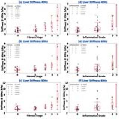 |
A New MR Elastography Parameter for Diagnosing Hepatic Fibrosis and Inflammation: Shear Attenuation 
Meng Yin, Jun Chen, Kevin Glaser, Jayant Talwalkar, Richard Ehman
Liver attenuation was investigated in comparison to the well-established liver stiffness for detecting hepatic fibrosis in 108 patients with histology-proven chronic liver diseases. Both liver stiffness and attenuation successfully detected varying fibrosis and inflammation with equivalent accuracy. At 40 and 60Hz, both have excellent accuracy for distinguishing clinical significant fibrosis or inflammation from others; moderate accuracy were obtained in distinguishing mild abnormalities from patients without abnormalities. Steatosis extent had no significant effect on liver stiffness and attenuation measurements. Our findings indicate that shear attenuation has equivalent diagnostic performance to that of liver stiffness for detecting liver disease progression.
|
17:27
|
0908.
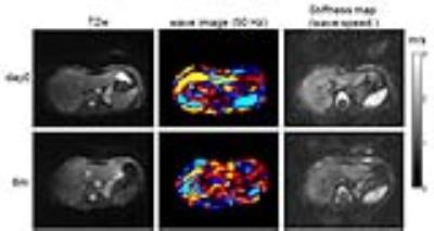 |
Treatment response of hepatic and renal stiffness in patients with chronic HCV infection monitored by multifrequency MR elastography 
Jing Guo, Stephan Marticorena Garcia , Florian Dittmann, Thomas Fischer, Jürgen Braun, Ingolf Sack
Multifrequency abdominal MRE was applied to monitor stiffness of the liver and kidney in renal transplant recipients with chronic HCV infection after antiviral therapy. Within a 6-months period after medication, hepatic stiffness was significantly reduced while renal stiffness in kidney transplants was not altered. Wave speed obtained by high-resolution multifrequency MRE can serve as a quantitative imaging maker to assess the treatment response and monitor abdominal organ stiffness longitudinally.
|
17:39
|
0909.
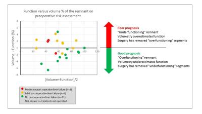 |
Dynamic Gadoxetate Enhanced MRI measurement of segmental liver function – A novel imaging biomarker for predicting Post-Hepatectomy Liver Failure 
David Longbotham, Daniel Wilson, Ashley Guthrie, Ernest Hidalgo, Raj Prasad, Steven Sourbron
Current risk assessment for post-hepatectomy liver failure (PHLF) is based on the volume of the future liver remnant (FLR). This is inaccurate when liver function is inhomogeneously distributed. We investigated whether the function of the FLR measured with DCE-MRI improves outcome predictions in 28 patients who had curative resection for colorectal liver metastases. We found the difference in preoperative estimates of FLR function and volume predicted PHLF with a sensitivity of 83% and specificity of 91%, indicating that: (1) inhomogeneous distribution of function is a major risk factor for PHLF; (2) DCE-MRI can improve patient outcome by correcting for the bias caused by volumetry.
|
17:51
|
0910.
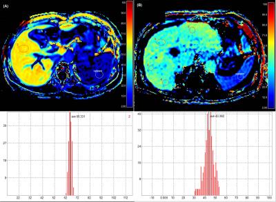 |
Liver function estimation using hepatocyte fraction map at gadoxetic acid enhanced liver MRI in patients with chronic liver disease 
Jeong Hee Yoon, Eunju Kim, Tomoyuki Okuaki, Jeong Min Lee
Hepatocyte fraction derived from gadoxetic acid enhanced liver MRI may provide quantitative surrogate marker of liver function for ICG R15 in patients with chronic liver disease.
|
18:03
|
0911.
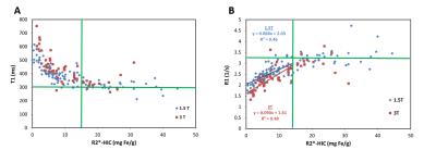 |
Does T1 Mapping Provide Additional Information in the Context of Hepatic Iron Overload? - permission withheld
Aaryani Tipirneni-Sajja, Eric Kercher, Ralf Loeffler, Ruitian Song, Matthew Robson, M. Beth McCarville, Jane Hankins, Claudia Hillenbrand
Hepatic iron content (HIC) is linearly correlated with R2*. Currently, there is little data on in vivo human liver iron assessment via longitudinal relaxation T1 (or R1) although an animal study previously suggested linear association, too. This study investigates hepatic T1 quantification in a breathhold by using ShMOLLI. T1 and T2* liver mapping were performed in 124 iron loaded patients. We found linear association between R1 and R2*/HIC values for mild-moderate HIC at 1.5T and 3T. No association between T1 and T2* was found for high HIC, which is most likely due to technical limits of ShMOLLI for short T2*.
|
|


