Monday, 24 April 2017
| Room 313A |
13:45 - 15:45 |
Moderators: Berkin Bilgic, Stefan Skare |
Slack Channel: #s_diffusion
Session Number: O73
13:45
 |
0172.
 |
Diffusion Weighted Imaging using a Dixon based Single Shot Turbo Spin Echo 
Xinzeng Wang, Holger Eggers, Marco Pinho, Ivan Pedrosa, Robert Lenkinski, Ananth Madhuranthakam
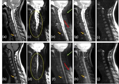
Diffusion weighted imaging using single-shot turbo spin-echo (DWI-SShTSE) is increasingly used due to its robustness to geometric distortions, but often suffers from incomplete fat suppression at 3T using spectrally-selective fat suppression methods (SPIR/SPAIR etc.) in challenging areas with large field inhomogeneities. STIR can improve the fat suppression but at the expense of reduced SNR. In this work, we developed a multi-echo Dixon DWI-SShTSE sequence with shared field map between lower and higher b-values for uniform fat suppression without using image navigator and increasing scan times. We also demonstrated its robustness to the phase variations due to diffusion gradients.
|
13:57
 |
0173.
 |
Kt-dSTEAM: high resolution diffusion-weighted imaging of the ex vivo human brain using B1+ homogenized STEAM at 9.4T 
Francisco J. Fritz, Desmond H Y Tse, Shubarthi Sengupta, Tim Loderhose, Bram Kraaijeveld, Svenja Caspers, Benedikt Poser, Alard Roebroeck
The investigation of entire human brains post mortem with diffusion MRI is an important research tool. However, the achievable resolutions and contrast are limited by gradient performance, RF-field inhomogeneity and strongly reduced T2 and diffusivity. Here, a diffusion-weighted STEAM sequence was modified to enable the use of kT-points B1+ homogenization and 3D segmented EPI readout. The resulting kT-dSTEAM sequence allows for high resolution (1000μm, 500μm and 400μm isotropic) diffusion-weighted imaging the entire human brain with homogenous contrast at 9.4T.
|
14:09
|
0174.
 |
High resolution whole brain diffusion MRI at 7 Tesla using RF parallel transmission - permission withheld
Xiaoping Wu, Edward Auerbach, An Vu, Steen Moeller, Christophe Lenglet, Sebastian Schmitter, Pierre-Francois Van de Moortele, Essa Yacoub, Kamil Ugurbil
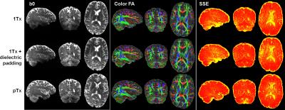
A major component of the Human Connectome Project (HCP) in the WU-Minn consortium is multiband (MB)-accelerated whole-brain diffusion MRI (dMRI) at both 3T and 7T. Although having some advantages over 3T dMRI in inferring connectivity, the 7T acquisition suffers from RF nonuniformity and is limited to MB2 acceleration because of SAR. Here, we demonstrate the utility of RF parallel transmission (pTx) for 7T HCP-type dMRI with ~1-mm isotropic resolution. Our results demonstrate that pTx can significantly improve RF uniformity across the entire brain and enable higher slice acceleration relative to single transmit configurations, thereby holding great potential for acquiring high quality, high resolution and high efficiency dMRI data.
|
14:21
|
0175.
 |
Fast high-resolution diffusion MRI using gSlider-SMS, interlaced subsampling, and SNR-enhancing joint reconstruction 
Justin Haldar, Kawin Setsompop
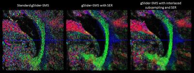
We describe a new approach that enables in vivo whole brain diffusion MRI with simultaneously high spatial resolution (660 Ám isotropic voxels) and high angular diffusion encoding resolution (64 orientations at b=1500 s/mm2 and 4 b=0 s/mm2images) in only 15 minutes. This is achieved by combining the gSlider-SMS acquisition strategy with constrained image reconstruction techniques that enable denoising (exploiting the fact that the diffusion images are smooth with correlated edge locations) and interlaced data subsampling (achieved by exploiting the same correlated edge constraints used for denoising, as well as through the use of q-space smoothness constraints).
|
14:33
 |
0176.
 |
Faster Diffusion-Relaxation Correlation Spectroscopic Imaging (DR-CSI) using Optimized Experiment Design 
Daeun Kim, Justin Haldar
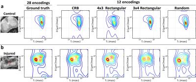
We propose a new experiment design method to accelerate the recent novel diffusion-relaxation correlation spectroscopic imaging (DR-CSI) experiment. DR-CSI acquires imaging data across a range of different b-value and echo time combinations. This enables new insights into tissue microstructure, but the contrast encoding can be slow. Our experiment design approach selects a small subset of the most informative observations to acquire using results from estimation theory. We demonstrate with ex vivo mouse spinal cord MR data that the new experiment design approach enables DR-CSI to be accelerated by a factor of more than 2 without a substantial loss in quality.
|
14:45
|
0177.
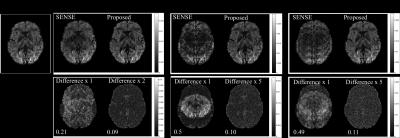 |
Accelerated k-q diffusion MRI reconstruction using Gaussian processes 
Wenchuan Wu, Peter Koopmans, Jesper Andersson, Karla Miller
Diffusion MRI commonly acquires multiple diffusion volumes (directions), which shares plentiful common features. In this work, we propose integrating Gaussian Processes into image reconstruction to utilize the shared information between diffusion volumes to reduce image artefacts associated with parallel imaging.
|
14:57
|
0178.
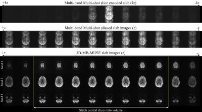 |
3D Multi-Band, Multi-Slab, and Multi-Shot High-Resolution Diffusion MRI - permission withheld
Iain Bruce, Hing-Chiu Chang, Nan-Kuei Chen, Allen Song
When diffusion MRI data is acquired with 3D multi-slab and/or multi-shot imaging techniques, scan times are often lengthy and phase variations between the acquired shots and/or slice-encoding planes of 3D slabs introduce severe motion artifacts in slice images. To accelerate the acquisition of high spatial resolution diffusion MRI volumes with high SNR and fidelity, we outline a 3D image reconstruction model that simultaneously accounts for both in-plane and through-plane motion artifacts in 3D multi-band, multi-slab and multi-shot diffusion data. Diffusion data acquired and reconstructed in this fashion can be acquired at sub-millimeter spatial resolution with high SNR in ~1-2min.
|
15:09
 |
0179.
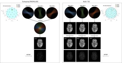 |
Improving angular resolution in multi-shot turbo spin-echo diffusion imaging using rotating single-shot acquisition (RoSA) 
Qiuting Wen, Mark Graham, Ivana Drobnjak, Hui Zhang, Yu-Chien Wu
The rotating single-shot acquisition (RoSA) technique is proposed to accelerate multi-shot diffusion imaging acquisitions by acquiring one shot per diffusion direction. The RoSA approach utilizes similarity existing in diffusion-weighted contrast for image reconstruction. It has been successfully implemented with echo planar imaging (EPI). In this study, we use the RoSA approach to improve turbo spin-echo (TSE) based multi-shot sequences. In particular, we will demonstrate that with the same acquisition time, RoSA increases the diffusion angular sampling resolution by 3-fold compared to a Turboprop sequence.
|
15:21
|
0180.
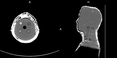 |
Combination of integrated dynamic shimming and readout-segmented echo planar imaging for diffusion-weighted MRI of the head and neck region at 3 Tesla. 
Sven Walter, Alto Stemmer, Berthold Kiefer, Konstantin Nikolaou, Petros Martirosian, Mike Notohamiprodjo, Sergios Gatidis
The purpose of this study was to evaluate possible improvements in EPI-based DWI of the head/neck at 3 Tesla using a combination of readout-segmented EPI and dynamic shimming. We assessed ADC quantification in an anthropomorphic phantom and evaluated the presence of geometric distortions, signal losses, ghosting artifacts, and overall image quality in both, phantom and in-vivo data from 10 volunteers. We found that combining integrated shimming with readout-segmented EPI significantly improves images quality of EPI based DWI of the head/neck at 3 Tesla compared to the single techniques alone or conventional single-shot EPI.
|
15:33
|
0181.
 |
Reduced Distortion in Diffusion Tensor MRI with Eddy Current Nulled Convex Optimized Diffusion Encoding (EN-CODE) 
Eric Aliotta, Kevin Moulin, Daniel Ennis
Eddy currents distort images and confound diffusion tensor reconstruction. In this work, convex optimized diffusion encoding (CODE) was extended to include an eddy current nulling term (EN-CODE) to achieve minimum TE diffusion tensor imaging (DTI) without eddy current distortions. EN-CODE was evaluated in simulations and through imaging in phantoms and healthy subjects. EN-CODE achieves distortion reduction on par with the existing twice refocused spin echo (TRSE) technique with a substantially shorter TE.
|
|


