Poster: Best of Cardiovascular MR: Myocardial Tissue Characterization
Electronic Power Pitch Poster
Cardiovascular
Tuesday, 25 April 2017
| Exhibition Hall |
17:15 - 18:15 |
| |
|
Plasma # |
|
0540.
 |
1 |
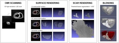
Three-dimensional holographic visualization of high-resolution myocardial scar on HoloLens 
Jihye Jang, Gifty Addae, Warren Manning, Reza Nezafat
We present a framework for 3D holographic visualization of high-resolution 3D late gadolinium enhancement (LGE) of myocardial scar on augmented-reality glass HoloLens via two approaches; 1) voxel-wise 3D scar rendering and 2) surface projection of the scar. Holographic visualization of high-resolution 3D LGE data will provide a true 3D perception of the complex scar architecture with an immersive experience to explore the clinical standard LGE images in a more interactive and interpretable way, which may facilitate MR-guided scar-related ventricular tachycardia ablation.
|
|
0541.
 |
2 |
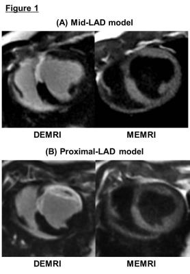
Inducibility of ventricular arrhythmia correlates with the indices of myocardial viability using manganese enhanced MRI (MEMRI) in a porcine ischemia reperfusion model 
Atsushi Tachibana, Junaid Zaman, Yuko Tada, Michelle Santoso, Phillip Yang
Peri-infarct region (PIR), containing the viable but injured myocardium, has been related to the occurrence of ventricular arrhythmia. Reliable in vivo detection of arrhythmogenic region presents significant challenge. While delayed enhanced MRI (DEMRI) with gadolinium (Gd) detects the myocardial infarction, the non-specific property does not detect the viable but injured cardiomyocytes in the PIR. Manganese (Mn) enters the live cells via L-type calcium channel, and enables dual enhancement technique to identify the overlapping viable region in PIR. We measured the correlation between the volume of the PIR and inducibility of ventricular arrhythmia using porcine ischemia reperfusion model.
|
|
0542.
 |
3 |
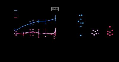
In Vivo Hyperpolarized MRS Study Showing Improved Cardiac Metabolism in Type 1 Diabetes with Daily L-Carnitine Treatment. 
Dragana Savic, Kerstin Timm, Vicky Ball, Lisa Heather, Damian Tyler
Carnitine acts as a buffer of acetyl-CoA units in the mitochondria, as well as facilitating transport of fatty acids, and carnitine levels are decreased in the diabetic heart. The purpose of this study was to investigate the effect of L-Carnitine supplementation on cardiac function and metabolism in the diabetic rat heart. We show that daily injections of L-Carnitine can alter cardiac metabolism in the in vivo diabetic rat heart, and can increase flux through pyruvate dehydrogenase. Such studies will allow a better understanding of the interactions between metabolism and function in the diabetic heart and may provide new insight into novel therapeutics.
|
 |
0543.
 |
4 |
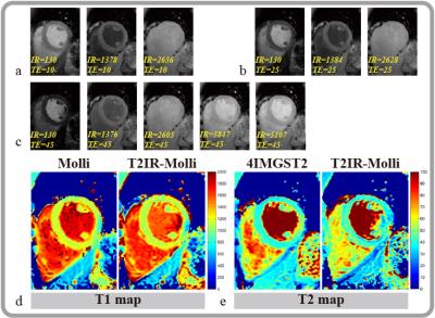
Integrated T2 preparation and Inversion Recovery pulse (T2IR) for combined myocardium T1 and T2 mapping 
Rui Guo, Zhensen Chen, Jianwen Luo, Haiyan Ding
In this study we designed a novel hybrid T1 and T2 magnetization preparation pulse (T2IR) and implement a combined T1 and T2 mapping sequence based on MOLLI scheme. Phantom experiments showed that the proposed sequence has high consistency with reference methods for both T1 (R2= 0.99) and T2 (R2=0.97) measurements. In vivo results showed that high quality T1 and T2 maps of myocardium could be obtained by the proposed sequence.
|
|
0544.
 |
5 |
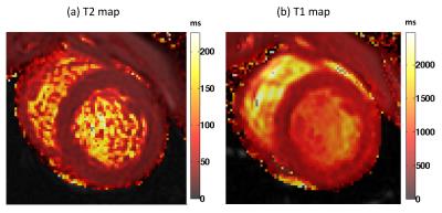
Non-contrast assessment of vasodilator response using native myocardial T1 and T2 mapping and Arterial Spin Labeled CMR 
Nilesh Ghugre, Hung Do, Kenneth Chu, Venkat Ramanan, Krishna Nayak, Graham Wright
Myocardial vasodilator response is an important indicator of microvascular function and integrity in ischemic injury. The objective of our study was to systematically compare myocardial stress response with native contrast mechanisms involving quantitative T2, T1 and Arterial spin labeled (ASL) imaging. Our findings suggest that oxygenation (T2 BOLD effect), blood volume (T1 effect) and perfusion (ASL) taken together could offer a complementary contrast-free framework to identify vasodilator dysfunction in heart disease. These could potentially offer insights into the myocardial remodeling process particularly in the remote territory, which develops hypertrophy and fibrosis in the high-risk patients in the chronic stage.
|
|
0545.
 |
6 |
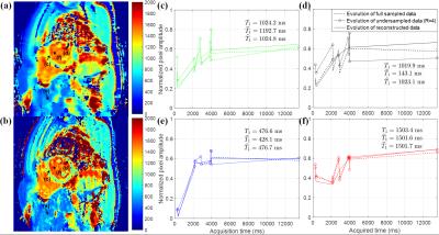
Dictionary-based Reconstruction for Free-Breathing Myocardial T1 Mapping 
Jinkyu Kang, Jihye Jang, Vahid Tarokh, Reza Nezafat
In this study, we propose a novel reconstruction framework for myocardial T1 mapping based on a dictionary-based reconstruction algorithm that simultaneously reduces scan time while compensating for respiratory and cardiac-induced motions between different T1-weighted images of T1 mapping sequence.
|
|
0546.
 |
7 |
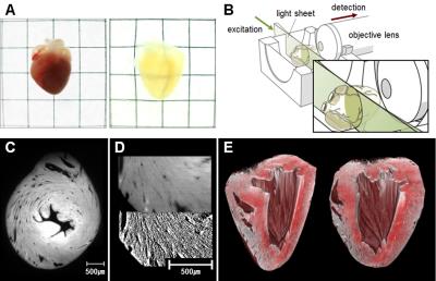
Validation of Cardiac Diffusion Tensor MRI using Transparent Tissue Preparation (CLARITY) with 3D Optical Microscopy 
Christopher Nguyen, Sang-Eun Lee, Jongjin Yoon, Hyuk-Jae Chang, Sekeun Kim, Chul Hoon Kim, Debiao Li
The myocardium consists of a complex 3-dimensional (3D) microstructure has been shown to be perturbed in the presence of myocardial ischemia. Recently, diffusion tensor magnetic resonance imaging (DT-CMR) was introduced which can characterize the 3D tissue microstructure in intact myocardium. However, past histologic validation of DTI has been limited since traditional pathology allows only 2D optical microscopy after potentially destructive tissue sectioning. We present a novel approach to validate the derivation of the myocardial fiber orientation (MFO) using DT-CMR with 3D histology using a non-destructive, transparent-tissue preparation technique (CLARITY). Results indicate MFO derived from 3D histology and DT-CMR are strongly concordant.
|
 |
0547.
 |
8 |
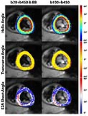
Free-breathing Black-blood Prepared Cardiac Diffusion Tensor Imaging - permission withheld
Constantin von Deuster, Georg Spinner, Robbert van Gorkum , Christian Stoeck, Sebastian Kozerke
In vivo cardiac Diffusion Tensor Imaging (DTI) can be influenced by myocardial perfusion. To address this limitation, reference images with moderate diffusion weighting can be employed, which, however, reduce diffusion contrast and require the acquisition of three orthogonal diffusion encoding directions. In this work, blood suppression was implemented using black blood preparation to reduce contribution of perfusion to the diffusion weighted signal. This allows for the use of a marginally weighted reference image resulting in a reduction in scan time by about 17% with improved or comparable accuracy of diffusion metrics relative to previous methods.
|
 |
0548.
 |
9 |
First-Pass Nitroxide-Enhanced MRI for Imaging Myocardial Perfusion without Gadolinium 
Sophia Cui, Frederick Epstein
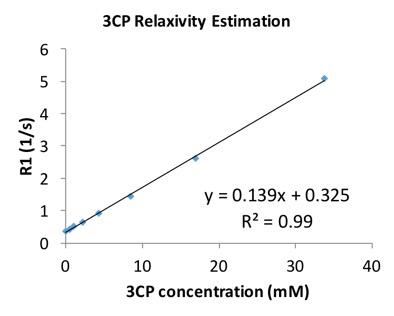
First-pass MRI using gadolinium-based contrast agents is widely used to image myocardial perfusion. However, gadolinium is contraindicated for patients with severely impaired renal function (a substantial portion of heart disease patients), and methods that do not employ gadolinium are needed. Nitroxide stable free radicals are non-metallic compounds with an unpaired electron and, correspondingly, are paramagnetic and T1-shortening. We investigated first-pass nitroxide-enhanced perfusion MRI of the heart as an alternative to first-pass gadolinium-enhanced MRI. Five C57Bl/6 mice underwent first-pass imaging with the nitroxide agent 3CP and the results showed that nitroxide-enhanced MRI can quantify regional myocardial blood flow, as the average myocardial perfusion was 7.0±1.3 ml/g/min, a value in the normal range for mice.
|
 |
0549.
 |
10 |
Cardiac fMRI - A Novel Approach for Reliably Detecting Myocardial Oxygenation Changes with Precise Modulation of Arterial CO2 
Hsin-Jung yang, Ilkay Oksuz, Michael Klein, Olivia Sobczyk, Damini Dey, Jane Sykes, John Butler, Xiaoming Bi, Behzad Sharif, Ivan Cokic, Debiao Li, Piotr Slomka, Frank Prato, Joseph Fisher, Sotirios Tsaftaris, Rohan Dharmakumar
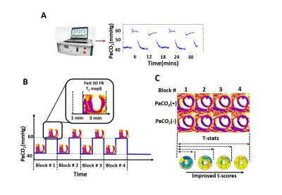
Although cardiac BOLD MRI can detect ischemic heart disease without ionizing radiation and contrast agents, its reliability remains poor due to the low sensitivity and specificity. We propose a novel strategy to overcome these barriers through: (i) an improved MRI strategy with free gas-exchange capability; (ii) repeat stimulation of heart using a validated prospective arterial CO2 targeting technique; and (iii) a statistical framework to increase the confidence measure of BOLD signal changes. Our results show that the proposed approach can be used to significantly increase the confidence in detecting myocardial BOLD response in conditions of health and disease.
|
|
0550.
 |
11 |
High-fat diet feeding in mice may partially protect the heart from pressure overload induced heart failure - a longitudinal study of cardiac metabolism and function 
Emmy Manders, Desiree Abdurrachim, Miranda Nabben, Klaas Nicolay, Jeanine Prompers
Obesity increases the risk of heart failure, but obese heart failure patients have a better prognosis and survival. Altered cardiac energy metabolism is proposed to be an important contributor to this discrepancy. With an in vivo longitudinal approach measuring cardiac function (MRI), energetics (31P-MRS) and lipid content (1H-MRS) during the development of heart failure we have shown that cardiac function was less impaired in obese mice compared with lean mice after induction of pressure overload. This suggests that metabolic adaptations in obese mice are not detrimental and may even be beneficial in heart failure development.
|
 |
0551.
 |
12 |
Cardiac Magnetic Resonance Elastography for Quantitative Assessment of Elevated Myocardial Stiffness in Cardiac Amyloidosis 
Arvin Arani, Shivaram Arunachalam, Ian Chang, Francis Baffour, Kevin Glaser, Joshua Trzasko, Kiaran McGee, Armando Manduca, Martha Grogan, Angela Dispenzieri, Richard Ehman, Philip Araoz
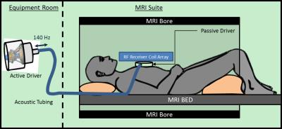
Myocardial stiffness plays an important role in cardiac function. The objective of this study is to evaluate if 3D high frequency cardiac MR elastography (MRE) can measure increased myocardial stiffness in cardiac amyloidosis patients compared to healthy volunteers. Twenty-two patients with cardiac amyloidosis and 16 healthy volunteers were enrolled. The myocardial stiffness of cardiac amyloid patients (median: 11.4 kPa, min: 9.2, max: 15.7) was found to be significantly stiffer (p < 0.01) than healthy controls (median: 8.2 kPa, min: 7.2, max: 11.8). These results motivate future investigation of 3D high frequency cardiac MRE in different patient cohorts.
|
|
0552.
 |
13 |
Ungated myocardial perfusion imaging with complete left ventricular coverage using radial simultaneous multi-slice imaging 
Ganesh Adluru, Jason Mendes, Ye Tian, Brent Wilson, Edward DiBella
Myocardial perfusion imaging is a promising tool to determine the downstream effects of blocked coronary arteries. Ungated perfusion imaging is a promising alternative to conventional ECG-gated acquisitions. Existing ungated methods can acquire only 4-5 short-axis slices often with large slice gaps. Continuous and complete coverage is desired in order to avoid missed regions of perfusion deficit. Here we use an undersampled radial simultaneous multi-slice (SMS) acquisition to obtain complete coverage of the left ventricle. We use a block matching constrained reconstruction that is robust to inter-time frame cardiac and respiratory motion. We obtain 12 short axis slices in ~250 msec.
|
|
0553.
 |
14 |
As Easy as Echo: Interactive Fetal Cardiac MR Imaging 
Davide Piccini, Jérôme Yerly, Jérôme Chaptinel, Milan Prsa, Yvan Mivelaz, Leonor Alamo, Yvan Vial, Gregoire Berchier, Chantal Rohner, Peter Speier, Tobias Kober, Matthias Stuber
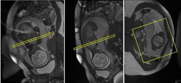
Although fetal echocardiography remains the gold standard for prenatal detection of congenital heart disease, mainly due to its ease-of-use, availability, and high diagnostic performance, MRI is occasionally used as a complementary and safe modality. However, MRI workflow is problematic as bulk fetal motion can occur anytime during acquisition, but can only be identified retrospectively, after image reconstruction. We describe an acquisition scheme that changes this workflow and allows interactive real-time planning of the fetal cardiac scan. The operator can easily find the desired scan plane even in a moving imaging target. This technique is applied and tested in two pregnant patients.
|
 |
0554.
 |
15 |
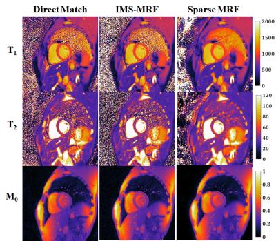
Low Rank Compressed Sensing Reconstruction for More Precise Cardiac MRF Measurements - permission withheld
Jesse Hamilton, Yun Jiang, Dan Ma, Yong Chen, Shivani Pawha, Wei-Ching Lo, Joshua Batesole, Mark Griswold, Nicole Seiberlich
A low rank compressed sensing and parallel imaging reconstruction termed Sparse MRF is introduced to improve the precision of mapping myocardial T1 and T2 with MR Fingerprinting. Sparse MRF enforces data consistency while also constraining the temporal signal evolutions using a low dimensional subspace derived from the SVD of the dictionary along time. Different reconstruction parameters are investigated in simulations with a cardiac phantom. Results from phantom and in vivo cardiac scans indicate that Sparse MRF yields approximately the same mean T1 and T2 measurements as other MRF matching techniques but with smaller standard deviations.
|
|
 Power Pitches Video


