ISMRM 25th Annual Meeting & Exhibition • 22-27 April 2017 • Honolulu, HI, USA

Traditional Poster Session: General Cancer Imaging
2888 -2910 Cancer Diffusion, Perfusion & Other
2911 -2930 Cancer Treatment Response & Preclinical
 
Cancer Diffusion, Perfusion & Other
Traditional Poster
General Cancer Imaging

 
Thursday, 27 April 2017
Exhibition Hall  13:00 - 15:00


2888.  
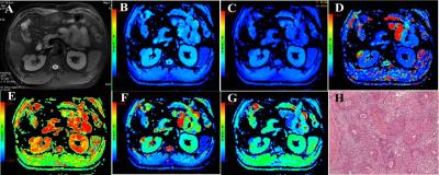
The Uesfulness of Intravoxel Incoherent Motion Diffusion-weighted Imaging for Predicting the Prognosis of Patients with Acute Myeloid Leukemia
JinLiang Niu, HongWei Wang, XiaoLi Song
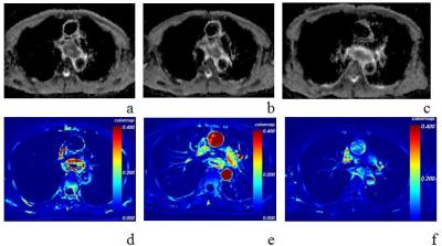
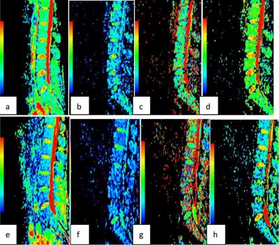
Intravoxel Incoherent Motion (IVIM) is capable of providing both diffusion and perfusion quantification using a single imaging study at the same time, without the need for intravenous contrast injection. acute myeloid leukemia (AML) patients with complete remission and non-complete remission could exhibit different characterizations before treatment in perfusion and tissue cellularity of the lumbar bone marrow. This study was to investigate the usefulness of IVIM parameters in evaluation of prognosis in AML patients. 


2889.   
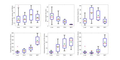
Repeatability of perfusion parameters in free-breathing dynamic contrast-enhanced MRI of malignant hepatic tumors
Bohyun Kim, Kyung Won Kim, Chang Kyung Lee, Nieun Seo, Seung Soo Lee, Jeong Kon Kim
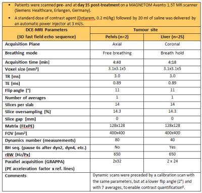
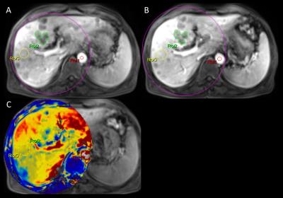
In dynamic contrast-enhanced (DCE)-MRI, establishing a high level of repeatability is essential to ensure reliability of changes in perfusion parameters over longitudinal studies in an individual subject. To assess the repeatabilities of perfusion parameters and identify the effect of T1 map on them, we performed two sequential free-breathing DCE-MRI studies in patients with solid hepatic malignancies. Our results showed that Kep had the highest repeatability, and the repeatabilities of all perfusion parameters were boosted when fixed T1 value (800 ms) was used instead of values obtained from T1 mapping.


2890.   
Quantitative evaluation of the effect of temporal resolution and acquisition duration on the accuracy of DCE-MRI measurements in a prostate phantom
Silvin Knight, Jacinta Browne, James Meaney, Andrew Fagan
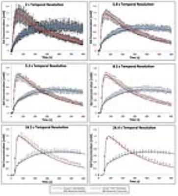
A novel anthropomorphic prostate phantom test device was used to investigate the effects of temporal resolution (Tres) and acquisition duration (AD) on the accuracy of contrast time-intensity curves measured using dynamic contrast-enhanced MRI.  When quantitatively compared to ground truth values, large errors in derived pharmacokinetic (PK) parameters (up to 230%) were found for Tres values > 8.1 s and AD values < 360 s.  The data demonstrate the critical and sensitive dependence of the accuracy of measured PK output parameter values on the acquisition protocol used, while such phantom studies can help identify optimal acquisition parameters for DCE-MRI scans


2891.  
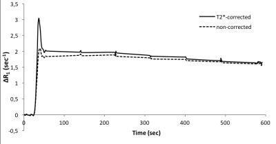
Improved sensitivity for rectal cancer differentiation by T2*-correction in DCE-MRI
Endre Grřvik, Kathrine Redalen, Tryggve Storĺs, Anne Negĺrd, Stein Holmedal, Anne Ree, Sebastian Meltzer, Atle Bjřrnerud, Kjell-Inge Gjesdal
Quantitative pharmacokinetic analysis from DCE-MRI often neglects CA-induced T2*-effect when converting signal intensity (SI) changes in tissue to CA concentration. The acquired signal may thus be significantly affected by T2* signal attenuation, which in turn results in errors in the DCE-derived parameters. This work utilized a dynamic multi-echo acquisition for extrapolation back to TE=0, thus providing both T2*-corrected and uncorrected measurements of DR1, and investigates the how the correction for T2*-attenuation effects may affect the sensitivity for rectal cancer differentiation by DCE-MRI. The T2*-corrected data showed improved differentiation of rectal cancer.


2892.   
Enhancement of Lesion Detection through Feature Projection and Classification of Perfusion Parameters
Ramin Jafari, Yi Wang, Martin Prince, Pascal Spincemaille
Perfusion analysis is a powerful tool to quantitatively characterize lesions. This can further be extended to differentiate lesions through comparison perfusion parameters. Typically there is overlap in perfusion parameters which makes it difficult to distinguish lesions. We demonstrate perfusion analysis combined with feature projection and classification enhances lesion detection by increasing separability and extracting features of the lesions simultaneously which can be used in automation of lesion detection. 


2893.   
Quantitative Ultra-short Time-to-Echo Contrast-Enhanced Magnetic Resonance Imaging (qUTE-CE MRI) technique use Ferumoxytol as a Positive-Contrast Agent to Delineate Nanoparticle Accumulation in Tumors
Ju Qiao, Codi Gharagouzloo, Liam Timms, Paraveen Kulkarni, Craig Ferris, Srinivas Sridhar, Anne van de Ven
Here we demonstrate quantitative ultra-short time-to-echo contrast-enhanced magnetic resonance imaging (qUTE-CE MRI) technique, which employs FDA-approved ferumoxytol as a positive-contrast agent to delineate nanoparticle accumulation in tumors. 


2894.   
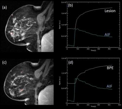
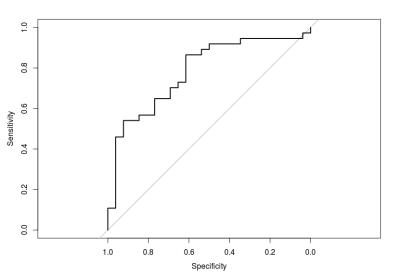
Evaluation of DCE-MRI Exam with High-Temporal Resolution for Breast Cancer Diagnosis in a Biopsy Cohort
Oliver Stewart, Laura Heacock, Linda Moy, Sungheon Gene Kim
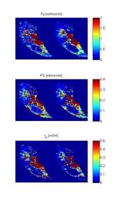
The purpose of this study was to assess the feasibility of using GRASP DCE-MRI for contrast kinetic analysis to determine lesion malignancy. This IRB-approved retrospective study included 73 women who underwent MRI-guided biopsy scans. The plasma flow in the malignant group was significantly higher than that of the benign group with the area under the curve of 0.77. The results in this study successfully demonstrate that the GRASP DCE-MRI method can be used to acquire adequate high-temporal and high-spatial resolution images of the breast for contrast kinetic analysis.


2895.   
Dynamic Contrast-Enhanced MRI Using Spatial Prior Knowledge
Radovan Jirík, Michal Bartoš, Karel Soucek, Eva Dražanová, Lucie Krátká, Lenka Dvoráková, Torfinn Taxt, Zenon Starcuk, jr.
A method to improve reliability of DCE-MRI is presented. It is based on the assumption that neighboring voxels have similar physiological properties. It is formulated as a total variation regularization of the perfusion parameter maps. The method is applied to the adiabatic approximation of the tissue homogeneity model (ATH). Evaluation was done on data of five tumor bearing mice recorded using two contrast agents with different molecular weights. The resulting perfusion parameter maps were more spatially consistent with preserved edges.


2896.  
Are radiation-induced perfusion changes in normal appearing brain tissue a confounding factor in tumour response evaluation with DSC-MRI?
Markus Fahlström, Tufve Nyholm, Elna-Marie Larsson
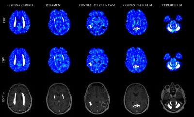
Radiation-induced brain perfusion changes after radiotherapy have not been studied extensively with DSC-MRI. Tumour perfusion is often normalised to contralateral normal-appearing white matter, why knowledge about radiation effects on perfusion in normal brain tissue is essential. Ten patients were examined with DSC-MRI at 1.5 T before and repeatedly after RT. Multiple volumes were drawn in normal brain tissue. Radiation dose for all volumes were available. Significant CBV and CBF decrease after radiotherapy was visible in grey matter volumes and showed tendency to recover. High radiation dose regions should be avoided when normalising DSC-MRI derived tumour perfusion values.  


2897.   
A population-based digital reference object (DRO) for optimizing dynamic susceptibility contrast (DSC) MRI methods for clinical trials
Natenael B Semmineh, Ashley M Stokes, Laura C Bell, Jerrold L Boxerman, C Chad Quarles
The standardization of DSC-MRI has been confounded by a lack of consensus on DSC-MRI methodology for preventing potential rCBV inaccuracies, including the choice of acquisition protocols and post-processing algorithms. To address these issues, we developed a digital reference object (DRO) aimed at validating image acquisition and analysis methods for accurately measuring rCBV in glioblastomas.  The DRO was developed using trained physiological and kinetic parameters derived from in vivo data, unique voxel-wise 3D tissue structures, and a validated MRI signal computational approach. The DRO’s ability to produce reliable signals was validated by comparison to separate cohort of patient data.


2898.   
Probing infiltration of the normal brain by glioblastoma using magnetic resonance perfusion and permeability techniques
Antoine Vallatos, Joanna Birch, Lindsay Gallagher, Haitham Al-Mubarak, Lesley Gilmour, Anthony Chalmers, William Holmes
MRI techniques probing brain perfusion and blood-brain barrier (BBB) permeability were assessed in their ability to detect low tumour invasion in mouse glioblastoma models. A multiple boli Arterial Spin Labeling technique was optimised, achieving high SNR perfusion imaging. Diffusion-weighted arterial spin labelling allowed to separate fast motion vascular components of the signal from slow motion tissue components, providing with BBB permeability weighted maps. Evaluation was performed by comparison with conventional MRI and immunohistochemistry sections (HLA) cut in the MRI plane . Both perfusion weighted maps and BBB permeability weighted maps allowed to identify low tumour regions not detected with conventional MRI techniques.


2900.   
DDC and a estimates from the stretched exponential model are more robust to variations in b-value selection than ADC estimates, in a cohort of 42 cervical tumours
Jessica Winfield, Nandita deSouza, Veronica Morgan, David Collins, Matthew Orton
The influence of b-value selection on fitted parameters from the stretched exponential model (distributed diffusion coefficient DDC, and ‘stretching parameter’ α) and mono-exponential model (apparent diffusion coefficient ADC) was investigated in a cohort of 42 patients with cervical tumours. Diffusion-weighted images images were acquired using 9 b-values between 0 and 800smm-2, and fitted voxel-by-voxel using all b-values (0 to 800smm-2) and b-values n-to-800smm-2 (where n is 20,40,60,80,100, or 300smm-2). ADC estimates are highly sensitive to b-value selection, with reduction in ADC when low b-values are excluded, whereas DDC and α are more robust to differences in b-value selection.


2899.   
Application of Monoexponential, biexponential and stretched exponential Diffusion-weighted MR Imaging for Differentiating between Minimal Fat Angiomyolipoma (MFAML) and Papillary Renal Cell Carcinoma (PRCC).
Haojie Li, Yao Hu, Daoyu Hu, Zhen Li
Accurate preoperative differentiation between MFAML and PRCC is important to determine appropriate treatment strategies.In this study, the preliminary date from our study show that different models of DWI may demonstrate various aspects of tissue properties.fp and α may provide additional information and could lead to improved differentiation with better sensitivity and specificity between MFAML and PRCC compared with conventional imaging parameters.


2901.   
Combined analysis of ten DW-MRI studies demonstrates excellent repeatability of apparent diffusion coefficient estimates in extra-cranial soft-tissue tumours
Jessica Winfield, Nina Tunariu, Mihaela Rata, Keiko Miyazaki, Neil Jerome, Michael Germuska, Matthew Blackledge, David Collins, Johann de Bono, Timothy Yap, Nandita deSouza, Simon Doran, Dow-Mu Koh, Martin Leach, Christina Messiou, Matthew Orton
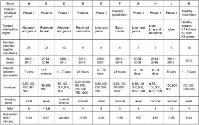
Repeatability of ADC estimates from ten diffusion-weighted MRI studies in extra-cranial soft tissues (nine patient studies and one healthy volunteer study, with a total of 111 subjects) were analysed in combination using the Radiological Society of North America (RSNA) Quantitative Imaging Biomarkers Alliance (QIBA) framework for assessment of technical performance of imaging biomarkers, in order to investigate factors affecting ADC repeatability. Coefficient of variation was between 2 and 7% for all studies, with no marked differences between imaging protocols or study populations, and better repeatability in large tumours compared with smaller tumours, indicating that ADC is a robust imaging metric with excellent repeatability in extra-cranial soft-tissue tumours.


2902.  
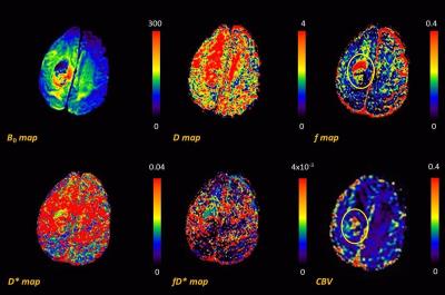
Correlation of Intra-Voxel incoherent motion (IVIM) imaging and Dynamic Susceptibility Contrast (DSC) MRI for high and low grade gliomas
Arush Honnedevasthana Arun, Neesha Nagaraj, Nithin Vajuvalli, Jitender Saini, Sairam Geethanath
The aim of this study was to validate the feasibility of Intravoxel Incoherent Motion (IVIM) imaging of human brain gliomas; and to differentiate between high grade and low grade gliomas using IVIM parameters (f, d, D, fD*). These parameters were then compared to CBV maps obtained through DSC-MRI to evaluate the role of non-contrast agent based perfusion imaging. Results obtained showed that IVIM perfusion fraction f could be used to differentiate between high and low-grade brain gliomas


2903.   
Differential diagnosis of Ocular adnexal lymphoma and idiopathic orbital inflammation:radiomics imaging features and their predictive performance
Guo Jian, Xian Junfang, Shen Chen, Liu Zhenyu, Tian Jie
Differential diagnosis of Ocular adnexal lymphoma (OAL) and idiopathic orbital inflammation (IOI) is very important due to their quite different therapeutic modalities and prognosis. However, it is not easy for both ophthalmologists and pathologists. Can the emerging radiomics enhance the diagnostic efficiency? This research devoted evidence for this question.


2906.   
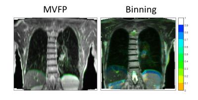
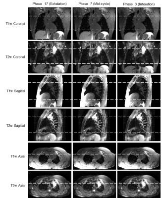
Evaluation of 4D-T2w MRI methods for lung radiotherapy treatment planning with application to an MR-linac
Joshua Freedman, David Collins, Christopher Rank, Hannah Bainbridge, Simeon Nill, Marc Kachelrieß, Martin Leach, Uwe Oelfke, Andreas Wetscherek
Due to enhanced soft-tissue contrast, 4D-T2w MRI could improve lung tumour target delineation in radiotherapy compared with 4D-CT; reducing planning margins and dose to healthy tissue for treatments on an MR-linac. We have developed a method for generating 4D-T2w MRI, where 3D-T2w MRI is warped using motion vector fields; calculated by image-registration between respiratory phases of 4D-T1w MRI (the MVFP method). Here, 4D-T2w MRI calculated with MVFP and with amplitude-binning of continuously acquired 2D-T2w MRI, are compared. 4D-T2w MRI from the MVFP method exhibited higher image quality and geometrical fidelity than the 2D-binning scheme, but displayed possible motion under-estimation.


2904.   
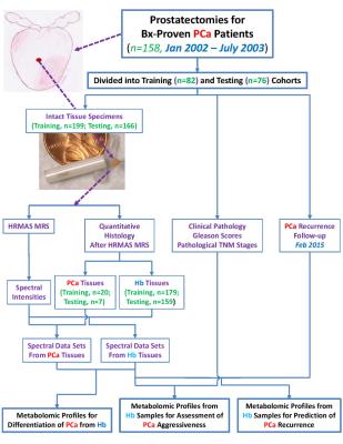
Prognostic Characterization of Prostate Cancer from Benign Tissue MRS
Leo Cheng, Emily Decelle, Johannes Kurth, Shulin Wu, Taylor Fuss, Lindsey Vandergrift, Elita DeFeo, Elkan Halpern, Matthias Taupitz, W. McDougal, Aria Olumi, Chin-Lee Wu
While serum prostate specific antigen (PSA) testing improved early detection of prostate tumors, implementation of this tool also created a large patient population in which identified cancer lesions were actually indolent. For patients who choose to undergo prostatectomy, cancer aggressiveness can only be determined by post-procedure pathology analysis of cancerous tissue. Additionally, cancer recurrence predictions are often unreliable. As an alternative method to aid decisions regarding treatment, we sought to develop MRS tools which can predict cancer aggressiveness based on the novel use of histologically benign (Hb) tissue. 


2905.  
Texture Analysis on Diffusion Weighted MRI in Osteosarcoma
Esha Baidya Kayal, Devasenathipathy K, Sameer Bakhshi, Raju Sharma, Amit Mehndiratta
Texture analysis (TA) uses mathematical approach to characterize the spatial distribution of signal intensity variations in an image, and extracts quantitative information that are otherwise imperceptible qualitatively. Good soft tissue contrast of MRI facilitates TA to produce promising results in diagnosis and prognosis. Diffusion Weighted (DW) MRI based TA might provide additional useful information as DWI captures early cellular changes in tumors in terms of corresponding intensity variations. We computed textural features in patients with Osteosarcoma. Experimental results revealed that textural features show good discrimination among normal and tumour ROI thus might be useful as diagnostic and prognostic marker.


2907.   
4D-T2 weighted MRI for lung radiotherapy treatment planning
Joshua Freedman, David Collins, Christopher Rank, Hannah Bainbridge, Simeon Nill, Marc Kachelrieß, Uwe Oelfke, Martin Leach, Andreas Wetscherek
Compared to 4D-CT, 4D-T2w MRI should better characterise respiratory motion in radiotherapy, due to its superior soft tissue contrast. However, 4D-T2w MRI is difficult to acquire due to the long echo and repetition time required. Here, 4D-T2w MRI is obtained by applying motion information from a 4D-T1w volume to a static 3D-T2w volume. For three patients, 4D-T2w MRI was compared to 4D-T1w MRI. Diaphragm positions were consistent to <1.5mm. A method to calculate 4D-T2w MRI, with potential application to an MR-linac, is presented and verified.


2908.  
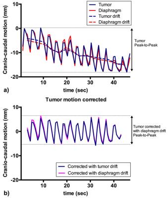
Can diaphragm motion function as a surrogate for motion of esophageal tumors during treatment?
Sophie Heethuis, Lucas Goense, Alicia Borggreve, Peter van Rossum, Richard van Hillegersberg, Jelle Ruurda, Stella Mook, Gert Meijer, Jan Lagendijk, Astrid van Lier
Esophageal tumors show large motion in cranio-caudal direction, with a Peak-to-Peak (P-t-P) range of 2.7 to 24.5mm. In case the motion of the tumor could be followed during radiotherapy treatment, this would enable treatment margin reduction. It is researched whether the motion of the diaphragm is correlated with the breathing motion and drift we can detect in esophageal tumors since it could function as surrogate for tumor motion during treatment. A high correlation was found between both motion patterns and correction of the tumor motion using the diaphragm drift resulted on average in a reduction in P-t-P motion in all patients.


2909.   
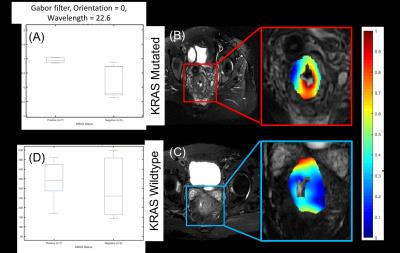
Radiomic features extracted from T2w MRI differentiates KRAS mutational status in rectal cancers: A pilot study
Shanker Raja, Jacob Antunes, Amrish Selvam, Anant Madabhushi, Sharad George, Mazen Alsagri, Adnan Hussain, Abdullah AlDosary, Musa Fageeh, Mohammed AlHarbi, Satish Viswanath
Contemporary management of rectal cancer includes consideration of adjuvant anti EGFR therapy; however, KRAS mutational status profiling is mandatory prior to initiation of this therapy.  KRAS mutational status is typically assayed on tissue samples (biopsy/surgical specimens). Radiomic features have demonstrated the ability to quantitatively characterize image phenotypic appearance associated with genotype expression in different tumors. We explored the utility of multi-scale, multi-oriented filter banks to define radiomics signatures that distinguish KRAS mutated vs wildtype tumors on T2W MRI.  Initial results indicate that macro-scale radiomic features depict significantly different responses between the 2 genotypic groups.


2910.   
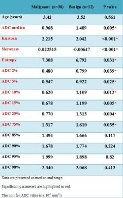
The role of ADC histogram analysis in discriminating between benign and malignant tumours in children
Karen Manias, Niloufar Zarinabad, Emma Meeus, Katharine Foster, Paul Davies, Jan Novak, Andrew Peet
We evaluated histogram analysis of ADC maps to differentiate benign from malignant solid paediatric tumours in children and distinguish tumour type. Fifty children (38 malignant, 12 benign tumours) were imaged using multi-b value diffusion-weighted MRI at diagnosis. Whole tumour regions-of-interest (ROIs) were drawn, ADC histograms constructed, and median, 2nd to 98th percentile ADC values, skewness, kurtosis and entropy calculated. ADC histogram parameters were significantly different between malignant and benign lesions, with skewness and kurtosis being the most predictive of malignancy. 
 
Cancer Treatment Response & Preclinical
Traditional Poster
General Cancer Imaging

 
Thursday, 27 April 2017
Exhibition Hall  13:00 - 15:00


2911.   
ADC and D from diffusion-weighted MRI correlate with histopathological assessment of nuclear-to-stromal ratio, and histology confirms Dixon fat fraction in retroperitoneal sarcomas
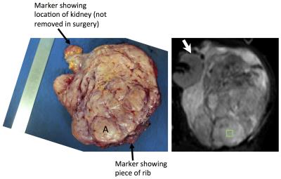
Jessica Winfield, Khin Thway, Aisha Miah, Dirk Strauss, David Collins, Martin Leach, Nandita deSouza, Sharon Giles, Christina Messiou
Soft-tissue sarcomas are often highly heterogeneous tumours. Clinical trials of non-surgical treatments, for example combined radiotherapy and systemic agents, require non-invasive methods for response assessment. Quantitative MRI provides non-invasive response assessment of the whole tumour volume, but metrics require validation against histopathology. In this study, 26 patients with retroperitoneal sarcoma were imaged prior to surgery, with written consent, as part of a prospective single-centre study. Diffusion-weighted MRI parameters (ADC, and D from IVIM) showed correlation with nuclear-to-stromal ratio, and were also related to stroma type and stroma grade. Dixon-derived fat fraction correlated strongly with histopathological assessment of fat fraction.


2912.   
Label-free CEST MRI detection of albumin and albumin-based nanoparticles
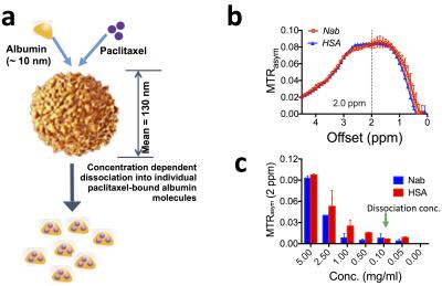
Yuguo Li, Dexiang Liu, Jiadi Xu, Peter van Zijl, Shibin Zhou, Guanshu Liu
Albumin is emerging as one of the most attractive drug carriers for the targeted delivery of therapeutic peptides and drugs. Herein, we developed a label-free MRI approach for monitoring albumin-based drug delivery systems by directly utilizing the inherent CEST signal of albumin. Our in vitro study showed that both human serum albumin (HSA) and albumin-based nanoparticle Nab-paclitaxel (Abraxane) could generate a strong CEST signal at ~2 ppm. Using this inherent CEST signal, we successfully demonstrated the label-free CEST MRI detection of the tumor uptake and distribution HSA and Nab-paclitaxel in a xenograft mouse model.  


2913.  
Quantitative ADC as an early imaging biomarker of response to chemoradiation in esophageal cancer
Benjamin Musall, Jingfei Ma, Brett Carter, Penny Fang, Amy Moreno, Jong Bum Son, Brian Hobbs, Bryan Fellman, Steven Lin
The goal of the study was to investigate if quantitative ADC can be used as an early imaging biomarker for predicting the treatment response to chemoradiation in esophageal cancer.  Using pathological findings as the gold standard, our study demonstrated that the change in quantitative ADC from baseline (before treatment) to interim (two weeks after the initiation of treatment) was highly predictive of whether patients had residual tumors at the end of their treatment.


2914.   
Non-gaussian IVIM imaging biomarkers can non-invasively predict the aggressiveness of small papillary thyroid cancers
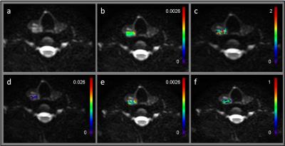
David Aramburu Nuńez, Yonggang Lu, Vaios Hatzoglou, Andre L Moreira, Hilda E Stambuk, Ramesh Paudyal, Yousef Mazaheri, Mithat Gonen, Joseph O Deasy, Ronald A Ghossein, Ashok R Shaha, Michael Tuttle, Amita Shukla-Dave
There is a need for non-invasive imaging to identify patients with aggressive tumors in papillary thyroid carcinoma (PTC). This study evaluates whether non-gaussian intravoxel incoherent motion (NG-IVIM) DW-MRI has the potential to stratify tumor aggressiveness in PTC. Twenty-four PTC patients underwent pretreatment NG-IVIM DW-MRI at 3T. The apparent diffusion coefficient (ADC), perfusion factor (f), diffusion (D), pseudo diffusion (D*) and diffusion Kurtosis (K) coefficients were calculated from the NG-IVIM model. All patients underwent surgery. Tumor aggressiveness was defined at pathology. ADC and D may be used to distinguish tumors with and without aggressive features in tumor size between 1-2 cm.


2915.   
Reproducibility of Quantitative DW-MRI and DCE-MRI of the Breast in the Community Setting: Preliminary Results
Anna Sorace, Jack Virostko, Stephanie Barnes, Jeffrey Luci, Debra Patt, Boone Goodgame, Sarah Avery, Thomas Yankeelov
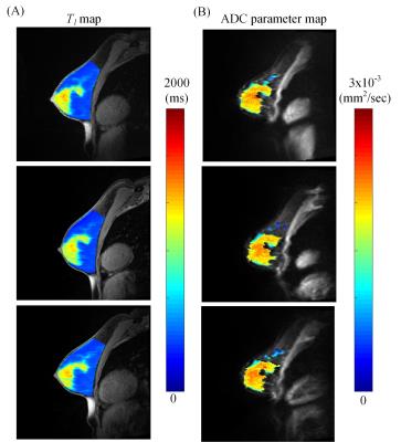
Implementation of quantitative DCE-MRI and DW-MRI in the community setting has the potential to impact patient care for a large number of breast cancer patients. Quantitative DW-MRI and DCE-MRI was assessed in phantoms and normal subjects across three sites. In normal subject fibroglandular tissue, the average percent difference in ADC across sites for all subjects was 1.8%, while the average percent difference in the inversion recovery scan and B1- corrected T1 map were 14% and 7.3%, respectively. Overall, the results from the phantom and normal subject scans reveal that quantitative MRI can be successfully implemented in the community setting.


2916.  
Ultra-early ADC (apparent diffusion coefficient) footprint successfully detects tumor irradiation and predicts radiotherapy outcome
Faisal Mahmood, Helle Hjorth Johannesen, Poul Geertsen, Rasmus Hvass Hansen
If ultra-early stratification of radiotherapy response was possible it could potentially reduce unnecessary irradiation of normal tissue and improve disease management. In this study repeated diffusion weighted magnetic resonance imaging was conducted along with fractionated radiotherapy in brain metastases patients. It was found that the decrease in the apparent diffusion coefficient (ADC) observed 24 hours after the first radiotherapy fraction may be an indicator of irradiation. Responding patients versus non-responding patients could be differentiated by their corresponding change in ADC seen 48 hours after start of radiotherapy. These findings may have great impact for the emerging hybrid MR – linear-accelerator systems.


2917.  
Providing improved reliability of plasma volume fraction estimates for monitoring anti-angiogenic effects in liver metastases with DCE-MRI
Mihaela Rata, Khurum Khan, David Collins, Nina Tunariu, Dow-Mu Koh, James d'Arcy, Maria Bali, Ian Chau, Nicola Valeri, David Cunningham, Martin Leach, Matthew Orton
Pharmacokinetic modelling of DCE-MRI data allows characterisation of tumour response to anti-angiogenic therapies by estimating the volume transfer constant Ktrans and the plasma volume fraction Vp. This work assesses the impact on Ktrans and Vp of treatment changes in an early clinical trial cohort by comparing 4 models: Kety, extended Kety (with two lower limits on Vp), and a new model with Vp proportional to Ktrans. When Ktrans is the endpoint of interest, use of the Kety model is sufficient to depict cohort response. If Vp is also of interest, the new model improves the significance of Vp treatment effects.


2918.   
Quantitative Evaluation of Treatment Response to Early Concurrent Chemoradiotherapy with Multi-parametric MRI in Esophageal Cancer
Tieming Xie, Guoliang Shao, Lulu Liu, Jun Yang, Peipei Pang
The best dose of concurrent chemoradiotherapy for esophageal cancer exists individual difference because of tumor heterogeneity. The treatment will be more predictable if we can assess the chemoradiotherapy earlier, which will be helpful in early intervention for optimization of treatment plan, treatment time and improvement of overall survival. Multi-parametric MRI is gradually used for evaluating tumor treatment, and provides earlier and more information than conventional MRI. In this study, we used Ktrans value (derived from DCE-MRI) and ADC value (derived from DWI) to assess treatment response after the fifth concurrent chemoradiotherapy in esophageal cancer.  


2919.   
In vivo OE-MRI quantification and mapping of response to hypoxia modifying drugs Banoxantrone and Atovaquone in Calu6 xenografts
Ross Little, Victoria Tessyman, Muhammad Babur, Susan Cheung, Yvonne Watson, Roben Gieling, Katherine Finegan, Thomas Ashton, Geoff Parker, W Mckenna, Geoffrey Higgins, Kaye Williams, James O'Connor
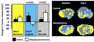
Oxygen-enhanced MRI (OE-MRI) has shown promise as a technique for quantifying and spatially mapping tumour hypoxia. Here we report the first evidence that OE-MRI signals in perfused tumour can non-invasively track therapy-induced changes in hypoxia in vivo in a tumour model. We show that OE-MRI detects (1) reduction in hypoxia and increase in necrosis induced by the hypoxia-activated cytotoxic prodrug Banoxantrone; and (2) reduction in hypoxia and increase in well oxygenated tumour induced by Atovaquone due to increased oxygen availability. These data support first-in-man use of OE-MRI biomarkers in clinical trials of hypoxia-modifying agents. 


2920.  
Combining DW- and DCE-MRI for treatment response assessment in patients with esophageal cancer undergoing neoadjuvant chemoradiotherapy
Sophie Heethuis, Lucas Goense, Peter van Rossum, Alicia Borggreve, Stella Mook, Francine Voncken, Richard van Hillegersberg, Jelle Ruurda, Gert Meijer, Jan Lagendijk, Astrid van Lier
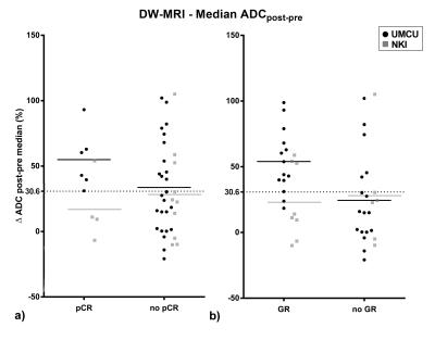
Neoadjuvant chemoradiotherapy prior to surgery is often used for treatment of patients with esophageal cancer. Potential benefit could be gained developing a patient tailored treatment, especially for the 29% of the patients who show a pathologic complete response. In this prospective multicenter study it was investigated whether combining DCE- and DW-MRI, which both showed potential for response prediction in previous studies, yields complementary information. It was found that the combination of DCE- and DW-MRI can increase the predictive values and reaches higher ROCAUC.


2921.   
MRI biomarkers for PEGPH20-enhanced treatment of pancreatic ductal adenocarcinoma
Ezekiel Maloney, Christopher DuFort, Ravneet Vohra, Markus Carlson, Navid Farr, Paolo Provenzano, Joshua Park, Sunil Hingorani, Donghoon Lee
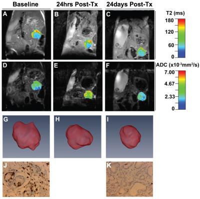
Pancreatic cancer is a devastating disease with poor prognosis. Pancreatic tumor therapy has been ineffective in part because pancreatic tumors have high interstitial fluid pressure (IFP), driven by high hyaluronan concentration, that inhibits penetration of drugs into the tumor. We performed multi-parametric MRI at high resolution to non-invasively assess tumor response in a KPC mouse model to pegylated recombinant hyaluronidase in isolation as well as combined with Gemcitabine. T1 and T2 relaxation as well as diffusion, and 3 dimensional volume measurements were used to characterize the tumors. MR measurements were compared with invasive IFP measurements and histopathological results.


2922.  
MRI Efficacy Evaluation of AvastinTM in Combination Temozolomide Therapy using GL261 Tumor Bearing Mice
Min-Kyoung Kang, Sang-Woo Kim, Ig-Jun Cho, Joo-Young Kim, Zhi Fang, Byung-Hwa Hyun, Jae-Jun Lee*
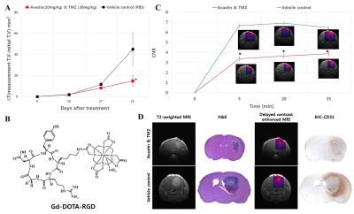
The aim of this study was to evaluate of the drug efficacy used by MRI. A mouse were randomly divided into the control and therapy groups for treatment. MRI were performed to compare with two groups and significant differences were observed in the two groups. The volume transfer constant (Ktrans), flux rate constant (kep) and contrast agent (Gd-DOTA-RGD) enhancement were decreased in the therapy group. Apparent diffusion coefficient (ADC) was lower in the control group. Furthermore, histopathologic assessments were in accord with MRI. Based on these results, efficacy evaluation used by MRI can be helped the development of new bio-drug.


2923.  
Estimation of Contributions to Z-Spectra from CEST and Magnetization Transfer Contrast from Active and Necrotic/Apoptotic Regions of MDA Tumour Xenografts
Wilfred Lam, Jonathan Klein, Farah Hussein, Christine Tarapacki, Gregory Czarnota, Greg Stanisz
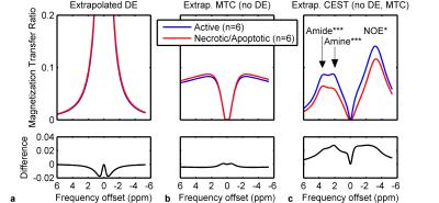
Chemical exchange saturation transfer (CEST) MR imaging has been demonstrated to be able to differentiate active from necrotic/apoptotic regions in MDA tumour xenografts. However, the Z-spectrum reflects not only CEST from dissolved proteins, but also contributions from water saturation and magnetization transfer contrast from semisolid macromolecules. We estimated their individual effect sizes and calculated which significantly contribute to the difference in Z-spectrum amplitude between the two tumour regions. The difference in Z-spectrum amplitude between active and necrotic/apoptotic regions in MDA tumour xenografts is due to the CEST effect with minimal contribution from magnetization transfer contrast and direct water saturation.


2924.  
In Vivo 3T Clinical Magnetic Resonance Imaging with a Biologically Specific Contrast Agent in Prostate Cancer: A Nude Mouse Model
Christopher Abraham, Prashant Jani, Roxanne Turuba, Michael Campbell, Ingeborg Zehbe, Laura Curiel
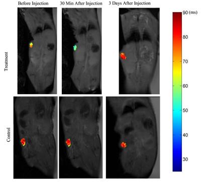
In this study we characterized in vivo a functional superparagmagnetic iron-oxide magnetic resonance contrast agent that effects the T2 relaxation time in MRI. The agent was developed by conjugating Molday Ion Carboxyl-6 (MIC6), with a de-immunized mouse monoclonal antibody (muJ591) targeting prostate-specific membrane antigen (PSMA). We propose this functional contrast agent as a non-invasive method to detect prostate cancer cells that are PSMA positive to provide increased differentiability from surrounding tissues for treatment. PSMA-positive prostate tumours were induced into 20 immunocompromised mice. The functional contrast agent was injected into 14 mice leaving 6 mice as controls. MR imaging was performed on a clinical 3T scanner using different parameters on a MESE sequence to obtain T2 relaxation time values. Tumour size, signal intensity, and T2 relaxation time were obtained pre and post injection and were found to have a lower value for treated mice compared to controls. ICP confirmed the increased level of elemental iron in treated mice tumours compared to controls. H&E staining showed healthy morphology of all tissues collected. The reduction in T2 relaxation time for the functional contrast agent, combined with its specificity against PSMA suggest its potential as a biologically-specific MR contrast agent. 


2925.   
Tumor Progression Monitoring with Hyperpolarized 13C Exchange Spectroscopy in Transgenic Adenocarcinoma of Mouse Prostate Treated with Androgen Deprivation Therapy
Zihan Zhu, Robert Bok, Hsin-Yu Chen, John Kurhanewicz, Daniel Vigneron
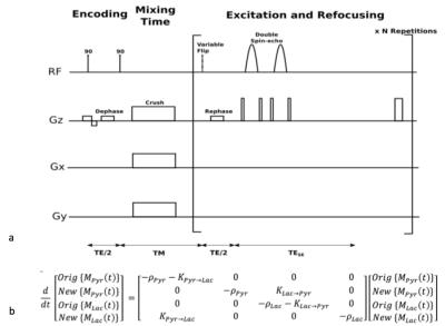
Hyperpolarized 13C MR imaging provides valuable enzyme-kinetic information for investigating disease metabolism. In this work, a new exchange spectroscopic imaging method was applied to a preclinical cancer treatment study and the metabolic information provided by this new method agreed with and proved additional quantitative kinetic metrics over traditional Response Evaluation Criteria in Solid Tumors (RECIST) measures. 


2926.   
In vivo follow-up of colorectal cancer on mice model using endoluminal MRI
Hugo Dorez, Raphaël Sablong, Hélčne Ratiney, Laurence Canaple, Hervé Saint-Jalmes, Sophie Gaillard, Driffa Moussata, Olivier Beuf
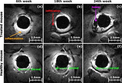
For 6 months, 32 mice, chemically treated to induce colorectal cancer, were followed with endoluminal MRI using dedicated endorectal coils. Based on high spatial resolution T1-weigthed images and T1-maps, quantified parameters (colon wall thickness and T1 relaxation time) were measured at each stage of the pathology from healthy tissues to cancer through inflammation. The colon wall thickness was found to be reliable in assessing early stages of the pathology (inflammation from infiltration), where the intrinsic contrast T1 time parameter was reliable for discerning infiltration from tumors. The two biomarkers provide complementary information in the characterization and staging of colorectal cancer.


2927.  
Combined DCE-MRI and immunohistochemical analyses of cervical cancer xenografts reveal differences in the physiological background of prognostic image parameters derived from the Tofts and Brix model
Tiril Hillestad, Tord Hompland, Anja Nilsen, Trond Stokke, Heidi Lyng
The Tofts and Brix parameters Ktrans and ABrix , derived from dynamic contrast-enhanced MRI (DCE-MRI), have been suggested as potential markers of hypoxia. This  phenotype is associated with poor outcome in cervical cancer treated with chemoradiotherapy. In this study, DCE-MRI was combined with immunohistochemical analysis of cervical cancer xenografts, to better understand the physiological background and prognostic potential of the parameters. Through correlations with hypoxic fraction, vascular and cellular densities, derived from pimonidazole, CD31 and hematoxylin staining of tumor sections, respectively, it was shown that ABrix could be the preferred DCE-MRI parameter for predicting hypoxia related treatment resistance in cervical cancer.


2928.   
Unraveling osteosarcoma tumor heterogeneity using MRI-defined tumor habitats
SUNING HUANG, William Dominguez-Viqueira, Epi Ruiz, Mikalai Budzevich, Bruna Jardim-Perassi, Robert Gillies, Gary Martinez
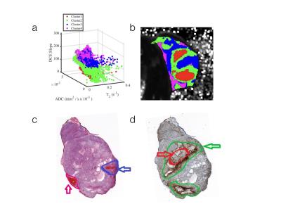
We explore heterogeneity in osteosarcoma using imaging “habitats”, which identify different physiological subregions by MRI. We propose a method to establish the relationship between the microenvironmental of a habitat by relating histology to MRI. Computational image analysis was used to cluster tumor habitats with a 3D printing approach to co-register MR images with histology and immunohistochemistry. Compared with H&E and the CA-9, we found that cellular morphology and density were in concordance with the clustered habitats, although there are subtle differences between histology and MRI slices. Thus, identifying tumor habitats in osteosarcoma using multiparametric MRI is feasible and promising.


2929.  
Multi-parametric MRI of glioblastoma invasion quantitative evaluation using histological stacks
Haitham Al-Mubarak, Antoine Vallatos, Lindsay Gallagher, Joanna Birch, Lesley Glmour, John Foster, Anthony Chalmers, William Holmes
We perform a quantitative histological evaluation of a range of MRI techniques in their ability to probe glioblastoma invasion in a mouse model. Using 3-D histological stacks co-registered with MRI slices allows to achieve high values in Dice, sensitivity and specificity tests (>90%). This approach enables to go beyond the standard evaluation tests, performing direct voxel-to-voxel comparison between MRI and histology, and facilitating the development of multi-parametric analysis models. We also identified promising methods for detecting low tumour concentration regions at the invasion limits.


2930.   
Multi-parametric MRI assessment of tumor progression in a mouse model of pancreatic cancer
Ravneet Vohra, Yak-Nam Wang, Joshua Park, Kayla Gravelle, Stella wHang, Joo-Ha Hwang, Donghoon Lee
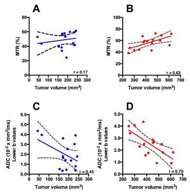
Pancreatic ductal adenocarcinoma (PDAC) is one of the most common forms of lethal human cancers with poor prognosis. Diagnosis is made usually late in tumor development. There is a dire need to develop sensitive non-invasive biomarkers to diagnose and monitor tumor progression and its associated pathological features. We propose to use multi-parametric MRI (mpMRI) to monitor tumor progression in a genetically engineered KPC mouse model that recapitulates human PDAC. Using mpMRI, we demonstrate that there is a significant correlation between increase in pancreatic tumor volume, Magnetization transfer ratio (MTR), apparent diffusion coefficient (ADC), and chemical exchange saturation transfer (CEST) imaging. 
 

The International Society for Magnetic Resonance in Medicine is accredited by the Accreditation Council for
Continuing Medical Education to provide continuing medical education for physicians.