|
Electronic Poster Session
Neuro |
Wednesday, 20 June 2018
Electronic PosterNeuro
4796 -4818 Neurovascular Imaging
4819 -4842 Ischemia & Stroke
4843 -4866 Brain Tumours
4915 -4937 Blood Brain Barrier & CSF Flow
4938 -4961 Traumatic Brain Injury
4962 -4985 Neuroimaging: Animal Studies |
| |
Neurovascular Imaging
Electronic Poster
Neuro
Wednesday, 20 June 2018
| Exhibition Hall |
13:45 - 14:45 |
| |
|
Computer # |
|
4796.
 |
49 |
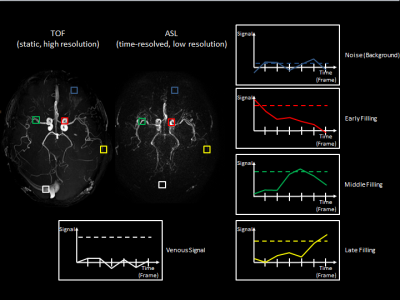
 4D high-resolution Angiography maps obtained by combining low-resolution time-resolved ASL with static high-resolution Time-of-Flight data 4D high-resolution Angiography maps obtained by combining low-resolution time-resolved ASL with static high-resolution Time-of-Flight data
Thomas Lindner, Naomi Larsen, Olav Jansen, Michael Helle
In this study a method to post-process Arterial Spin Labeling (ASL) and Time-of-Flight (TOF) data is presented. The time-resolved low-resolution images of ASL are mapped onto static high-resolution TOF images to create high-resolution time-resolved angiograms.
|
|
4797.
 |
50 |
Improving Cerebrovascular Reactivity Assessment Using High-Resolution MB-EPI Multi-Delay PCASL Imaging
Xiufeng Li, Nicholas Evanoff , Lynn Eberly , David Tupper, Anne Murray , Gregory Metzger , Donald Dengel
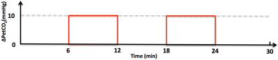
Arterial spin labeling (ASL) imaging with a respiratory challenge can provide both quantitative baseline cerebral blood flow and the assessment of the cerebrovascular reactivity (CVR), an index for cerebrovascular function. However, to date, low-resolution and single-delay ASL imaging is primarily applied to assess CVR, and therefore limited. We proposed and successfully applied high-resolution multi-delay pseudo-continuous ASL (PCASL) imaging using a slice accelerated EPI readout for respiratory challenge studies. The study results suggest that the respiratory challenge can induce significant changes in arterial transit time (ATT), and that the estimates of ATT from the multi-delay imaging protocol are critical to achieve unbiased CVR measurements.
|
|
4798.
 |
51 |
Reduced cerebral blood flow and oxygen consumption in asymptomatic unilateral carotid stenosis patients assessed by arterial spin labeling and multi-parametric quantitative BOLD imaging
Jens Goettler, Stephan Kaczmarz, Claus Zimmer, Christian Sorg, Christine Preibisch, Fahmeed Hyder
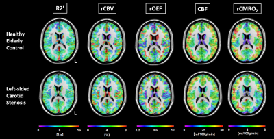
Assessing cerebral hemodynamics and oxygen consumption in patients with clinically asymptomatic, high-grade internal carotid artery stenosis is crucial to estimate risk of stroke and cognitive impairment. Here, 23 patients with unilateral high-grade carotid stenosis and 24 age-matched healthy controls underwent a range of MRI scans, including quantitative BOLD, DSC and pCASL imaging to assess relative oxygen extraction (OEF), blood flow (CBF) and oxygen consumption (CMRO2). Both CBF and CMRO2 were reduced on the stenosis side, indicating that the proposed easily applicable MR-protocol is useful to estimate even subtle CBF and CMRO2 changes in carotid stenosis patients.
|
 |
4799.
 |
52 |
4D Flow MRI analysis of cerebral blood flow before and after high-flow EC-IC bypass surgery for ICA aneurysm
Erika Orita, Tetsuro Sekine, Yasuo Murai, Ryo Takagi, Yasuo Amano, Takahiro Ando, Kotomi Iwata, Makoto Obara, Yoshio Matsumura, Shin-ichiro Kumita
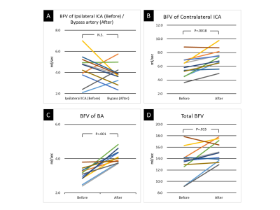
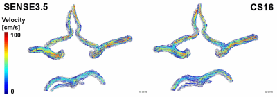
The purpose of this study was to clarify the change of hemodynamics after high-flow extracranial-intracranial (EC-IC) bypass surgery for ICA aneurysm by using time-resolved 3D-phase contrast (4D Flow) MRI. We enrolled 11 patients who underwent high-flow EC-IC bypass surgery. They underwent 4D Flow MRI before and after the surgery. We evaluated the blood flow direction of the circle of Willis. We measured blood flow volume (BFV) of bilateral ICAs, BA, and bypass artery. Seven of 11 patients exhibited collateral retrograde flow in the circle of Willis after surgery. The BFV of contralateral ICA and BA, and total brain BFV statistically increased after surgery. While, there was no evidence of post-operative hyperperfusion in any cases. 4D Flow MRI could quantify the change of hemodynamics after the high-flow bypass surgery.
|
|
4800.
 |
53 |
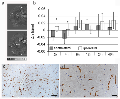
Prominent vessels on quantitative susceptibility maps indicate microvascular pathology after experimental cerebral ischemia and reperfusion
Markus Vaas, Andreas Deistung, Jürgen Reichenbach, Annika Keller, Anja Kipar, Jan Klohs
We tested the utility of quantitative susceptibility mapping (QSM) to assess vascular abnormalities in a mouse model of experimental stroke. We acquired high resolution gradient echo data of mice at different time points after ischemia/reperfusion for computation of susceptibility maps. Prominent vessels with increased magnetic susceptibility values were detected surrounding the ischemic lesion at all times, indicating an increase in oxygen extraction. Immunohistochemistry revealed narrowed capillaries and dilated larger vessels. Thus, prominent vessels are an important indicator of underlying microvascular pathology and may by pivotal for diagnosis and therapeutic decision making in stroke patients.
|
|
4801.
 |
54 |
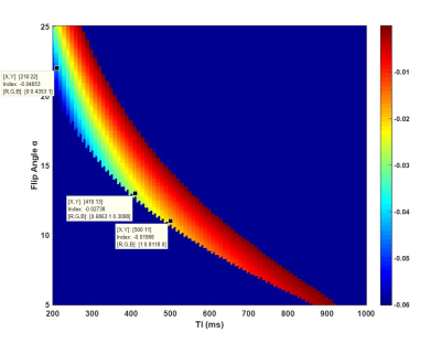
Analyzation and Optimization of Simultaneous Non-Contrast Angiography and intraPlaque Hemorrhage (SNAP) Magnetic Resonance Angiography in Intracranial Vascular Imaging
Yuhui Xiong, Le He, Yu Ma, Xihai Zhao, Chun Yuan, Hua Guo
In this study, we used theoretical simulation and in-vivo scan validation to explore the relationship between intracranial SNAP-MRA signal and blood flow velocity to explain why the performance of SNAP-MRA may vary greatly between healthy people and patients with neurodegenerative diseases. We also found the way to re-optimize the scan parameters (TI and flip angle) to enhance the SNAP-MRA signal and acquire high quality intracranial SNAP-MRA.
|
|
4802.
 |
55 |
3D high resolution Black Blood(BB) Multiple Echo(ME) T2* Imaging Technique for Quantitative Ferumoxytol Imaging on Delayed Scans
Seong-Eun Kim, J Scott McNalley, Adam de Havenon , Dennis Parker, Gerald Treiman
The purpose of this work was to develop a 3D BB ME T2* Imaging technique to to allow quantitative ferumoxytol imaging on delayed scans by measuring T2* in intracranial atherosclerotic plaque(ICAD). Post-gadoliniun enhancement in ICAD may be related to endothelial dysfunction or breakdown or secondary to plaque inflammation. Delayed ferumoxytol imaging allows intravascular clearance with retention in the macrophages present in vulnerable atherosclerotic plaque. We developed a 3D BB ME T2* imaging technique to allow quantitative ferumoxytol imaging on delayed scans by measuring T2* in ICAD.
|
|
4803.
 |
56 |
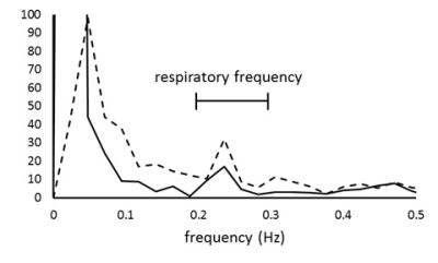
Influences of smoking on cerebral arteriolar vasomotor function: evaluation by using magnetic resonance signal fluctuation
Yusuke Nitanda, Minghui Tang, Toru Yamamoto
The vasodilation and vasoconstriction properties of cerebral arterioles (arteriolar vasomotor function) would be a biomarker of early diagnosis of dementia. Although the vasodilation ability has been studied by using vasodilators such as Diamox, these vasodilators cause non-natural extreme vasodilation. Focusing on the natural arteriolar vasomotion induced by respiratory variation of blood CO2, we have reported a method to evaluate cerebral arteriolar vasomotor function by spectral analysis of fluctuation of venous MRI signal. In this study, we improved our method and applied it to young smokers, and demonstrated the degeneration of arteriolar vasomotor function after a few years of chronic smoking.
|
|
4804.
 |
57 |
Dynamic Susceptibility Contrast and Dynamic Contrast Enhanced Perfusion Performed in a Single Acquisition Using mDIXON Quant
Brian Johnson, Sandeep Ganji, Ivan Dimitrov
This work investigates the use of mDIXON Quant as a perfusion technique to acquire dynamic susceptibility contrast (DSC) and dynamic contrast enhanced (DCE) scans in a single acquisition. The use of multi-echo mDIXON Quant for assessment of perfusion can be used to eliminate the need for split dosing and allows for acquiring DCE and DSC in a single acquisition. Elimination of a split contrast dose will remove contamination from changes in T1, T2, and T2*. This technique can potentially simplify the workflow for the DCE- based perfusion MRI imaging and reduce overall scan time.
|
 |
4805.
 |
58 |
Initial experience using combined quantitative susceptibility mapping and quantitative bold oxygen level dependent imaging (QSM+qBOLD) oxygen extraction fraction for evaluation of acute ischemic stroke
Shun Zhang, Junghun Cho, Thanh Nguyen, Pascal Spincemaille, Wenzhen Zhu, Yi Wang
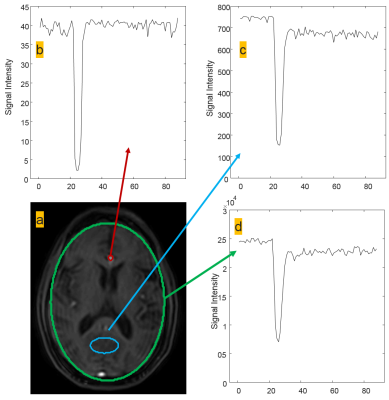
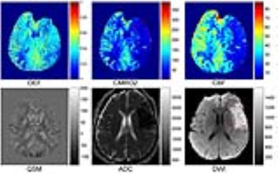
Oxygen extraction fraction (OEF) reflects tissue oxygen consumption, which is very useful to predict the outcome of ischemic stroke in metabolic level. In this work, we evaluate a combined quantitative susceptibility mapping (QSM) and quantitative bold oxygen level dependent (qBOLD) method for measuring OEF based on MRI multi echo gradient echo (GRE) imaging in 11 acute ischemic stroke patients. OEF maps displayed various patterns both in the lesion and in the ASL-CBF/DWI mismatch area, consistent with previous PET studies. OEF with a heterogeneous increase within the lesion or in the CBF/DWI mismatch area may represent salvageable ischemic tissue, while OEF decrease may suggest irreversible infarct.
|
|
4806.
 |
59 |
Myelin imaging may reveal ischemic microstructural damage correlated with neurocognitive dysfunction in patients with moyamoya disease
Shoko Hara, Masaaki Hori, Yasuaki Tsurushima, Yoji Tanaka, Taketoshi Maehara, Shigeki Aoki, Tadashi Nariai
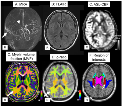
To investigate myelin structural damage caused by chronic ischemia, we applied myelin imaging using magnetization transfer saturation (MTsat) method to 15 patients with moyamoya disease (36.6±11.6-year-old) and 10 normal volunteers (28.4±5.2-year-old). Although many patients received bypass surgery in the past and currently had good hemodynamic status, we found myelin volume fraction (MVF) was significantly lower and g-ratio was significantly higher in the patient group compared to normal controls. Moreover, regional MVF values showed some correlation with neurocognitive tests. This finding suggests myelin damage occurs in moyamoya disease, is associated with neurocognitive dysfunction, and is perhaps irreversible.
|
|
4807.
 |
60 |
The relationship between advanced perfusion MRI and measurements of vessel size in human gliomas using image-guided stereotactic biopsies and quantitative immunohistochemistry
Ararat Chakhoyan, Kevin Leu, Robert Harris, Mitra Harati, William Yong, Albert Lai, Phioanh Nghiemphu, Linda Liau , Noriko Salamon, Whitney Pope, Timothy Cloughesy, Benjamin Ellingson
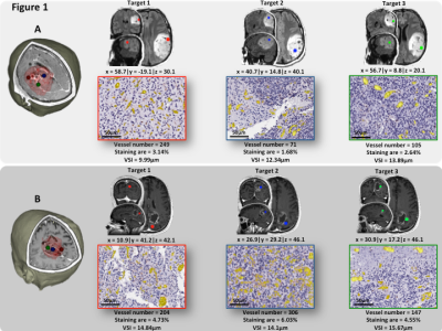
Following an accurate sampling of glioma tissues with 3D T1w-MRI coordinates, we quantified VSI from multi-echo spin-and-gradient echo DSC perfusion as well as from CD31 staining. Eleven patients were included in this retrospective study with in total 30 evaluated targets. We demonstrated the robustness of VSI quantification by MRI. These maps showed a high sensibility and specificity for tumor grading. Finally, in comparison with classical DSC approaches for rCBV estimations, the quantification of VSI could be automated in clinical settings and enhance our understanding of micro and macro-vessel evolution in glioma patients.
|
|
4808.
 |
61 |
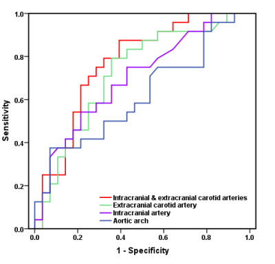
Atherosclerotic Diseases in Entire Craniocervical Arteries and Aortic Arch and Stroke Risk: A 3D Multicontrast MR Vessel Wall Imaging Study
Dongye Li, Wei Dai, Ying Cai, Yongjun Han, Guoen Yao, Huijun Chen, Chun Yuan, Xihai Zhao
Vulnerable atherosclerotic plaque in intracranial and extracranial carotid arteries and aortic arch is one of major causes of ischemic stroke. This study investigated the characteristics of atherosclerotic plaques in the craniocervical arteries and aortic arch and their relationships with stroke risk using 3D multicontrast MR vessel wall imaging. We found that high risk atherosclerotic plaques were most prevalent in intracranial arteries among three vascular beds. Combination of the maximum wall thickness in intracranial with extracranial carotid arteries might be a stronger predictor for cerebral acute ischemic lesions than that in each vascular bed alone.
|
|
4809.
 |
63 |
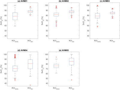
Magnetic Susceptibility Mapping Reveals Altered Vein Oxygenation in Patients with Brain Arteriovenous Malformations: A Preliminary Study
Emma Biondetti, Alvaro Rojas Villabona, Hans Rolf Jäger, David Thomas, Karin Shmueli
Arteriovenous malformations (AVMs) are vascular anomalies characterised by arteriovenous shunting with the lack of a capillary bed. Since the veins that drain an AVM contain arterialised blood, they would be expected to have a higher venous oxygen saturation (SvO2) than normal veins. Due to the paramagnetic properties of deoxyhaemoglobin, SvO2 can be calculated using magnetic susceptibility mapping (SM). Here, we calculated SM-based SvO2 in five patients with a brain AVM. We found higher SvO2 in the AVM draining veins compared to normal veins, showing that SM might be a valuable tool to study AVM physiology.
|
|
4810.
 |
64 |
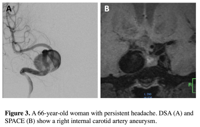
Surveillance of unruptured intracranial saccular aneurysms using non-contrast 3D black blood MRI: comparison of 3D TOF and CE-MRA with DSA
Chengcheng Zhu, Xinrui Wang, Bing Tian, Qi Liu, Christopher Hess, David Saloner, Jianping Lu
Patients with unruptured intracranial aneurysms (UIAs) routinely undergo surveillance imaging to monitor the growth. CTA and CE-MRA provide good accuracy in measuring size relative to gold standard 3D DSA, but require contrast agent and/or have radiation, which is undesirable for repeated imaging. We compared three MRI techniques on 58 aneurysms: 1) 3D non-contrast black blood MRI (SPACE), 2) 3D TOF 3) CE-MRA, against gold standard 3D DSA. SPACE was in excellent agreement with DSA, better than CE-MRA and TOF. Our results support the use of non-contrast SPACE for surveillance of UIA in the clinical setting.
|
|
4811.
 |
65 |
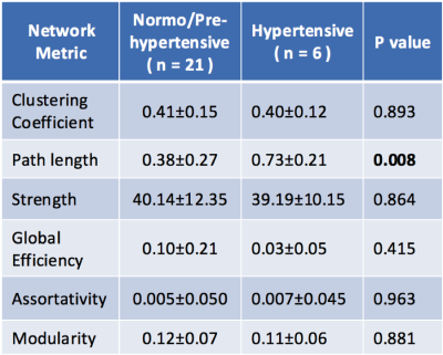
Hypertension induces changes in brain network organization
Guixiang Ma, Bokai Cao, Philip Yu, Ann Ragin
Hypertension is a risk factor for dementia and age-related neurological disorders. Analysis of resting state fMRI for brain network organization may capture early changes induced by hypertension. This investigation examined characteristic network metrics in young, otherwise asymptomatic adults (n=27; mean age 34) classified for hypertension. Path length was the most discriminating global metric. Differences in node clustering were identified using machine learning, including for subcortical regions that have been identified as brain network hubs (thalamus, hippocampus and putamen). These are critical structures for memory, supporting a potential role in cognitive deterioration and dementia and the premise of hub vulnerability.
|
|
4812.
 |
66 |
Oxygen Extraction Fraction and R2* Mapping in Cerebrovascular Disease using an Acetazolamide Challenge.
Christopher Leatherday, Seena Dehkharghani, Fadi Nahab, Jason Allen, Junjie Wu, Ranliang Hu, Deqiang Qiu
Increased cerebral oxygen extraction fraction (OEF) in cerebrovascular disease is linked with a greatly elevated risk of recurrent ischemic stroke. The current gold standard for OEF imaging is Oxygen-15 PET; which is less widely available and more expensive than MRI, and includes an ionizing radiation dose. We studied quantitative susceptibility mapping derived OEF maps and R2* mapping combined with an Acetazolamide challenge in a group of unilateral CVD patients, and found increased OEF and reduced cerebrovascular reactivity in the disease-affected hemisphere using these methods. With further refinement, these techniques may provide a clinical alternative to 15O-PET for OEF imaging.
|
|
4813.
 |
67 |
Effects of physical exercise on hippocampal volume and vasculature in young adults
Antonia Kaiser, Michelle Solleveld, Linda Knutsson, Matthias van Osch, Liesbeth Reneman, Paul Lucassen, Anouk Schrantee
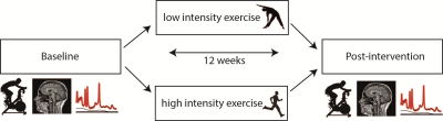
The underlying neurobiological changes of exercise-induced hippocampal volume increases are poorly understood, but a substantial role for vascular plasticity, such as perfusion and angiogenesis, has been suggested. We here studied the effect of a high and low intensity exercise intervention on the hippocampal volume and vasculature. Exercise did not induce hippocampal volume changes, despite a baseline association between fitness and volume. Interestingly, improved fitness resulted in increased hippocampal cerebral blood flow (CBF) (p=0.01) and gray matter CBF (p=0.07). No effect on cerebral blood volume was found. This may suggest that perfusion effects are not hippocampus-specific.
|
|
4814.
 |
68 |
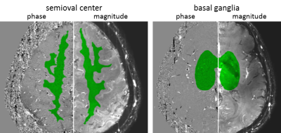
New vision of tuberous sclerosis complex on 7-Tesla MRI
Kaibao Sun, Jianfei Cui, Bo Wang, Tao Jiang, Zhongwei Chen, Fei Cong, Yan Zhuo, Rong Xue, Shuli Liang, Lin Chen
Tuberous sclerosis complex is a multisystem genetic disorder characterized by the growth of numerous tuberous lesions in brain. However, few in vivo studies on TSC have focused on venous structure changes, their association with TSC lesions, and iron accumulation in basal ganglia. 7T susceptibility weighted imaging was performed on eleven TSC patients in comparison with fifteen age- and sex-matched healthy controls. The tubers might develop along penetrating veins. There might be coexistence of iron deposition and calcification in basal ganglia. These in vivo 7T MRI findings provided new perspectives for better understanding the brain pathology in patients with TSC.
|
|
4815.
 |
69 |
Increased intracellular volume fraction, orientation dispersion and diffusion kurtosis in the brain are associated with poor functional outcome in comatose cardiac arrest patients
Ona Wu, Eric Rosenthal, Brittany Mills, Gaston Cudemus-Deseda, Brian Edlow, W. Kimberly, Ming Ming Ning, William Copen, Pamela Schaefer, Joseph Giacino, David Greer
Cardiac arrest patients who are comatose after restoration of spontaneous circulation were prospectively studied to determine whether changes to intracellular volume fraction (ICVF), orientation dispersion and diffusion kurtosis imaging (DKI) can be used to discriminate patients likely to recover consciousness. Subjects who failed to wake up had greater median ICVF, and DKI compared to subjects who woke up. Increases in ICVF, and DK are associated with more severe acute ischemic brain injury. Multi-shell diffusion imaging may help identify patients that may recover consciousness.
|
|
4816.
 |
70 |
Hemodynamic Biomarkers to Assess Disease Severity in Patients with Intracranial Atherosclerotic Disease using Dual-Venc 4D Flow MRI
Alireza Vali, Maria Aristova, Sameer Ansari, Ayesha Muzaffar, Shyam Prabhakaran, Michael Markl, Susanne Schnell
To conduct a comprehensive assessment of hemodynamics in patients with intracranial atherosclerotic disease (ICAD), an automated analysis tool was developed to quantify 4D flow MRI data, including extraction of pressure gradient and flow resistance across the ICAD stenosis and flow and peak velocity asymmetry indices. For three ICAD cases with identical degree of stenosis, the results demonstrated variability in both flow resistance and flow asymmetry indices. With the inclusion of more patients spanning a spectrum of stenosis degrees, it may be possible to demonstrate the utility of flow resistance as a new metric for characterizing the hemodynamic impacts of ICAD.
|
|
4817.
 |
71 |
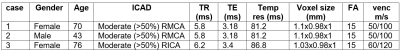
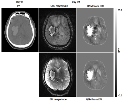
Rapid Quantitative Susceptibility Mapping of Intracranial Hemorrhage using Echo Planar Imaging
Ashmita De, Hongfu Sun, Ahmed Elkady, Derek Emery, Kenneth Butcher, Alan Wilman
Intracranial hemorrhage(ICH) accounts for about 20% of strokes. Quantitative Susceptibility Mapping (QSM) may be valuable for tracking iron changes in ICH. Here we apply Echo Planar Imaging (EPI) for rapid QSM at 3.0 T in hemorrhage patients. The acquisition time is only 9 sec without parallel imaging and 27 sec if parallel imaging is considered to maintain high resolution, appropriate echo time and minimize blurring effects. High correlation was observed for ICH area and mean susceptibility between standard QSM and EPI-QSM in hemorrhage. Hence EPI-QSM method has potential for clinical ICH studies when time is a limiting factor.
|
|
4818.
 |
72 |
A comparison of cerebrovascular reactivity at 1.5 and 3T in cerebral small vessel disease patients
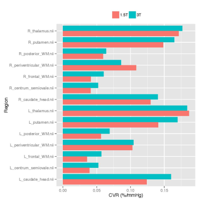
Michael Stringer, Gordon Blair, Yulu Shi, Iona Hamilton, Ian Marshall, Fergus Doubal, Michael Thrippleton, Joanna Wardlaw
Cerebrovascular reactivity can be measured using blood oxygen level dependent (BOLD) MRI and is a potential mechanism in cerebral small vessel disease (SVD). Investigations of the effect of field strength on CVR have been limited, particularly in patient groups. In this study CVR measurements within a series of preselected regions in SVD patients were assessed at 1.5 and 3T. Mean CVR was greater at 3T in 12 of the 14 regions, however differences, as assessed with Bland-Altman plots, were within reasonable limits. These results point to the importance of considering other scanner specific factors beyond field strength when measuring CVR.
|
|
Ischemia & Stroke
Electronic Poster
Neuro
Wednesday, 20 June 2018
| Exhibition Hall |
13:45 - 14:45 |
| |
|
Computer # |
|
4819.
 |
73 |
Motor recovery after initial severe stroke: confronting kinematics with brain activations
Liesjet van Dokkum, Isabelle Laffont, Denis Mottet, Jerome Froger, Alain Bonafe, Nicolas Menjot-de Champfleur, Emmanuelle le Bars
To maximize motor recovery of the upper-limb post-stroke, rehabilitation should be adapted to the individual patient. This requires the identification of motor recovery markers in relation to corresponding brain activations. During elbow flexion/extension, kinematic analysis was confronted with corresponding fMRI activations, comparing 21 participants post-stroke with 13 controls. This provided insight into the underlying functioning and organisation of motor control, switching between ‘automatic’ feed-forward and ‘conscious’ feedback control. Post-stroke, the latter strategy was applied with an additional role for visualisation and the contralesional hemisphere, whereby different kinematic profiles were related to different brain activations, opening doors to personalized rehabilitation.
|
|
4820.
 |
74 |
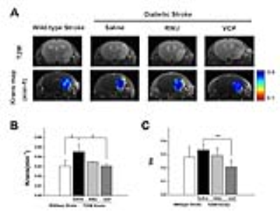
Monitoring diabetic stroke response to novel p38 MAPK inhibitor therapy using dynamic contrast enhanced magnetic resonance imaging (DCE-MRI)
Did Not Present
Yu Cai, Shenghong Ju
We monitored the increased disruption of blood brain barrier (BBB) by DCE-MRI at acute-stage of ischemic stroke in T2DM mice non-invasively. Furthermore, administration of novel P38 inhibitor is a promising way to promote BBB recovery in diabetic stroke and the therapeutic efficacy can be monitored by DCE-MRI.
|
|
4821.
 |
75 |
Prognostication of stroke recovery using structural connectivity
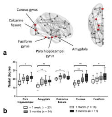
Xiaopei Xu, Kui Kai Lau, Leonard Li, Yuen Kwun Wong, Christina Yau, Henry Mak, Queenie Chan, Edward Hui
We aim to investigate the longitudinal changes in the structural brain network of patients with acute subcortical ischemic infarct in the motor system, and the relation between motor recovery and network measures. Our results showed that the nodal degree of parahippocampus, amygdala, calcarine fissure, cuneus and fusiform gyrus increased with time after stroke, and that network topology measured at acute phase was associated with the recovery of motor function at 6 months after stroke. These findings suggested that network topology could potentially be a prognostic indicator of motor recovery for patients with acute subcortical ischemic infarct in the motor system.
|
|
4822.
 |
76 |
A cost effective, 3D printed vasculature phantom for MR imaging of stroke
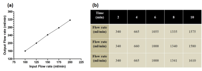
Rashmi Rao, Priyanka Harish, Nithin Vajuvalli, Arush Arun, Ashwini Kumnoor, Sairam Geethanath
Stroke is mainly caused due to hemorrhage or ischemia due to infarct. Current work aims to develop a phantom which can mimic structure and function of a human brain stroke using 3D print technology. The normal human brain vasculature was printed using Poly Lactic Acid. An ischemic infarct was mimicked using Poly Vinyl Alcohol and a cerebral aneurysm was integrated to the vasculature to demonstrate simultaneous onset of hemorrhagic and ischemic stroke. Waterflow to the phantom was introduced by integrating the peristaltic pump. T1, T2 , DW images and T 1and T2maps were generated which depict the stroke vasculature
|
|
4823.
 |
77 |
Reproducibility of BOLD delay perfusion measurements in acute stroke patients
Ahmed Khalil, Ayse Ceren Tanritanir, Ulrike Grittner, Arno Villringer, Jochen Fiebach, Ralf Mekle
To assess perfusion in ischemic stroke is an important task in clinical diagnosis. In this context, a technique sensitive to the delay of blood-oxygenation-level-dependent (BOLD) oscillations at rest called BOLD delay has been proposed. In this study, the reproducibility of this technique in acute stroke patients was examined. Magnitude differences between perfusion measurements from two timepoints were calculated and evaluated in a statistical model. In particular, the effect of head motion was considered. Reproducibility was found to be limited by motion, but the magnitude of the observed variations was small compared to delays observed due to hypoperfusion in stroke patients.
|
|
4824.
 |
78 |
Multi-parametric MR Microscopy of Cerebral Thrombi as a Tool for Prediction of Thrombectomy Procedure Times in Stroke Therapy
Did Not Present
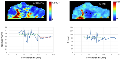
Franci Bajd, Jernej Vidmar, Eduard Kralj, Andrej Fabjan, Fajko Bajrovic, Igor Kocijancic, Zoran Miloševic, Miran Jeromel, Igor Serša
In this study human cerebral thrombi were quantitatively characterized after their acquisition by mechanical thromectomy. The characterization was based on multi-parametric MRI using 3D T1-weighted imaging and ADC and T2 mapping. In the study it was shown that thrombi complex structure can be assessed by ADC and T2 mapping MRI mapping techniques and that the MRI maps of thrombi can be used for prognosis of the mechanical thrombectomy procedure times prior to the interventions.
|
|
4825.
 |
79 |
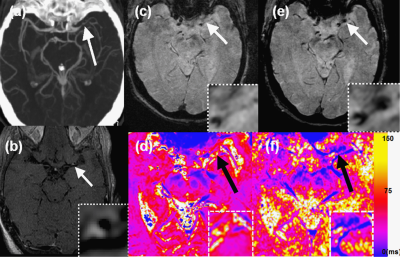
Can susceptibility weighted imaging indicate the ischemic penumbra in patients with acute infarction in middle cerebral artery?
Yu Luo, Linglei Meng, Yongming Zhou, Shuang Xia, E.Mark Haacke
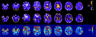
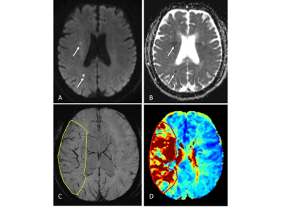
1.Purpose to evaluate the penumbra in acute ischemic stroke by quantitative mismatch between susceptibility weighted imaging(SWI) and diffusion weight imaging(DWI) in comparison with perfusion weighted imaging(PWI) and diffusion weight imaging(DWI) mismatch. 2.Method 85 eligible patients were enrolled with acute ischemic stroke who underwent MR scan including DWI, SWI and PWI before treatment within 12 hours after symptom onset. SWI-DWI mismatch was demarcated by the volume of asymmetrical prominent cortical veins(APCV)region in SWI MIP extending beyond the volume of infarct core segmentation of ADC maps. PWI-DWI mismatch was determined by using infarct core and perfusion deficits segmented from ADC and Tmax maps.
3.Result 41 cases have SWI-DWI mismatch,while 43 cases have PWI-DWI mismatch in totally 85 patients. 42 cases have neither SWI-DWI mismatch nor PWI-DWI mismatch. Only 2 cases have PWI-DWI mismatch without SWI-DWI mismatch. None has SWI-DWI mismatch without PWI-DWI mismatch. There is no a significant difference between SWI-DWI and PWI-DWI in showing mismatch with MCA stroke(P<0.01).The NIHSS of patients with SWI-DWI mismatch was statistically higher compared to the patients without SWI-DWI mismatch (t=-4.956, P<0.01). The NIHSS of patients was also statistically higher with PWI-DWI mismatch in comparison with none PWI-DWI mismatch(t=-4.481, P<0.01).
4.Conclusion APCV in SWI might to be a good instrument to indicate the ischemic penumbra as well as PWI. SWI may be an alternative to PWI in some stroke cases.
|
|
4826.
 |
80 |
Oxygen extraction fraction is elevated in acute stroke with evidence of preserved metabolism: a quantitative susceptibility MRI study
Audrey Fan, Ahmed Khalil, Jochen Fiebach, Arno Villringer, Greg Zaharchuk, Kersten Villringer, Claudine Gauthier
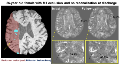
We utilized a novel MRI susceptibility method to quantify oxygen extraction fraction (OEF) in cortical vessels of 22 patients with acute stroke. The observed OEF ratio between affected and contralateral hemispheres depended on patient perfusion status, and tended to normalize (decrease) in follow-up scans on average 3 days later. Stroke cases with substantial perfusion-diffusion mismatch (indicative of potentially salvageable penumbra) showed the greatest OEF elevation (OEF ratio = 1.2 ± 0.1). Patients with large mismatch also showed an inverse relationship between OEF and relative cerebral blood flow (from dynamic susceptibility contrast), suggestive of mechanisms to maintain tissue oxygen metabolism even in ischemic tissue.
|
|
4827.
 |
81 |
Unruptured Intracranial Aneurysms: Relationship between Wall Enhancement and Rupture Risk Factors Based on High-resolution Magnetic Resonance Imaging
Chengcheng Zhu, Xinrui Wang, Qi Liu, Christopher Hess, David Saloner, Jianping Lu
Wall enhancement (AWE) of intracranial aneurysms (IAs) on high-resolution black blood MRI has been described in ruptured aneurysms. This study investigated 103 unruptured saccular IAs and aims to assess the association between AWE and traditional risk factors and estimated one-year and five-year rupture risk estimated from previous large trails. We found aneurysms with AWE had more than 3 times higher estimated rupture risk (one-year and 5-year, 2.2% and 6.7%) than aneurysms without AWE (0.6% and 2.0%), and AWE was associated with traditional risk factors (size, location, symptoms). Identifying AWE may improve the risk assessment of IAs.
|
|
4828.
 |
82 |
Quantifying dynamic CBF changes in 4-day follow up after carotid endarterectomy using 3D pCASL
Video Permission Withheld
Ting Wang, Bo Bu, Bing Wu, Lin Ma, Xin Lou
Hyperperfusion syndrome was a severe complications of carotid endarterectomy (CEA), the routine measurement for preventing this was to take antihypertensive drugs for at least one week after operative. But there were no definite criteria to assess whether this method was reasonable and appropriate; our objective was to detect dynamic CBF changes in 4-day follow up after CEA using 3D pCASL technique. It is seen that compared to patients before CEA, both the ipsilateral and opposite CBF values were increased in various time points after CEA (P<0.05), whereas, the bilateral CBF values shows no significant differences (P>0.05). This shown that 3D pCASL is sensitive to temporal hemodynamic changes after CEA and may provide a quantitative criterion for assessing antihypertensive drugs taken. Also the observed increases shortly in the first and the second day after operations may suggest for potential drug intervention in case of post-CEA complications.
|
|
4829.
 |
83 |
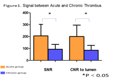
The Value of Magnetic Resonance Black-blood Imaging in Differentiating Acute and Chronic Cerebral Venous Thrombosis
Did Not Present
Xiaoxu Yang, Fang Wu, Ye Wu, Tianyi Qian, Xianggong Duan, Xiangying Du, Xunming Ji, Qi Yang
This study aims to demonstrate the value of magnetic resonance black-blood imaging (MRBTI) for differentiating acute and chronic cerebral venous thrombosis (CVT) as well as the diagnosis accuracy of CVT in segment levels. The SNR and CNR of the acute CVT group were significantly higher than that of the chronic group. The sensitivity and specificity of MRBTI were 95.6% (152 /159) and 98.0% (352 /359), respectively. Furthermore, the sensitivity of MRBTI in detecting acute thrombus is up to 100%, compared with 88.5% in the chronic group, which means MRBTI has high sensitivity for early diagnosis.
|
|
4830.
 |
84 |
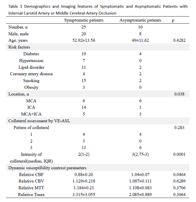
Vessel Encoded Arterial Spin Labeling Evaluation of Collateral Circulation in Symptomatic and Asymptomatic Patients with Internal Carotid Artery or Middle Cerebral Artery Occlusion
Jinhao Lyu, Xiaoxiao Ma, Yina Lan, Lin Ma, Xin Lou
Symptomatic and asymptomatic patients with internal carotid artery (ICA) or middle cerebral artery (MCA) occlusion have the different prognosis. The present study had evaluated collateral circulation by Vessel Encoded Arterial Spin Labeling (VE-ASL) and cerebral vasoreactivity by mean transit time (MTT) obtained by Dynamic susceptibility contrast in symptomatic and asymptomatic patients with unilateral ICA and/or MCA occlusion. The study had found that the intensity of collateral in asymptomatic patients was significantly better than symptomatic patients while MTT showed no significant differences, which indicate that hemodynamic impairments in symptomatic patients with ICA/MCA occlusion may be mainly induced by the insufficiency of collateral circulation.
|
|
4831.
 |
85 |
Proximal Internal Carotid Artery Stenosis Associates with Diffuse Wall Thickening in Petrous Arterial Segment of Moyamoya Disease Patients: A 3D MR Vessel Wall Imaging Study
Xiaoyi Chen, Jian Wang, Bing Zhang, Dongye Li, Huiyu Qiao, Shuai Liu, Yongjun Han, Hualu Han, Yongbo Yang, Fei Zhou, Xueping Li, Xihai Zhao
It has been shown that rapid reduction of lumen diameter at the proximal ICA can be seen in moyamoya disease (MMD) patients. The arterial wall thickness at downstream of proximal ICA may aggravate cerebral ischemia and affect the outcome of revascularization. This study sought to investigate the association between proximal ICA stenosis and diffuse wall thickening in ipsilateral petrous ICA in MMD patients. We found that proximal ICA stenosis was significantly associated with wall thickness (r = 0.434, p<0.001) and presence of diffuse wall thickening (odds ratio=4.433, 95% confidence interval 1.980–9.925, p<0.001) in ipsilateral petrous ICA in MMD patients.
|
|
4832.
 |
86 |
Cerebrovascular resistance responses to CO2 improve after revascularization surgery
Larissa McKetton, Olivia Sobczyk, Julien Poublanc, Kevin Sam, Adrian Crawley, Lakshmikumar Venkat Raghavan, James Duffin, Joseph Fisher, David Mikulis
The cerebral hemodynamics of patients undergoing revascularization surgery for intracranial steno-occlusive disease (IC-SOD) were assessed by deriving an estimate of their cerebrovascular resistance response to CO2 from their BOLD response to CO2. Significant improvements were found in the sigmoid parameters describing their resistance responses.
|
|
4833.
 |
87 |
Predicting Hyperperfusion syndrome by measurement of leptomenigeal collateral and preoperative cerebral blood flow in patients with unilateral internal carotid artery stenosis
Yina Lan, Jinhao Lyu, Jianxun Qu, Lin Ma, Xin Lou
Hyperperfusion syndrome (HPS) is a rare but potentially fatal postoperative complication after revascularization, while preoperative predictor of HPS had not been fully established. We used pseudo continuous arterial spin labeling (pCASL) to investigate the correlation between the collateral flow proportion and the elevated cerebral blood flow (CBF) ratio relative to the preoperative CBF in patients with unilateral internal carotid artery (ICA) stenosis. A significant correlation was observed between the collateral flow proportion and the elevated CBF ratio . (r =0.588, P =0 .01). As an indication, HPS are likely to occur in patients with low preoperative CBF and good collateralization.
|
|
4834.
 |
88 |
Carotid stenosis: a risk factor for white matter disease even at presymptomatic stage
Pedro Henrique Rodrigues da Silva, Ana Paula Afonso Camargo, Antonio Carlos Santos Senra Filho, Luiz Otavio Murta Junior, Octávio Marques Pontes Neto, Renata Ferranti Leoni
Studies have suggested that cerebral white matter hyperintensity (WMH) is due to hypertension and is associated with carotid artery stenosis (CAS). However, it is unclear whether this association is attributable to effects on WM and how asymptomatic CAS contributes to it. Therefore, we aimed to assess the association between ACAS and WMH lesions and its relationship with cognitive decline using MRI to provide information that may help predicting cases at risk of brain ischemia. Our data showed that ACAS is associated with WMH lesions and cognitive decline, indicating that ACAS, in addition to age, is likely to cause WM lesions.
|
|
4835.
 |
89 |
Brain morphometric changes and functional connectivity alterations in post-stroke fatigue
Milanka Visser, Thomas Lillicrap, Carlos Garcia-Esperon, Bénédicte Maréchal, Mark Parsons, Christopher Levi, Andrew Bivard
Debilitating fatigue is the most common consequence of stroke, however there are no known clinical or radiological biomarkers associated with post-stroke fatigue. We assessed differences in regional brain volumes obtained from T1-weighted, high-resolution structural scans between stroke survivors with and without severe fatigue. Differences were observed in the volume of the globus pallidus and putamen, as well as the ipsilesional temporal, parietal and frontal lobe. The mentioned morphological differences between stroke survivors with and without severe fatigue have also been reported in multiple sclerosis and Parkinson’s-related fatigue, suggesting a possible common mechanism.
|
|
4836.
 |
90 |
Preoperative predictors of hyperperfusion after CEA: a study using vessel selective ASL
Tianye Lin, Zhichao Lai, Yuelei Lyu, Zhentao Zuo, Bing Wu, Jianxun Qu, Hui You, Bo Hou, Changwei Liu, Feng Feng
To identify preoperative predictors for cerebral hyperperfusion (CHS) after CEA based on vessel selective ASL. The perfusion volume of each brain feeding artery and the corresponding mean CBF in each perfusion volume before and after CEA were calculated. It was found that the sum of perfusion volumes corresponding to LICA, RICA and VBA (TotalPerVol) and the preoperative territory perfusion of the surgery side (preCBF_surg) was inversely correlated with the degree of CBF increase. The result indicated that tASL and ASL were useful in predicting cerebral hyperperfusion.
|
|
4837.
 |
91 |
Evaluation of Tranvascular Water Exchange Index (WEI) in post Thrombectomy Patients
Young Kim, Jerold Boxerman
The dynamic evolution of vascular impairment following thrombotic stroke has important clinical implications for designing effective management and treatment strategies. In the current study, we quantified the rate of water exchange across the blood-brain barrier (BBB) via water exchange index (WEI) in clinical patients undergone thrombectomy. Although extravasation of Gd-DTPA was not observed in most of the patients, the WEI was significantly elevated in both infarct and peri-infarct areas, revealing compromised integrity of the BBB.
|
|
4838.
 |
92 |
Correlation between Intracranial Artery Atherosclerotic Disease and the Integrity of Circle of Willis in Symptomatic Patients: A 3D MR Vessel Wall Imaging Study.
Did Not Present
Yilan Xu, Dongye Li, Zhuozhao Zheng, Chun Yuan, Xihai Zhao
This study investigated the correlation between intracranial artery atherosclerotic disease and the integrity of communicating arteries in circle of Willis in symptomatic patients using MR imaging. We found that the intracranial artery stenosis was significantly associated with presence of anterior and posterior communicating arteries. Our findings suggest that the intracranial artery stenosis might be an independent indicator for the integrity of circle of Willis. Our data also suggest that, with the progression of intracranial artery stenosis, collateral circulation tends to be integrated from the anterior to posterior communicating arteries.
|
|
4839.
 |
93 |
Brain Perfusion and Structure Variation of Patients with Unilateral Middle Cerebral Artery Stenosis
Did Not Present
Shuang Yan, Tianyi Qian, Mingli Li, Li Zhang, Zhengyu Jin
To investigate the brain structure variation of patients with chronic unilateral middle cerebral artery (MCA) stenosis, a volume-based quantitative segmentation method was used to measure the morphological changes of brain structure. A correlation analysis was performed to find the relationship between the morphometry and cerebral blood perfusion. The results showed that the patients had atrophy in many brain regions within the MCA territory. And the perfusion status may have impact on the atrophy process.
|
|
4840.
 |
94 |
Quantitative Cerebral Blood Flow Measured with Arterial Spin Labeling MRI in the Unaffected Contralateral Brain Hemisphere Predicts Outcome in Acute Ischemic Stroke
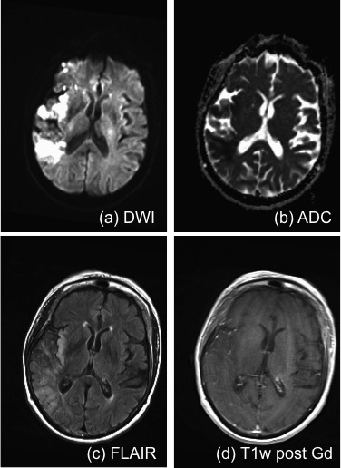
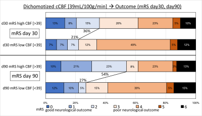
Thoralf Thamm, Jia Guo, Jarrett Rosenberg, Tie Liang, Michael Marks, Soren Christensen, Huy Do, Stephanie Kemp, Emily Ryan, Tudor Jovin, Bart Keogh, Jenny Chen, Maarten Lansberg, Greg Albers, Greg Zaharchuk
During acute stroke, perfusion of the ischemic penumbra might be sustained by both collaterals and elevated systemic blood pressure. Arterial Spin Labeling (ASL) is an MR imaging tool to quantify Cerebral Blood Flow (CBF) non-invasively. We focused on the non-affected brain hemisphere and utilized this contralateral CBF (cCBF) as an imaging biomarker for late neurological outcome prediction. Stroke patients were dichotomized by the median cCBF into high (>39 mL/100g/min) and low (<39 mL/100g/min) cCBF. Our analysis revealed that high cCBF predicts good neurological outcome at day 90 after stroke.
|
|
4841.
 |
95 |
Association between Diffusion Weighed Imaging Measured Pretreatment Ischemic Volume and Functional Outcome in Ischemic Stroke
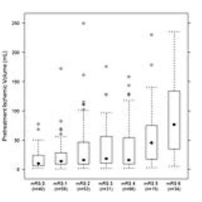
Yu XIE, Catherine Oppenheim, Francis Guillemin, Vincent Gautheron, Benjamin Gory, Hélène Raoult, Sébastien Soize, Bailiang CHEN, Jacques Felblinger, Gabriela Hossu, Serge Bracard
The association between pretreatment ischemic volume (PIV) measured on diffusion weighted images (DWI) and functional outcome after mechanical thrombectomy is of great clinical importance but has yet to be determined. We analyzed 298 ischemic stroke patients from the multicentric study THRACE. Our results showed that increased PIV was an independent predictor for a lower probability of functional independence, a less favorable degree of disability, and a higher mortality rate. PIV measured on DWI is a valuable early predictor for functional outcome in ischemic stroke patients, and thus can contribute to patient selection for optimal therapeutic intervention.
|
|
4842.
 |
96 |
Comparison of ASL-MRA and 3D TOF-MRA in patients with transit ischemic attack
Did Not Present
Yan Wang, Jing Chen, Chuanchen Zhang, Jianxun Qu, Mingzhen Wu
A comparison of ASL-MRA and TOF-MRA was performed for patients suffered from TIA. TOF-MRA is superior to ASL-MRA in morphological assessment of the stenosis arteries. However, ASL-MRA can reflect more hemodynamic information of the blood supply arteries for the hypoperfusion area.
|
|
Brain Tumours
Electronic Poster
Neuro
Wednesday, 20 June 2018
| Exhibition Hall |
13:45 - 14:45 |
| |
|
Computer # |
|
4843.
 |
97 |
MRI-derived Oxygen Metabolism and Neovascularization Characterization for Grading and IDH Gene Mutation Detection of Gliomas
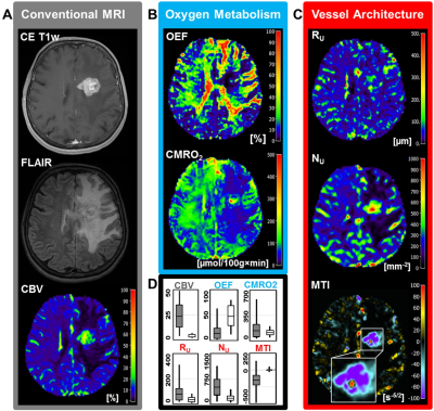
Andreas Stadlbauer, Max Zimmermann, Arnd Dörfler, Stefan Oberndorfer, Michael Buchfelder, Gertraud Heinz, Karl Rössler
The purpose was to explore the diagnostic performance of combined physiological MRI of oxygen metabolism and neovascularization for glioma grading and characterization of isocitrate-dehydrogenase-1 (IDH1) gene mutation status. 83 patients with glioma WHO°II-IV were examined using vascular architecture mapping (VAM) and multiparametric quantitative BOLD (mp-qBOLD). Neovascularization correlated with increasing WHO° and microvessel type indicator (MTI) had the best diagnostic performance (AUC=0.782) for differentiation between glioma WHO°III and IV. IDH1-mutation was associated with significantly decreased cerebral metabolic rate of oxygen (CMRO2; P=0.037) in glioma WHO°II and significantly increased (P=0.013) MTI in glioma WHO°III, resulting in best diagnostic performance for IDH1-mutation detection.
|
|
4844.
 |
113 |
Vascular Hysteresis Loops and Vascular Architecture Mapping in Patients with Glioblastoma treated with Antiangiogenic Therapy
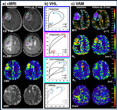
Andreas Stadlbauer, Max Zimmermann, Stefan Oberndorfer, Arnd Dörfler, Michael Buchfelder, Gertraud Heinz, Karl Rössler
Glioblastoma are among the most vascularized of all solid tumors and attractive targets for antiangiogenic therapies. Antiangiogenic therapy response assessment in glioblastoma is challenging due to decreased vessel permeability and diminished contrast agent extravasation. Here, we investigated the variability of vascular hysteresis loop (VHL) shapes and the spatial heterogeneity of neovascularization using vascular architecture mapping (VAM) in patients with recurrent glioblastoma during bevacizumab mono-therapy. Responding, non-responding, progressive, and remote-progressive tumor areas were observed. Analysis of VHLs in combination with VAM biomarkers may lead to a new perspective on investigating the spatial heterogeneity of neovascularization in glioblastoma during antiangiogenic therapy.
|
|
4845.
 |
98 |
Diagnostic Accuracy of 2-Hydroxyglutarate Magnetic Resonance Spectroscopy in Newly-Diagnosed Brain Mass and Suspected Recurrent Gliomas
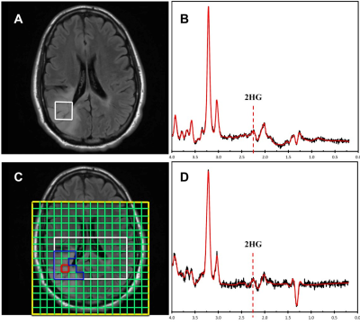
Alexander Lin, Min Zhou, Huijun Liao, Raymond Huang
Previous studies have reported the utility of 2-hydroxyglutarate magnetic resonance spectroscopy (2HG MRS) in diagnosing isocitrate dehydrogenase (IDH) status, which is of great value for patient management. We determined the optimal thresholds of single voxel spectroscopy (SVS) and chemical shift imaging (CSI) 2HG MRS in differentiating IDH-mutant gliomas from non-IDH-mutant controls, and then determined the diagnostic accuracy in two prospective cohorts of patients. We show that 2HG MRS provided diagnostic utility for IDH-mutant gliomas both preoperatively and at time of suspected tumor recurrence. Our findings may provide guidance for devising optimal MRS imaging protocol tailored to specific clinical settings.
|
|
4846.
 |
99 |
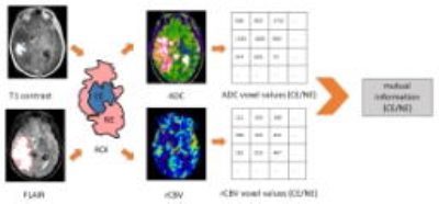
Mutual Information: Depicting the Interdependence of Perfusion and Diffusion Magnetic Resonance Imaging in Glioblastoma Patients
Chao Li, Shuo Wang, Turid Torheim, Florian Markowetz, Stephen Price
The mismatch between energy demands of tumor growth and heterogeneous blood supply may cause variations is associated with tumor aggressiveness. Multi-parametric imaging may enable incorporation of complementary imaging modalities. However, finding validated surrogates to depict the interrelation between imaging modalities remains a challenge. We used the mutual information to describe the interrelation between the perfusion and diffusion imaging. The results showed that the higher values of mutual information may contribute to a worse patient survival. The chemical shift imaging results suggested that the higher mutual information may be correlated with a more migratory phenotype.
|
|
4847.
 |
100 |
Radiomics of MRI at Diagnosis is Predictive of Extreme Survival in Glioblastoma Multiforme
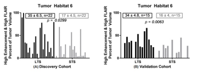
Olya Stringfield, Mahmoud Abdalah, Sandra Johnston, Nicolas Rognin, Yoganand Balagurunathan, John Arrington, Kristin Swanson, Kathleen Egan, Robert Gatenby, Natarajan Raghunand
We retrospectively analyzed pre-treatment MR scans in two cohorts diagnosed with Glioblastoma. The Long-Term Survival (LTS) group survived >36 months post-diagnosis, while the Short-Term Survival (STS) group survived ≤18 months. The discovery cohort included 22 LTS patients and 22 STS patients and the validation cohort consisted of 15 patients, each. Tumor voxels were clustered on post-contrast T1w and FLAIR sequences into 6 distinct “habitats”. Radiomic features were extracted from both sequences. The enhancement value on T1w and fraction of Habitat 6 (high signal on T1w and FLAIR) were significantly higher in the LTS groups compared to the STS groups.
|
|
4848.
 |
101 |
Radiological Assessment of Vascular Supply of Intracranial Meningeomas by Superselective Arterial Spin Labelling.
Ulf Jensen-Kondering, Michael Helle, Thomas Lindner, Arya Nabavi, Olav Jansen
Detailed information on the extent and ratio of blood supply is required to assess the feasibility of presurgical embolisation procedures of intracranial meningeomas. This study investigates the feeding vasculature to intracranial meningeomas using superselective ASL. In 31 prospectively included patients harboring a total of 42 meningeomas, superselective ASL was performed to visualize the contribution of blood supply to the tumor and rated by two readers. We demonstrated that superselective ASL is capable of identifying and quantifying the contribution of feeding arteries in intracranial meningeomas. Agreement with gold standard DSA is also demonstrated.
|
|
4849.
 |
102 |
Autopsy analysis of the overlap of radiomic profiles associated with poor overall survival and predictive maps of tumor cellularity in glioblastoma patients
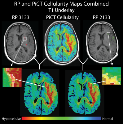
Sarah Hurrell, Sean McGarry, Elizabeth Cochran, Jennifer Connelly, Scott Rand, Wade Mueller, Peter LaViolette
Multiparametric MRI radiomic profiles (RPs) of de novo glioblastoma (GBM) brain tumors have been shown to predict patient prognosis prior to treatment. This study compares prognostic RPs to predictive maps of tumor cellularity derived from radiological-pathological (rad-path) correlation to determine the convergence of both imaging biomarkers. We find that RPs associated with poor prognosis co-localize with high cellularity, both predicted and pathologically confirmed in 6 patients assessed at autopsy.
|
|
4850.
 |
103 |
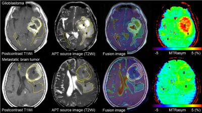
Amide proton transfer-weighted imaging of glioblastoma and metastatic brain tumor: histogram analysis in enhancing tumors and peritumoral regions
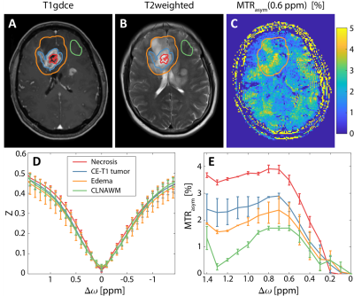
Kiyohisa Kamimura, Masanori Nakajo, Tomohide Yoneyama, Hirofumi Hirano, Takashi Iwanaga, Yuta Akamine, Jochen Keupp, Takashi Yoshiura
To determine whether amide proton transfer-weighted imaging (APTWi) is useful for distinguishing glioblastomas (GBMs) from metastatic brain tumors (Mets), we compared APT-related signal intensity (APTSI) between the two tumor types in the areas of enhancing tumor and peritumoral high signal intensity areas (PHAs) using histogram analysis. In the enhancing tumor, the mean and 90,75,50,25 and 10 percentiles of APTSI histogram were significantly higher in GBMs than in Mets, whereas no APTSI histogram parameters in PHA showed significant difference between GBMs and Mets. APTSI in the areas of enhancing tumor, not in PHA is useful for differentiation between GBMs and Mets.
|
|
4851.
 |
104 |
The Influence of Heterogenous Subregions on Predicting MGMT Methylation Status of Glioblastomas: A Radiomics Analysis on Multimodal MRI
Did Not Present
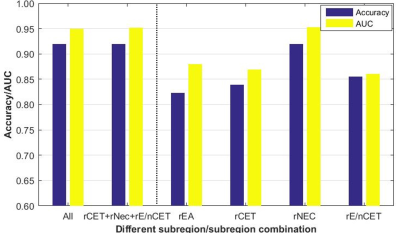
Qiang Tian, Xi Zhang, Lin-feng Yan, Yu-chuan Hu, Yu Han, Ying-zhi Sun, Wen Wang, Guang-bin Cui
MGMT promoter methylation is associated with longer survival and better treatment response of GBM patients. Intratumor heterogeneity is partly responsible for inaccurate detection of MGMT status. Therefore, assessing the effect of different heterogenous subregion of GBM on MGMT status would be critical. In this study, a radiomics approach integrated optimal features of heterogenous subregions in multimodal MRI and machine learning model was proposed for effectively predicting MGMT methylation, and meanwhile assessing the prediction efficiency of subregions or subregion combinations. The proposed approach achieved a promising MGMT methylation detection performance and indicated that rNEC may play a role in this issue.
|
|
4852.
 |
105 |
High-resolution Deuterium MR Spectroscopic Imaging of the Warburg Effect in Brain Tumor
Video Permission Withheld
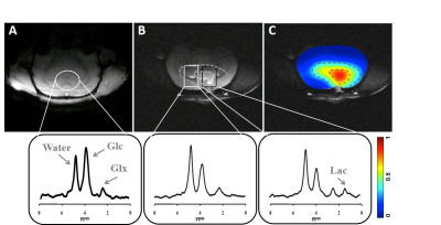
Ming Lu, Xiao-Hong Zhu, Yi Zhang, Walter Low, Wei Chen
The best-known metabolic abnormality in brain cancer is the Warburg effect, which shifts the fuel consumption from oxidation towards glycolysis. Recently, we developed a novel in vivo Deuterium (2H) MR spectroscopic imaging (DMRSI) approach for simultaneously assessing brain glycolysis and oxidation at 16.4 T. In this study, we aimed to image the Warburg effect in a rat model with gliosarcoma using DMRSI with improved resolution. High-resolution quantitative image using the ratio of [lactate] to [glutamate/glutamine] showed a huge contrast between brain tumor and intact tissue and promise to study the decoupling relationship between glycolysis and oxidation in tumor.
|
|
4853.
 |
106 |
Edge Contrast of the FLAIR Hyperintense Region Predicts Survival in Patients with High Grade Gliomas Following Treatment with Bevacizumab
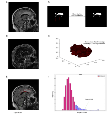
Naeim Bahrami, David Piccioni, Roshan Karunamuni, Nate White, Yu-Hsuan Chang, Tyler Seibert, Rachel Delfanti, Jona jhattangadi-gluth, Nikdokht Farid, Anders Dale, Carrie McDonald
Treatment with bevacizumab is standard of care for recurrent high grade gliomas (HGGs) and the level of border distinctness is a major parameter to monitor the therapy. Previously, the level of border distinctness was defined qualitative. In this study, we calculated the distinctness of the fluid-attenuated inversion recovery (FLAIR) hyperintense border—edge contrast (EC)—and showed it improves the evaluation of response to bevacizumab in patients with HGG. We showed that after bevacizumab, lower EC of the FLAIR hyperintense region was associated with poorer survival among HGG patients. We developed a quantitative parameter to characterize the border of the tumor.
|
|
4854.
 |
107 |
True-Diffusion Coefficient Retracted from Intra-Voxel Incoherent Motion (IVIM) Can Stratify Biopsy-Approved Infiltrative Edema from Normal Tissue and Active Tumor in Diffuse Brain Gliomas
Anahita Fathi Kazerooni, Nima Gilani, Mahnaz Nabil, Mehdi Zeinalizadeh, Kavous Firouznia, Farid Azmoudeh-Ardalan, Mohammad Peikari, Mohammadreza Alviri, Mehrdad Hadavand, Hamidreza Saligheh Rad
Infiltration of tumorous cells in the normal brain parenchyma is an intrinsic characteristic of diffuse gliomas and is a determinant factor in tumor recurrence, transformation into malignant form, and poor prognosis. The objective of this study was to investigate the role of intra-voxel incoherent motion (IVIM) imaging in characterizing tumor infiltration through localized biopsies. Histopathologically-approved regions of active tumor, infiltrative glioma (edema), and normal tissues were accurately discriminated by true (perfusion-free) diffusion coefficient (D).
|
|
4855.
 |
108 |
Characterization of Active and Infiltrative Tumorous Subregions from Normal Tissue in Brain Gliomas Using Multi-Parametric MRI
Anahita Fathi Kazerooni, Mahnaz Nabil, Mohammadreza Alviri, Mehdi Zeinalizadeh, Kavous Firouznia, Farid Azmoudeh-Ardalan, Hamidreza Saligheh Rad
In this preliminary work, a variety of MRI techniques, including conventional high-resolution T1-weighted, T2-weighted, and T2-FLAIR, as well as quantitative techniques comprising of T2-relaxometry, DWI, DTI, DSC-MRI, and IVIM derived features were acquired from patients with gliomas. The features extracted from the mentioned images were explored for their potential in stratification of histopathologically-approved samples, labelled as active tumor, infiltrative glioma (edema) and normal brain tissue. Furthermore, the most accurate combination of the features for discrimination of tissue subregions was generated through a machine learning technique.
|
|
4856.
 |
109 |
Combined Diffusion Kurtosis Imaging and Dynamic Susceptibility Contrast Magnetic Resonance Perfusion Imaging for In Vivo Molecular Profiling of Human Glioma.
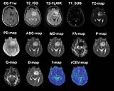
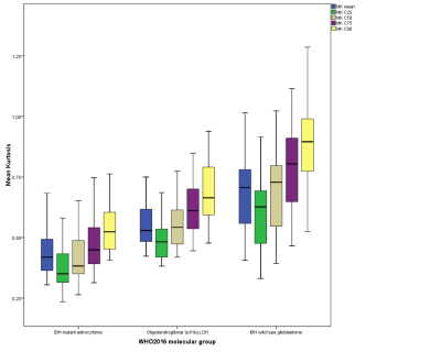
Johann-Martin Hempel, Jens Schittenhelm, Nils Nüssle, Cornelia Brendle, Benjamin Bender, Ghazaleh Tabatabai, Marco Skardelly, Salvador Castaneda Vega, Ulrike Ernemann, Uwe Klose
The purpose of this study was to assess the diagnostic performance of combined DKI and DSC-MRI maps for in vivo assessment of the 2016 WHO integrated glioma grades. Histogram parameters of DKI show a higher diagnostic performance than those of DSC-MRI in stratifying gliomas according to the integrated molecular approach of 2016 CNS WHO. However, DSC-MRI may provide additional insight into the MGMT methylation profile of primary IDH wild-type GBM. Thus, combined DKI and DSC-MRI provide promising potential biomarkers for glioma.
|
|
4857.
 |
110 |
Deep-learned 3D black-blood imaging using automatic labeling technique and 3D convolutional neural networks for detection of metastatic brain tumors
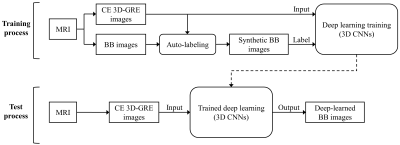
Yohan Jun, Taejoon Eo, Taeseong Kim, Hyungseob Shin, Dosik Hwang, Sohi Bae, Yaewon Park, Hojoon Lee, Byoungwook Choi, Sungsoo Ahn
Black-blood (BB) imaging has complementary roles in addition to contrast-enhanced 3D gradient-echo (CE 3D-GRE) imaging for detection of brain metastases. We proposed deep-learned 3D BB imaging with an auto-labeling technique and 3D convolutional neural networks (CNNs) for detecting metastatic brain tumors. On deep-learned BB imaging, vessel signals of the brain were effectively suppressed in all patients. According to per lesion analysis, overall sensitivities were 90.3% for deep-learned BB and 100% for original BB. There were eight false positive nodules on original BB and only one on deep-learned BB. Deep-learned 3D BB imaging can be effectively used for detecting metastatic tumors in the brain.
|
|
4858.
 |
111 |
High Resolution T1-Perfusion using Compressed-SENSE for Glioma Grading
Rakesh Kumar Gupta, Indrajit Saha, Anup Singh, Pradeep Kumar Gupta, Rupsa Bhattacharjee , Anandh Ramaniharan , Rana Patir, Sunita Ahlawat, Jitender Saini, Marc Cauteren
T1-perfusion MRI derived relative cerebral blood volume (rCBV) is a key bio-marker for pre-surgical grading of gliomas; however, acquiring clinically relevant higher resolution T1-perfusion data with whole brain coverage is challenging due to possible loss in temporal resolution. This study takes the advantage of combining compressed-sensing with SENSE parallel-imaging i.e., Compressed-SENSE (CSENSE), to develop high-resolution whole brain T1-perfusion with improved temporality. The CSENSE enabled T1-perfusion derived rCBV values successfully differentiated high and low grade gliomas and matched with the histopathological grading. The rCBV cut-off value from CSENSE assisted T1-perfusion was similar to the routine T1-perfusion without-CSENSE of histopathology-matched gliomas.
|
|
4859.
 |
112 |
Lactate-weighted Chemical Exchange Saturation Transfer (Lactate-CEST) Imaging in Glioma Patients at 7 Tesla
Daniel Paech, Jan-Eric Meissner, Markus Wennmann, Andreas Korzowski, Alexander Radbruch, Martin Bendszus, Wolfgang Wick, Andreas Unterberg, Peter Bachert, Mark Ladd, Heinz-Peter Schlemmer
Non-invasive imaging of lactate is of enormous significance, particularly in oncologic diseases or metabolic disorders. In this work, we applied Lactate-weighted Chemical Exchange Saturation Transfer (Lactate-CEST) magnetic resonance imaging (MRI) at 7 Tesla (7T) to newly-diagnosed glioma patients. Lactate-CEST MRI revealed increased levels of lactate production in brain tumors of patients with glioma and could therefore serve as an additional imaging biomarker in diagnostic oncology with implications for biopsy targeting, patient therapy and response monitoring.
|
|
4860.
 |
114 |
Advanced MRI and MRS precursors to progression in Grade II and III Glioma
Tracy Luks, Yan Li, Marisa LaFontaine, Angela Jakary, Michael Wahl, Susan Chang, Sarah Nelson
The goal of this project is to identify serial advanced imaging markers that reveal tumor progression in grade II and III gliomas prior to the Response Assessment in Neuro-Oncology Criteria (RANO) criteria for progression. Serial advanced imaging demonstrated significant changes associated with tumor activity prior to the clinical determination of tumor progression. In diffusion imaging, there were declines in nFA, and an increase in nADC in the contrast enhancing lesions. In spectroscopic imaging, there were declines in nNAA and nCRE, and increases in nCho, nLIP and nLAC.
|
|
4861.
 |
115 |
Volumetric Amide Proton Transfer-Weighted (APTw) Image Metrics as Biomarkers for the Identification of Tumor Progression in Patients with Post-treatment Glioblastoma
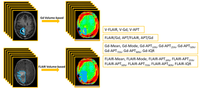
Shanshan Jiang, Hye-Young Heo, Yi Zhang, Jinyuan Zhou
We quantified the accuracy of volumetric APTw image derived metrics in identifying recurrent malignant glioma. 31 patients with suspected recurrent glioblastoma underwent a volumetric APTw imaging sequence at 3T. Volumes with Gd-enhancing, FLAIR abnormality and APTw hyperintensity were drawn as regions of interest (ROIs). Ratios and APTw histogram parameters of volumetric ROIs were calculated and analyzed. There were significant differences in multiple parameters between treatment effects and recurrent tumor. APT/Gd and Gd-APT10% showed the highest diagnostic performance. FLAIR-Mean showed reasonable diagnostic performance with great operation simplicity.
|
|
4862.
 |
116 |
Predicting efficacy of a novel immune stimulator in an animal model of glioblastoma with ferumoxytol cell tracking MRI
Runze Yang, A. Hamilton, Susobhan Sarkar, Reza Mirzaei, V. Wee Yong, Jeff Dunn
Treatment of malignant gliomas with immunotherapy has become an important area of exploration. However, one of the major problems in glioma immunotherapy is the lack of sensitive imaging techniques to differentiate tumor progression (detrimental) from pseudoprogression (beneficial, caused by stimulated macrophages). We hypothesized that tracking macrophages is a sensitive way to detect immunotherapy treatment response. We used a novel drug that stimulates the innate immune system and showed that ferumoxytol based cell tracking MRI is a sensitive way to detect monocyte infiltration and predict tumor growth. Ferumoxytol is used clinically, so this method has high potential for clinical translation
|
|
4863.
 |
117 |
Late-delayed perfusion decrease following radiochemotherapy in glioblastoma patients
Jan Petr, Henri Mutsaerts, Ivan Platzek, Vera Keil, Frank Hofheinz, Iris Asllani, Annekatrin Seidlitz, Marketa Petrova, Esther Troost, Mechthild Krause, Jörg van den Hoff
Temozolomide-based radiochemotherapy (RCT) is a treatment standard for glioblastoma patients. However, RCT is associated with risks of neurocognitive decline. Perfusion is a possible early marker of tissue damage and has been shown to correlate with cognitive changes in many diseases. Perfusion decrease at 3 to 6 months after RT was recently reported in glioblastoma patients. However, it remains unclear whether the decrease is reversible and thus possibly a precursor of the late-delayed cognitive changes. In this study, we have measured perfusion changes up to 18 months following RCT. No further progress of perfusion deficits was found indicating that the early perfusion decrease is predictive of late perfusion decrease and might thus be connected with cognitive decline.
|
|
4864.
 |
118 |
THE CHALLENGE OF TRACTOGRAPHY APPLIED TO CRANIAL NERVES: OUR EXPERIENCE ON DESIGN OF REGIONS OF INTEREST
Timothée Jacquesson, François Cotton, Justine Bosc, Moncef Berhouma, Emmanuel Jouanneau, Arnaud Attye, Carole Frindel
Recent studies have demonstrated diffusion tensor imaging tractography of cranial nerves (CNs). Spatial and angular resolution, however, is limited with this imaging technique. In this study, we reported our experience in CNs tractography detailing the influence of ROI design. We demonstrated that understanding in detail the key role of ROI design and its influence helps to provide coherent tracts. We expect this work to enable a more reliable CNs tractography and made it a useful tool for surgical planning of complex skull base tumors.
|
|
4865.
 |
119 |
23Na MRI at 7 Tesla for Early Response Assessment in Patients with Glioblastoma and Skull Base Meningioma
Sebastian Regnery, Daniel Paech, Heiz-Peter Schlemmer, Mark Ladd, Armin Nagel, Stefan Rieken, Jürgen Debus, Sebastian Adeberg, Nicolas Behl
Radiotherapy is a cornerstone in the treatment of glioblastoma and skull base meningioma. Here, the first results of a prospective longitudinal study employing 23Na MRI for the response evaluation of glioblastoma and skull base meningioma patients during radiotherapy are presented. The study results show that radiation treatment of glioblastoma leads to considerable changes in sodium concentrations within the tumor and the surrounding edema that are dependent on treatment response.
|
|
4866.
 |
120 |
MRI Texture Analysis based on 3D tumor measurement in the identification and prognosis of Gliomas with IDH1 Mutations
Liang Han, Yanwei Miao, Junyi Dong, Xiaoxin Li, Yangyingqiu Liu, Shiyun Tian, Mame KEITA, Weiwei Wang, Yan Guo, Qingwei Song
The 2016 World Health Organization Classification of Tumors of the Central NervousSystem(CNS) used molecular parameters in addition to histology to define manytumor entities, thus formulating a concept for how CNS tumor diagnosis should bestructured in the molecular era.IDH1 is an important molecular marker which has important clinical significance. The prognosis of gliomas with IDH1 mutation is better than those without one. In general,IDH1is detected by pathological biopsy. In this study, gliomas with or without IDH1 mutation was distinguished by using non-invasive MRI texture analysis based on 3D tumor measurement, and then the relationship between MRI texture parameters with survival rate was further assessed.
|
|
Blood Brain Barrier & CSF Flow
Electronic Poster
Neuro
Wednesday, 20 June 2018
| Exhibition Hall |
14:45 - 15:45 |
| |
|
Computer # |
|
4915.
 |
49 |
Pulsatility and velocity in cerebral penetrating arteries in patients with carotid occlusive disease with 7T phase contrast: preliminary results
Tine Arts, Laurien Onkenhout, Jeroen Siero, Jaco Zwanenburg, Geert Jan Biessels
The direct contribution of hemodynamics to the development and progression of vascular cognitive impairment (VCI) is relatively unexplored due to technical challenges concerning the assessment of hemodynamic properties of small vessels. This ongoing study explores changes of hemodynamics by measuring the velocity and pulsatility of perforating arteries in patients with internal carotid artery disease and healthy controls. The preliminary results indicate that high resolution velocity and pulsatility measurements in patients are challenging, particularly due to motion related artefacts. Thus, future research will evaluate user independent analysis to reduce the influence of artifacts and assess test-retest agreement by repeated scanning.
|
|
4916.
 |
50 |
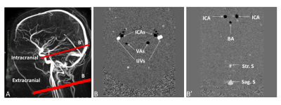
INVESTIGATION OF CEREBRAL BLOOD FLOW PULSATILITY IN AGING PROCESS USING PHASE CONTRAST MAGNETIC RESONANCE IMAGING
Armelle LOKOSSOU, Bader CHAARANI, Souraya ELSANKARI, Catherine GONDRY-JOUET, Olivier BALEDENT
No study already evaluated how arterial blood flow is transferred from extracranial to intracranial level and how venous outflow is transferred from intracranial sinuses to jugular veins. Healthy young and elderly volunteers were enrolled and underwent phase contrast magnetic resonance imaging to investigate intracranial and extracranial arterial and venous flows. In both groups, we found a significant decrease of arterial and venous flows pulsatilities inside the cranium. However, the intracranial and extracranial cerebral blood flow pulsatilities increased significantly with age.
|
|
4917.
 |
51 |
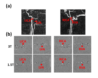
Quantification of total cerebral blood flow measurements using phase contrast magnetic resonance imaging: Comparison and validation at 1.5T and 3T
Yen-Chih Huang, Chun-Ming Chen, Shin-Lei Peng
The effect of field strengths of the total cerebral blood flow (TCBF) quantification using non-gated phase contrast magnetic resonance imaging (PC-MRI) was evaluated in this study. Our results show that, non-gated PC-MRI for TCBF quantification at 3T provided better inter-scan reproducibility when compared to that at 1.5T. Nevertheless, non-gated PC-MRI for TCBF measurements can be performed equally well at 1.5T and 3T. Findings of this study may facilitate data interpretation and comparison of TCBF between different field strengths.
|
|
4918.
 |
52 |
4D CSF Flow Measurement in Cervical Spine at 3T
Ruponti Nath, MJ Negahdar, Robert Bert, Amir Amini
Quantitative and Qualitative analysis of CSF flow was done on a Siemens Skyra 3T Scanner. Four normal Volunteers data shows highly accurate flow waveform, average distance travelled by particle along spinal axis and clear axial and coronal flow visualization between C3-C6 in the cervical spine. Particle tracking was performed in each axial slices. Distance travelled by particles along spinal axis was measured in all volunteers. The average distance travelled in all slices during systole in the head to foot direction varied from 0.1mm to 0.38 mm and during diastole varied from 0.14mm to 0.28 mm in the opposite directions.
|
|
4919.
 |
53 |
Pseudo spiral sampling and Compressed Sensing reconstruction provides high acceleration of intracranial 4D flow MRI at 7T
Lukas Gottwald, Johannes Töger, Eva Peper, Karin Markenroth Bloch, Qinwei Zhang, Bram Coolen, Gustav Strijkers, Pim van Ooij, Aart Nederveen
Long scan times limit the application of 4D flow in clinical practice. Even at 7T, considerable scan times are needed for modest spatiotemporal resolutions. This work demonstrates the advantage of Compressed Sensing acceleration of 4D flow MRI at 7T with a novel undersampling technique. Healthy subjects (n=5) were scanned using standard SENSE and a proposed undersampling technique with Compressed Sensing reconstruction. Flow analysis showed minor differences, and image quality improved for Compressed Sensing reconstructions with maintained resolution and reduced scan time. The method enables further increases of acceleration and spatiotemporal resolution, adding more physiological details beyond current resolution limitations.
|
|
4920.
 |
54 |
Influence of Signal Magnitude on Intracranial Flow Quantification using 4D flow MRI
Leonardo Rivera-Rivera, Tilman Schubert, Patrick Turski, Oliver Wieben, Kevin Johnson
Physiological parameters derived from quantitative flow MRI can potentially improve characterization of a large spectrum of vascular diseases if routinely used in a clinical setting. However, current barriers limit the use of quantitative flow MRI in a clinical setting, partially due to a lack of calibration tests, and concerns regarding accuracy and reproducibility. In this study we investigate the potential induced bias of flow measurements in a cranial 4D flow MRI acquisition due to signal magnitude heterogeneity, and the implications for comparing protocols with differing flip angle or contrast agent usage.
|
|
4921.
 |
55 |
Blood Brain Barrier Water Permeability in Non-Enhancing Multiple Sclerosis Lesion with Intrinsic Diffusivity Encoding of Arterial Labeled Spins (IDEALS)
Kenneth Wengler, Jason Ha, Patricia Coyle, Mark Schweitzer, Tim Duong, Xiang He
Persistent endothelial abnormalities and blood-brain barrier (BBB) disruption may play an important role in MS lesion formation and progression. In MS, BBB dysfunction occurs not only in active lesions with contrast-enhancement, but also in inactive (chronic) lesions and normal appearing white matter. In this study a novel method to map whole-brain BBB water permeability (IDEALS) was used to measure in non-enhancing MS lesions of 11 relapse-remitting MS patients. Although the permeability for MRI contrast in non-enhancing MS lesions is several orders of magnitude lower than that of enhancing MS lesions, our study demonstrated a robust reduction of lesion water permeability.
|
|
4922.
 |
56 |
Epidermal Growth Factor's lowers Blood Brain Barrier leakiness in an Alzheimer's disease mouse model as detected using Diffusion Weighted Arterial Spin Labeling MR Imaging
Frederick Damen, Riya Thomas, Rong-Wen Tain, Weiguo Li, Leon Tai, Kejia Cai
Blood-brain barrier (BBB) dysfunction is reemerging as a critical component of Alzheimer’s disease (AD). Higher BBB leakiness is one of the mechanistic pathways through which AD risk factors induce cognitive decline. We have previously demonstrated that epidermal growth factor (EGF) prevents BBB leakiness in a model of two important AD risk factors: APOE4 and female sex (female E4FAD mice). The goal of this study was to use Diffusion Weighted Arterial Spin Labeling MRI to determine whether post-symptomatic EGF treatment, (during 8-10 months) can reduce BBB leakiness in EGF treated mice compared to vehicle controls.
|
|
4923.
 |
57 |
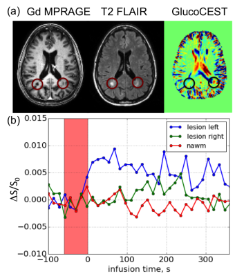
Imaging Blood Brain Barrier Disruption in Multiple Sclerosis using GlucoCEST MRI
Xiang Xu, Pavan Bhargava, Linda Knutsson, Martin Pomper, Peter Calabresi, Peter van Zijl
It has been demonstrated that glucose can be used as a contrast agent for chemical exchange saturation transfer (glucoCEST) and enhanced relaxation (T2 or T1ρ). In the present work we show the possibility of detecting blood brain barrier (BBB) disruption in multiple sclerosis (MS) using glucoCEST MRI at 7T and 3T. At both field strengths, glucoCEST enhancement was observed in some MS lesions that were not enhanced by Gd T1w images. Our results show that glucose may be more permeable than Gd to minor BBB disruptions, suggesting that glucose transport may also be more sensitive to MS disease activities.
|
|
4924.
 |
58 |
High Resolution UTE-MRAs for Longitudinal Visualization of Revascularization and Blood-Brain Barrier Disruption on a Rat Transient Middle Cerebral Artery Occlusion Model
MungSoo Kang, HyungJoon Cho
Longitudinal study of revascularization and BBB disruption after ischemic stroke provides prognostic and therapeutic information. In this work, high resolution UTE-MRAs with superparamagnetic iron oxide nanoparticles (SPION) were performed on a rat tMCAO model to visualize revascularization and BBB disruption longitudinally. UTE-MRAs before and after SPION injection clearly visualized arterial vessels and all vessels, respectively. Thickened and twisted vessels at the rat brain surface of ipsilateral hemisphere were highly resolved (59 μm3 isotropic).
|
|
4925.
 |
59 |
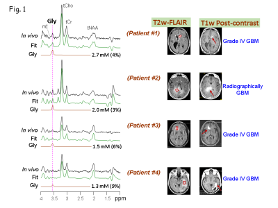
Glycine is an early 'biomarker' of blood brain barrier breakdown in Gliomas
Vivek Tiwari, Zhongxu An, Yiming Wang, Michael Levy, Marco Pinho, Elizabeth Maher, Edward Pan, Toral Patel, Bruce Mickey, Changho Choi
Cancer cells may use altered metabolic pathways with respect to their normal counterparts; this metabolic switch is necessary to support their rapid proliferation in oxygen- and nutrient-poor conditions. A few of the metabolites are up-regulated while some others are down-regulated or unaltered. High grade malignant tumors present with enhancement on T1-weighted (T1w) image. Enhancement on post-contrast T1w images is an indicative of breakdown of blood brain barrier (BBB). The increased number of tumor cells may stress the vessels and, thus BBB tend to rupture. A non-invasive bio-marker that can predict the disruption of BBB will be of great clinical significance for assessing the tumor aggressiveness. Here, we show elevated Gly can be a potential bio-marker for predicting the tumor’s potential to present with ruptured BBB.
|
|
4926.
 |
60 |
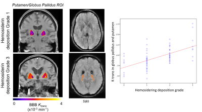
Increased vascular permeability in the lenticulostriate arteries results in increased hemosiderin deposition in the basal ganglia in aging and cognitive impairment
Axel Montagne, Giuseppe Barisano, Meng Law, Farshid Sepherband, Arthur Toga, Berislav Zlokovic
The intramural periarterial drainage pathway is critical for the elimination of metabolic waste products from the brain. In a number of neurological diseases such as Alzheimer’s Disease, blood-brain barrier damage and increased vascular permeability may play an important role in the pathogenesis. Leakiness of the blood-brain barrier allows fibrin(ogen), hemosiderin and metabolic wastes to exudate and deposit around the vessels in the basal ganglia. In order to test this hypothesis, we evaluated the relationship between blood-brain barrier permeability measured as ktrans and hemosiderin deposition in 76 subjects scanned at 3T MRI.
|
|
4927.
 |
61 |
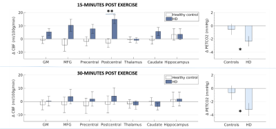
Exercise selectively increases cerebral blood flow in the postcentral gyrus in patients with Huntington’s disease
Jessica Steventon, Hannah Furby, Richard Wise, Kevin Murphy
The therapeutic potential of exercise - a potent trigger for both neurogenesis and vascular plasticity - is currently a hot research topic in neurodegenerative diseases. Here we tested whether a single 20-minute bout of aerobic exercise was sufficient to induce changes in cerebral blood flow (CBF) in people with Huntington’s disease (HD). We find a transient and selective increase in CBF in the postcentral gyrus in HD participants following exercise, a region with reduced intrinsic functional connectivity in HD patients, with no change in healthy controls. The CBF change was unrelated to clinical and genetic markers of disease.
|
|
4928.
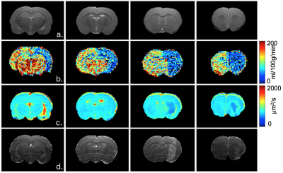 |
62 |
Impact of focused ultrasound on cerebral blood flow
Vasile Stupar, Anthony Delalande, Nora Collomb, Claire Rome, Emmanuel Barbier
One major application of FUS is the opening of the blood brain barrier (BBB) to target the brain cells with drugs such as in glioma. While the impact of FUS on brain disease is under intense evaluation, its impact of normal brain physiology has received less attention. In this study, we evaluate the impact of BBB opening using FUS on cerebral blood flow (CBF), assessed by pseudo-continuous arterial spin labeling (ASL). Following 0.6MPa FUS at 1.2Mhz, a small change in ADC was observed, suggesting a small vasogenic edema. In addition, there was a 50%CBF reduction in a large vascular territory.
|
|
4929.
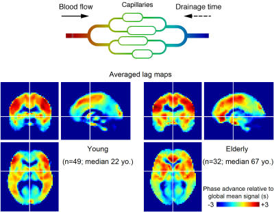 |
63 |
Age-related shift of cerebral venous outflow pathway detected by BOLD signal-based blood flow tracking
Toshihiko Aso, Shinnichi Urayama, Tsukasa Ueno, Naoya Oishi, Yukako Nakagami, Toshiya Murai, Hidenao Fukuyama
A novel blood tracking technique based on BOLD MRI signal was applied to two age groups of healthy subjects (n=81) to investigate age-related alterations in cerebral circulation. By mapping the phase of low-frequency component and between-subject regression analysis on this “BOLD lag map”, linear extension of venous drainage times with age was found in the deep venous system draining the periventricular region. Interestingly, age-related shortening of washout time was observed in the superficial system involving the major sinuses. This dissociation of deep and superficial venous systems may reflect focal inefficiency in the deep system as part of normal aging processes.
|
|
4930.
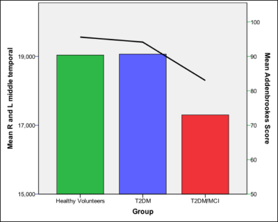 |
64 |
Cerebral Blood Flow, Intracranial Volumes and Cognitive Status in Type-2 Diabetes Mellitus
Leanne Hunt, Dinesh Selvarajah, Solomon Tesfaye, Iain Wilkinson
This study quantitates regional brain volumes and arterial cerebral perfusion characteristics (ASL) in patients with Type-2 Diabetes Mellitus (T2DM) who have Mild Cognitive Impairment, in patients with T2DM who have normal cognitive status and in non-diabetic healthy volunteers. Mean group differences were identified in both grey matter volume and cerebral blood flow in regions that include the medial temporal lobes. These findings may further our understanding of the mechanisms that lead to an increased risk of the development of cognitive impairment associated with diabetes.
|
|
4931.
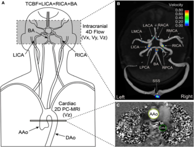 |
65 |
Aortic flow and cerebral hemodynamics in age-related brain volume loss
Ann Ragin, Can Wu, Guixiang Ma, Sameer Ansari, Michael Markl, Susanne Schnell
Concurrent cardiac and neurovascular (4D flow) MR imaging were used to quantify cardiac and cerebral hemodynamics in 30 healthy adults to determine the relationship to brain volumetric measures of regions considered vulnerable in aging. Cardiac index (p=0.001); ascending aorta (p=0.003) total cerebral blood flow (p=0.001) and flow for left internal carotid (p<0.001), basilar (p=0.04), and right anterior cerebral (p=0.03) arteries were significantly lower in midlife compared to younger adults. Lower cardiac index, total cerebral blood flow and left internal carotid flow were correlated with reduced gray matter, superior frontal cortical thinning and volume loss in putamen.
|
|
4932.
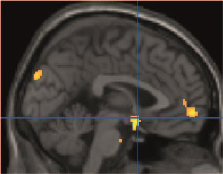 |
66 |
Altered cerebral blood flow before and after 4-weeks of neurostimulation in patients with episodic migraine
Lars Michels, Jessica Aschmann, Franz Riederer, Andreas Gantenbein, Roger Luechinger, Spyros Kolllias, Peter Sandor
Cerebral blood flow is altered in migraineurs but it is unknown if this can be changed by repetitive neurostimulation, leading to less migraine attacks. In a double blind and sham-controlled study, we used arterial spin labeling MRI and transcranial direct current stimulation (tDCS) to address this question. Four weeks of real tDCS diminished initial hyperperfusion in patients with episodic migraine in pain processing brain regions (p < 0.001). In addition, less migraine attacks occurred after real tDCS compared to baseline (p < 0.05). Our results indicate a regulating effect of tDCS on cerebral blood flow and clinical outcome in migraineurs.
|
|
4933.
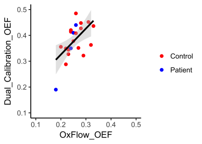 |
67 |
Quantifying brain oxygen extraction fraction: correlation between a global venous susceptibility method and calibrated fMRI mapping method.
Hannah Chandler, Michael Germuska, Thomas Okell, Ana Rodriguez-Soto, Erin Englund, Rachael Stickland, Catherine Foster, Jessica Steventon , Fabrizio Fasano, Kevin Murphy, Valentina Tomassini, Felix Wehrli, Richard Wise
Measuring oxygen extraction fraction (OEF) may prove a useful clinical tool for assessing brain oxygen consumption. Dual-calibrated fMRI is used to map voxel-wise OEF across grey-matter and requires respiratory challenge (hypercapnia and hyperoxia). However, this method presents challenges to clinical implementation. Conversely, OxFlow provides a global estimate of OEF, has a shorter acquisition time and does not require respiratory challenge. Here we examine the relationship between both approaches for measuring OEF, revealing a significant association between methods. While further investigation is required, OxFlow may offer additional clinical utility for measuring cerebral oxygen consumption.
|
|
4934.
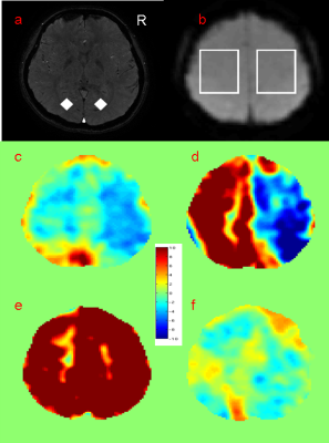 |
68 |
MR susceptometry based superior sagittal sinus venous oxygen saturation: effects of carbon dioxide based cerebral vascular reserve and anesthesia in Moyamoya patients
Chou-Ming Cheng, Tzu-Chen Yeh, Jen-Chuen Hsieh, Hsiao-Wen Chung
Venous oxygen saturation (SvO2) is measured on Moyamoya patients with previous proposed automatic methods based on MR susceptometry from the phase information obtained with multi-echo gradient-echo imaging. According to anesthesia condition and carbon dioxide based cerebral vascular reserve (CO2-CVR) using blood oxygen level dependence (BOLD) technique, 21 Moyamoya patients are classified into four groups. Results from them suggested that global SvO2 value may indicate global pathophysiological change supported by reduced SvO2 without obviously impaired CO2-CVR and elevated SvO2 under anesthesia.
|
|
4935.
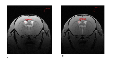 |
69 |
Assessment of Intravenous Immunoglobulin treatment on BBB function following inflammation
Oliviero Gobbo, Aislinn Mc Phillips, Tara Hamilton, Jerrard Hayes, Gavin Davey
The blood brain barrier (BBB) is a complex structure that separates brain interstitial fluid from blood. BBB dysfunction is a key feature of Central Nervous System diseases. This in vivo study examines the effects of Intravenous Immunoglobulin (IVIg), regularly used to treat immune and inflammatory diseases, on BBB function following LPS-induced BBB disruption. The change in permeability of the BBB was evaluated by MRI, using gadolinium. Our results suggest that LPS breach the integrity of the BBB with regional specificity and can be reversed by IVIg administration.
|
|
4936.
 |
70 |
Permeability of the blood-brain barrier predicts no evidence of disease activity at two years after natalizumab or fingolimod treatment in relapsing-remitting multiple sclerosis
Stig Cramer, Helle Simonsen, Ulrich Lindberg, Aravinthan Varatharaj, Ian Galea, Jette Frederiksen, Henrik Larsson
Dynamic contrast-enhanced MRI enables measurements of the permeability of the blood-brain barrier (BBB), possibly a marker of disease activity in multiple sclerosis (MS). In order to investigate if permeability predicts early suboptimal treatment response, defined as loss of no evidence of disease activity (NEDA) status after two years, we included 35 relapsing-remitting MS patients initiating either fingolimod or natalizumab, drugs with a common effect of decreasing lymphocyte influx into the CNS. We find that permeability measured after six months of treatment was a good predictor loss of NEDA status at two years and a surrogate marker of the state of health of the blood-brain barrier.
|
|
4937.
 |
71 |
Imaging correlates of the Blood Brain Barrier disruption in HIV associated neuro cognitive disorder and therapeutic implications
Did Not Present
Joga Chaganti, Bruce Brew
. Neurocognitive impairment in HIV infection, known as HIV associated neurocognitive disorder (HAND), in the context of suppressive combination antiretroviral therapy still occurs. We hypothesized that ongoing chronic systemic inflammation, a feature of HIV disease despite viral suppression, causes blood brain barrier (BBB) disruption thereby allowing entry of neurotoxic cytokines/chemokines or HIV itself (in very low levels because of suppression) thereby causing HAND or at least worsening it. To test this hypothesis, we have used dynamic contrast enhancement derived metrics (K-trans) to measure the capillary permeability as, a direct indicator for possible BBB disruption and correlated it with neuro inflammatory markers in the cerebrospinal fluid (CSF ) MR spectroscopy derived metabolites as well as with clinical indices of neurocognition (neuropsychological scores).
|
|
Traumatic Brain Injury
Electronic Poster
Neuro
Wednesday, 20 June 2018
| Exhibition Hall |
14:45 - 15:45 |
| |
|
Computer # |
|
4938.
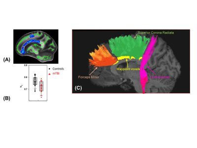 |
73 |
Effects of Track Length on White Matter Alterations in Mild Traumatic Brain Injury
Sourajit Mustafi, Jaroslaw Harezlak, Joaquin Goni, Laura Flashman, Thomas McAllister, Yu-Chien Wu
In the present study, we performed streamline tractography to characterize effects of track length on white-matter microstructural alterations after mild traumatic brain injury. Streamline length and counts were studied in involved white-matter fiber tracts that were found to have decreased axonal density at some points along the tracts using voxel-based analyses. The results suggested that long fibers in the brains of individuals who sustained mild traumatic brain injury are more vulnerable to the injury.
|
|
4939.
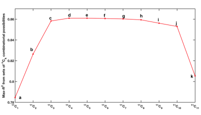 |
74 |
Regional fractional anisotropy and processing speed in diffuse traumatic brain injury: An exploratory analysis using a linear regression panel model
Sindhuja Govindarajan, David Ouellette, Junghoon Kim, Tim Duong
Recently, Ware and colleagues1 showed that FA-derived damage assessment in WM regions can accurately classify subjects with processing speed impairment in moderate to severe diffuse TBI at 3 months post-injury. Using a subset of the same cohort who completed follow-up DTI scans at 6 and 12 months post-injury, the goals of this study were 1) to investigate if regional FA remains associated with processing speed in a dataset including all time points and 2) to conduct an exploratory analysis to identify a subset of regions that can best explain such association. Four regions were identified that explained most variance in processing speed.
|
|
4940.
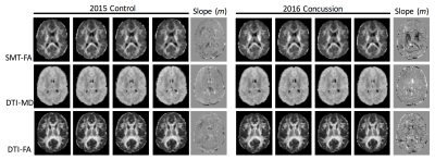 |
75 |
Within-Subject Characterization of Sports Concussion with Serial Diffusion MRI and Spherical Mean Technique
Video Permission Withheld
Matthew Budde, L. Tugan Muftuler, Andrew Nencka, Kevin Koch, Yang Wang, Timothy Meier, Michael McCrea
Diffusion tensor imaging is a promising biomarker of mild Traumatic Brain Injury and concussion, but it has not been refined sufficiently for diagnosis in individual subjects. In this study, we demonstrate that spherical mean technique (SMT) diffusion MRI can detect post-concussion changes in single subjects over the acute recovery period.
|
|
4941.
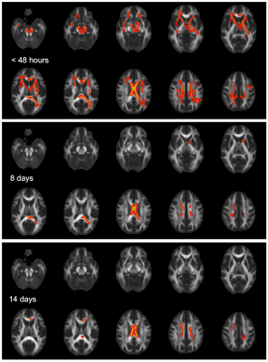 |
76 |
Characterizing acute, subacute and chronic changes in the brain following sport-related concussion using Diffusion Kurtosis Tensor Imaging
L. Tugan Muftuler, Timothy Meier, Daniel Olson, Michael McCrea
The aim of this study was to characterize acute, subacute and chronic changes in the brain following sport-related concussion in a group of young athletes. Diffusion kurtosis tensor parameters were compared between concussed athletes and controls. The concussed group demonstrated widespread increase in axial kurtosis compared to the controls at the acute time point, which gradually diminished over the course of 14 days. Conversely, alterations in radial kurtosis were mostly in the frontal white and gray matter and occipital gray matter regions. These findings may have important implications for the clinical management of sport-related concussions.
|
|
4942.
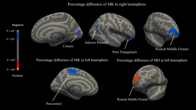 |
77 |
Evaluating Microstructural Integrity of Cortical and Deep Gray Matter over a Season of High School Football Using Diffusion Kurtosis Imaging and Quantitative Susceptibility Mapping
Did Not Present
Nan-Jie Gong, Samuel Kuzminski, Michael Clark, Melissa Fraser, Mark Sundman, Kevin Guskiewicz, Jeffrey Petrella, Chunlei Liu
Over the course of a single high school football season, QSM showed no evidence of microhemorrhage or iron-related changes; however, we observed significant microstructural alterations, as reflected by DKI metrics, in cortical and deep gray matter over the course of a single high school football season. These microstructural changes correlated with the frequency of head impacts, suggesting that DKI imaging of cortical and deep gray matter may yield valuable biomarkers for tracking subclinical traumatic brain injury in contact-sport athletes.
|
|
4943.
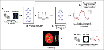 |
78 |
Exploring the effects of concussion history on quantitative estimation of temporal delays and cerebrovascular reactivity: A novel approach combining hypercapnia and hyperoxia in collegiate football players
ALLEN CHAMPAGNE, NICOLE COVERDALE, ALEX BHOGAL, DOUGLAS COOK
Recent evidence suggests that concussion history may be associated with long-term structural and functional changes in the brain. However, little is known about the effects of head injury on vascular reactivity. In this study, we combined hypercapnic and hyperoxic respiratory manipulations to explore the effects of concussion history on hemodynamic latencies and tissue cerebrovascular reactivity (CVR) in football players. We found that, although tissue CVR was not different between the groups (P>0.05), time delays to hypercapnia were shorter in subjects with a history of concussion (P<0.05), which may suggest some compensatory mechanism in the vasculature that persists beyond clinical recovery.
|
|
4944.
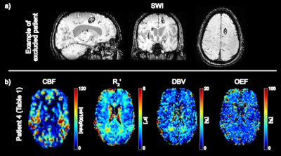 |
79 |
Investigating tissue oxygenation in acute Traumatic Brain Injury using Arterial Spin Labelling and quantitative-BOLD
Alan Stone, Tim Lawrence, Thomas Okell, Natalie Voets, Nicholas Blockley
Following a traumatic brain injury, oxygen metabolism may be disrupted by changes in oxygen supply and demand. This study aims to investigate global changes in oxygen metabolism in the hours immediately following the initial insult to the brain. Pseudo-continuous ASL (PCASL) and streamlined-qBOLD (sqBOLD) are used to measure blood flow and oxygenation in the acute stages of TBI (<24 hours post-injury). When compared with an age matched control group, blood oxygenation appears to increase during injury (decreased OEF (p=0.03)) at this acute time-point.
|
|
4945.
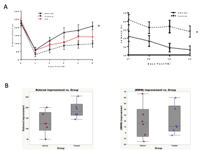 |
80 |
DTI Connectome Analysis in Moderate TBI: Parcellation Improves Detection of Relevant Injury and the Monitoring of Therapeutic Response
TALAIGNAIR VENKATRAMAN, RYAN PEARMAN, HAICHEN WANG, CHRIS PETTY, ALLEN SONG, DANIEL LASKOWITZ, CHRISTOPHER LASCOLA
Conventional DTI readouts such as fractional anisotropy (FA), axial diffusion (RD) and radial diffusion (RD) show distinct alterations following closed head injury but do not correlate well with measured functional and cognitive outcomes. 3D DTI with parcellated connectome analysis reveals neural fiber networks between brain regions that are most vulnerable to closed head (acceleration-deceleration) injury and also most relevant to measurable functional deficits. In this study, fiber tract number between hippocampus and cortex is the most sensitive marker of both motor and memory deficits and therapeutic improvement following administration of a novel neuroprotective therapeutic agent.
|
|
4946.
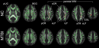 |
81 |
Working Memory And White Matter Microstructure In Mild Traumatic Brain Injury
Sohae Chung, Els Fieremans, Xiuyuan Wang, Charles Morton, Dmitry Novikov, Joseph Rath, Yvonne Lui
Working memory is a critical cognitive function implicated after mild traumatic brain injury (MTBI). Here, we investigate the association between white matter microstructure and working memory in normal controls (NC) and MTBI patients, using diffusion white matter tract integrity (WMTI) and WAIS-IV subtests, respectively. For the NC group, significant correlations were observed in axonal water fraction (AWF; higher axonal density/myelination) and mean kurtosis (MK; greater tissue complexity) with letter-number sequencing (LNS). However, such relationships were not present in the MTBI group.
|
|
4947.
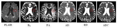 |
82 |
CNR comparison to identify the detectability rate and FA histogram analysis of FLAIR lesions in DTI metrics.
Ramtilak Gattu, Randall Benson, Ewart Haacke
There is a paucity of studies trying to investigate the sensitivity of DTI metrics like Fractional Anisotropy (FA), Apparent Diffusion Coefficient (ADC), Axial Diusivity and Radial Diffusivity in areas corresponding to the lesion sites detected in clinical MRI using FLAIR images. Aim of this study was to compare the utility of DTI with FLAIR by evaluating the contrast to noise ratio (CNR) in their detecting capacity of DAI lesions and to evaluate whether DTI is more accurate than FLAIR or vice versa.
|
|
4948.
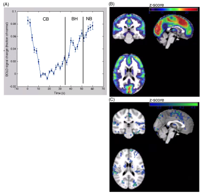 |
83 |
Using fMRI to assess cerebrovascular reactivity after acute concussion
Nathan Churchill, Michael Hutchison, Simon Graham, Tom Schweizer
Cerebrovascular reactivity (CVR) is an important biomarker of concussion, as brain activity and blood flow regulation are often impaired after brain injury. This study examines an fMRI breath hold paradigm as a probe of CVR in concussed athletes, showing significant variations with days post-injury.
|
|
4949.
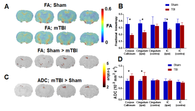 |
84 |
Functional correlates of microstructural damage in developmental traumatic brain injury: a multi-modal MRI study of neuroanatomy, cerebrovascular reactivity, and functional connectivity
Maxime Parent, Ying Li, Basavaraju Sanganahalli, Vijayalakshmi Santhakumar, D.S. Fahmeed Hyder, Sridhar Kannurpatti
The developing brain is particularly vulnerable to traumatic brain injury (TBI), with symptoms and co-morbidities lasting into adulthood. Animal models of developmental TBI are useful tools for investigating molecular pathways of TBI injuries and to assess novel therapeutics; however, validation of in vivo injury biomarkers in these models is important for the translational application of pre-clinical findings. Here, we demonstrate that the persistent dysfunctions of developmental TBI in an animal model can be measured at the level of microstructural damage (with diffusion MRI), functional interhemispheric connectivity (with resting-state fMRI) and systemic neurovascular coupling (with CO2 challenge fMRI).
|
|
4950.
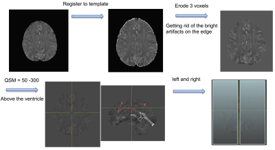 |
85 |
Military TBI Patients Demonstrate Increased Cerebral Venous Volume Using Quantitative Susceptibility Mapping
Wei Liu, Gerard Riedy, Ping-Hong Yeh, Dominic Nathan, Chihwa Song, Grant Bonavia, John Ollinger
Cerebral venous volume above the lateral ventricle was analyzed using quantitative susceptibility mapping in patients with severe (n = 7), moderate (n = 21) and mild (n = 27 for blast and n = 19 for nonblast) TBI. Severe and moderate TBI patients both demonstrated increased venous volume compared to control subjects without history of TBI. Blast injured mild TBI patients also showed increased venous volume compared to the control subjects. These results suggest that microvascular injury may present in TBI patients and blast related injury might have a more pronounced impact on the venous system.
|
|
4951.
 |
86 |
Transient neuronal dysfunction correlates with hypo-perfusion in patients with moderate post-traumatic brain injury
Video Permission Withheld
Eva-Maria Ratai, Suk-tak Chan, Maria Gabriela Figueiro Longo, Michael Wenke, Termara Parker, Jonathan Welt, Joyce van Loon, Emad Ahmadi, Anastasia Yendiki, Jacqueline Namati, Isabel Chico-Calero, Blair Parry, Can Ozan Tan, Jarone Lee, Michael Lev, Michael Hamblin, Benjamin Vakoc, Rajiv Gupta
We present a DOD funded study to assess the effects of low-level light therapy (LLLT) on metabolites measured by MRS and cerebral perfusion measured by ASL using a double-blind, placebo controlled study where patients with moderate TBI were randomized between LLLT and sham treatment. We report CBF and metabolic changes pertaining to neuronal injury and neuroinflammation for the entire cohort (i.e., both treated and control arms), before the blind is broken. Our findings suggest that transient neuronal dysfunction in the posterior cingulate cortex and neuroinflammation in the thalamus correlated with hypo-perfusion during the subacute phase of moderate TBI.
|
|
4952.
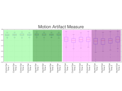 |
87 |
Eye motion artifacts in T2-weighted images, identified by deep neural networks, correlate with concussion
Andrew Nencka, John Bukowy, Robin Karr, Andrew Klein, Kevin Koch, Peter LaViolette, Sean McGarry, Timothy Meier, Brad Swearingen, Michael McCrea
Oculomotor deficits occur with traumatic brain injury, and eye motion yields artifacts in the phase encoding directions of common MRI acquisitions. Here we quantify motion artifacts in regions of interest of T2-weighted MRI head images in concussed and healthy high school and collegiate athletes. Regions of interest over eyes, and inner ear structures as a control, were automatically generated using a convolutional neural network. Acute and sub-acute injury was found to yield significantly increased motion artifact compared to controls in ROIs covering eyes. No differences in motion artifacts covering inner ear structures were found. These results indicate that anatomical MRI following traumatic brain injury may offer increased diagnostic or prognostic information through artifact resulting from eye motion associated with injury.
|
|
4953.
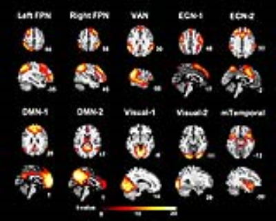 |
88 |
Alteration of Resting-state Functional Networks is Associated with Post Concussion Symptoms in Mild Traumatic Brain Injury
Nai-Chi Chen, Chia-Feng Lu, Li-Chun Hsieh, Sho-Jen Cheng, Yu-Chieh Kao, Ho-Fang Huang, Cheng-Yu Chen
A subgroup of patients with mild traumatic brain injury (mTBI) suffer from a series of post-concussion symptoms, such as headache, dizziness, and cognitive deficits. These patients also present altered neural connectivity compared to healthy controls (HC). In our study, we examined resting-state functional connectivity in 30 mTBI patients and 35 HC using independent component analysis. We found that patients showed lower functional connectivity in several resting-state networks, and the left frontoparietal network was associated with the level of dizziness in patients. We concluded that the alterations of resting-state neural networks may support post-concussion symptoms after mTBI.
|
|
4954.
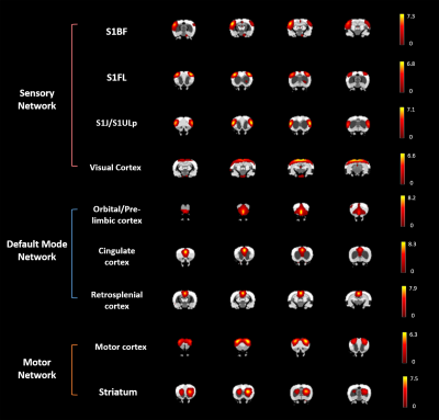 |
89 |
Low-frequency Fluctuations of Resting-state fMRI BOLD Signal after Experimental Mild Traumatic Brain Injury
Yu-Chieh Jill Kao, Chia-Feng Lu, Ping-Huei Tsai, Fei-Ting Hsu, Bao-Yu Hsieh, Cheng-Yu Chen
Regional amplitude of rsfMRI BOLD signal after mTBI was quantified using amplitude of low-frequency fluctuation (ALFF). The significant increase of ALFF around the impact regions at 24 h and renormalized at 50 days after mTBI may suggest the dynamics of neuronal activity and neurovascular response induced by the injury.
|
|
4955.
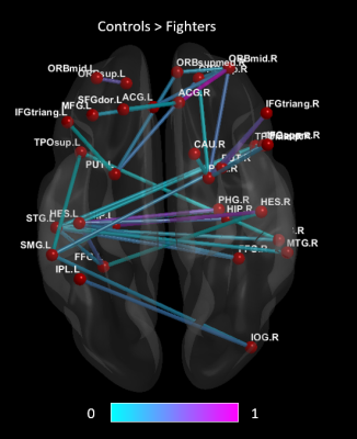 |
90 |
Altered Functional Connectivity and Network Topology in Active Profession Fighters
Karthik Sreenivasan, Zhengshi Yang, Virendra Mishra, Sarah Banks, Dietmar Cordes, Charles Bernick
Studies have shown that both active and retired athletes with repeated head trauma are more likely to suffer from cognitive decline and loss of executive and attention functions when compared to age- matched healthy controls. Our results show decreased functional connectivity in the active fighter group when compared to controls between regions known to be implicated in traumatic brain injury. Furthermore, we found a shift towards a less efficient network topology with altered integration and segregation in active professional fighters.
|
|
4956.
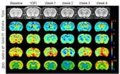 |
91 |
GlucoCEST weighted MRI detects metabolic abnormalities following experimental TBI on 7T and 9.4T
Tsang-Wei Tu, Jaclyn Witko, Joseph Frank
Delayed glucose hypometabolism has been reported in traumatic brain injury (TBI) patients from weeks to years posing a high risk for neurodegenerative diseases. This study shows the feasibility of glucoCEST weighted MRI (GWI) to detect metabolic abnormalities following TBI on 7T and 9.4T. The GWI results were compared to 2DG autoradiography and immunohistochemitry indicating that the injured brain needed immediate energy to restore nervous function and then entered a hypometabolism state in week 2. GWI affords the sensitivity to detect cerebral metabolic states following TBI and has potential to identify the treatment window to increase neuronal survival.
|
|
4957.
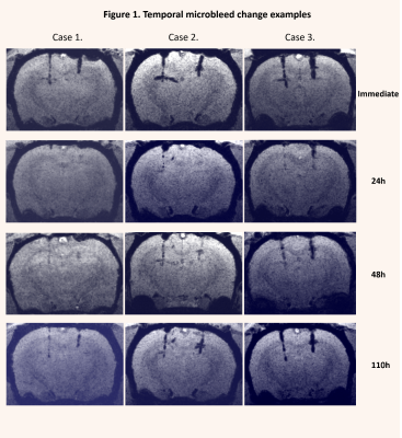 |
92 |
Transient disappearance of traumatic microbleeds on susceptibility weighted imaging in rats
Did Not Present
Arnold Tóth, Bálint Környei, Zoltán Berente, Endre Czeiter, Krisztina Amrein, Péter Bogner, Tamás Dóczi, András Büki, Attila Schwarcz
Traumatic microbleeds (TMBs) are regarded as markers of traumatic brain injury (TBI) related diffuse axonal injury (DAI). According to several recently published observations, TMBs depicted by susceptibility weighted imaging (SWI), seems to show temporal changes. This study aims to explore the temporal features of TMBs in a rat model.
|
|
4958.
 |
93 |
Gray matter volume changes following Cranial Nerve Non-invasive NeuroModulation in patients with traumatic brain injuries
Jiancheng Hou, Arman Kulkarni, Neelima Tellapragada, Veena Nair, Mitch Tyler, Yuri Danilov, Kurt Kaczmarek, Beth Meyerand, Vivek Prabhakaran
The main conventional approach for treating traumatic brain injury related gait and balance deficits has been through physical therapy, but few approaches have focused on brain based rehabilitation efforts that create direct neuroplastic changes. There remains a need for such an approach. Therefore, the goal of this study was: 1) to apply Cranial Nerve Non-Invasive NeuroModulation (CN-NINM) via the tongue in combination with multiple symptom-specific physical therapy exercises in patients with mild to moderate TBI, and 2) to investigate and quantify gray matter volume changes prior to and after intervention as well as their correlation with behavior.
|
|
4959.
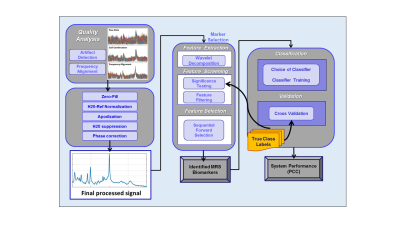 |
94 |
Novel Processing of Magnetic Resonance Spectroscopy Enables Biomarker Discovery for PTSD and mTB
John Irvine, Laura Mariano, Ben Rowland, Huijun Liao , Kristin Heaton, Alexander Lin
The objective of this study was to determine the neurochemical biomarkers for mild traumatic brain injury (mTBI) and posttraumatic stress disorder (PTSD) among members of the military. A sample of 100 participants were assigned to each cohort (mTBI only, PTSD only, mTBI and PTSD, military control, and civilian control). Analysis of metabolite concentrations in the mTBI and PTSD cohorts showed significant metabolite difference across 3 voxels in the brain (posterior cingulate gyrus (PCG), anterior cingulate cortex (ACC), and posterior white matter (PWM)), indicating that magnetic resonance spectroscopy provides objective biomarkers for distinguishing these conditions.
|
|
4960.
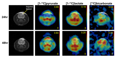 |
95 |
Assessment of metabolic changes in traumatic brain injury using hyperpolarized [1-13C]pyruvate: a longitudinal study
Edward Hackett, Laura Ingle, Brenda Bartnik-Olson, Jae Mo Park
In this study, we longitudinally assessed acute metabolic changes during 2-120hrs post traumatic brain injury in a controlled-cortical impact rat model using hyperpolarized [1-13C]pyruvate. We observed mildly increased pyruvate conversion to lactate and significantly reduced bicarbonate production in the injured site at 24hrs after impact. Conversely, lactate was reduced when measured at 48hrs-post injury. Bicarbonate production in the lesion remained low with significantly increased bicarbonate production in the contralateral normal-appearing brain
|
|
4961.
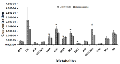 |
96 |
Hippocampal-Cerebellar metabolic investigation in TBI rats: NMR spectroscopy study
Did Not Present
Subash Khushu, Kavita Singh, Richa Trivedi, Sonia Gandhi, Poonam Rana
Cerebellar atrophy and dysfunction has been reported in clinical TBI. hippocampal-cerebellar cognitive collaborations and its manifestation in TBI is being reported. This study investigated TBI mediated metabolic irregularities in CB and HP in animal model of TBI. These results indicated that CB and HP are metabolically different regions. The models generated from the changes due to trauma could accurately discriminate CB and HP and injury severity. We speculate that reduced NAA and Cho levels may be due to substantial focal neuronal loss after weight drop TBI. Edema and excitotoxity following injury leads to altered levels of osmolytes and Glu/Gln.
|
|
Neuroimaging: Animal Studies
Electronic Poster
Neuro
Wednesday, 20 June 2018
| Exhibition Hall |
14:45 - 15:45 |
| |
|
Computer # |
|
4962.
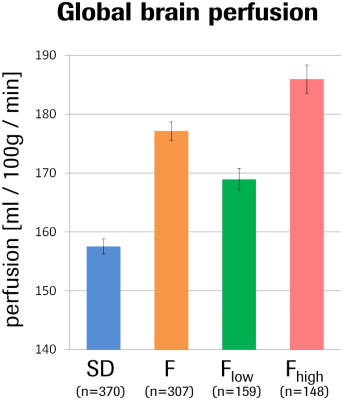 |
97 |
ASL-based fMRI phenotyping of high-anxiety Fischer rat strain reveals two unexpected sub-traits or -states: caveat for rodent studies
Andreas Bruns, Thomas Mueggler, Markus von Kienlin, Basil Künnecke
Fischer rats serve as a rodent model of high trait anxiety in neuroscience and drug discovery. Using ASL-based fMRI, we characterized Fischer rats with respect to their regional brain activity patterns and with reference to the standard Sprague-Dawley rat strain. Fischer rats clearly differed from Sprague-Dawley rats, but also split into two distinct subpopulations, with one showing a more deviant pattern than the other. Although it has remained elusive whether this is a trait or a state phenomenon, our data suggest that neuronal networks related to anxiety and/or depression are implicated.
|
|
4963.
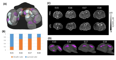 |
98 |
In utero monitoring of early embryonic mouse brain development and injury progression with localized diffusion MRI
Dan Wu, Jun Lei, Yan Zhu, Michael McLane, Irina Burd
In utero diffusion MRI (dMRI) of the embryonic mouse brain is challenging due to maternal and fetal motions and limited resolution. We have previously developed a localized imaging technique and achieved fast dMRI of the live embryos. Here we explored the capacity of this technique to image embryonic mouse brains from mid-to-late gestation stages during development. Furthermore, we monitored the progression of embryonic brain injury in a mouse model of intrauterine inflammation. Our results suggested in utero dMRI is an indicator of fetal outcome after inflammatory injury, and the embryonic brain injury correlates with placental injury in this animal model.
|
|
4964.
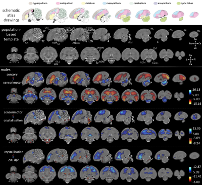 |
99 |
Longitudinal voxel-based morphometry in juvenile zebra finch brains reveals volume changes linked to distinct stages of the critical period for song learning
Julie Hamaide, Kristína Lukácová, Geert De Groof, Johan Van Audekerke, Marleen Verhoye, Annemie Van der Linden
We present an in vivo longitudinal MRI study that traces relative volume changes in the brain of juvenile male zebra finches from 20 to 200 days post hatching. In parallel, we also characterized song behavior to explore whether improvements in song performance can be linked to regional volume changes in the developing bird brain.
|
|
4965.
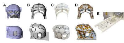 |
100 |
Towards the Macaque Connectome: 24-Channel 3T Multi-Array Coil, MR Sequences, and HCP-style Preprocessing
Joonas Autio, Takayuki Ose, Masahiro Ohno, Yoshihiko Kawabata, Yuta Urushibata, Kantaro Nishigori, Masataka Yamaguchi, Yuki Hori, Atsushi Yoshida, Katsutoshi Murata, Matthew Glasser, Takuya Hayashi
Macaque monkeys are an important neuroscientific model for understanding cortical organization of primates, yet non-human primate MRI applications have lagged behind those developed for humans. To address this issue, we developed a 24-channel receive coil for macaque brain imaging and adapted the Human Connectome Project (HCP)’s image acquisition protocols matched to monkey’s neuroanatomical resolution. By adapting HCP’s preprocessing methods for the macaque, we demonstrate that the resulting HCP-style monkey MRI data show great promise for multi-modal analyses of cortical architecture and connectivity, which have previously only been possible in humans.
|
|
4966.
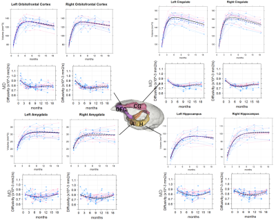 |
101 |
Orbitofrontal-limbic structural development maturation in non-human primates: a longitudinal study
Akiko Uematsu, Junichi Hata, Yuji Komaki, Fumiko Seki, Chihoko Yamada, Norio Okahara, Yoko Kurotaki, Erika Sasaki, Hideyuki Okano
We investigated typical orbitofront-limbic structural development of a non-human primate model, common marmoset, using longitudinal MRI data. Overall, robust volumetric growth was observed during infancy. This rapid brain volumetric growth was associated with the largest decrease in diffusivities of diffusion tensor imaging. That suggest that the volume increment result from an increase in the number and size of cells, dendrites, and spines in the regions. In addition, our result suggested familial factors contributed the development of the orbitofrontal-limbic brain regions. Overall, this study provides further data on the factors and timing important for typical brain development.
|
|
4967.
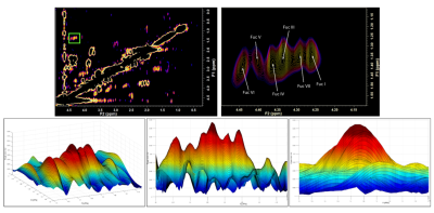 |
102 |
Assignment of Two More Fucose-a-(1-2)-Galactose Glycans in the Human Brain Linked in Animal Models to Learning and Memory
Nathan Tosh, Scott Quadrelli, Graham Galloway, Carolyn Mountford
Two-dimensional COSY scans at 3T using a 64 channel head and neck coil have enabled the inspection of fucose-α-(1-2)-galactose glycans that resonate in the region of F1=0.9-1.7ppm; F2= 4.1-4.5ppm. With accurate shimming and water suppression, two new peaks were identified (Fuc VI at 4.44-1.37 ppm and Fuc VII at 4.29-1.36 ppm). These cross-peaks were consistently visualised across 86 participants.
|
|
4968.
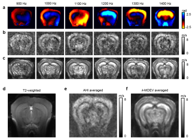 |
103 |
Tomoelastography of the mouse brain by multifrequency single-shot MR elastography
Gergely Bertalan, Jing Guo, Heiko Tzschätzsch, Charlotte Klein, Jürgen Braun, Ingolf Sack
The overall aim of this study was to introduce in-vivo multifrequency single-shot MR elastography (MRE) for full-field-of-view stiffness mapping of mouse brain and to compare in-vivo stiffness of neural tissues featuring different white-to-gray matter ratios. Tomoelastography retrieves mouse brain stiffness with greater detail. The measured order of shear vave speed values indicates that white matter in the mouse brain is softer than gray matter within the examined frequency range from 900 to 1400 Hz.
|
|
4969.
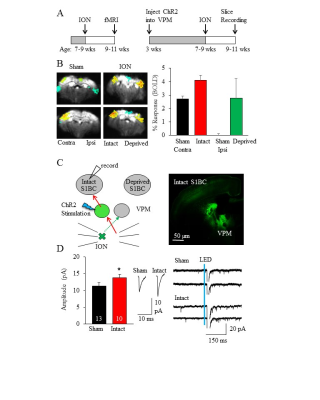 |
104 |
Synaptic Basis for Remodeling in the Mouse Somatosensory System Characterized by fMRI
Emily Petrus, Galit Saar, Zhiwei Ma, Steve Dodd, Alan Koretsky
Functional MRI (fMRI) is an effective tool to analyze brain activity, with research and clinical implications. Here we describe changes in fMRI stimulus evoked BOLD responses after unilateral whisker denervation in adult mice. These effects were mediated by specific synaptic remodeling in the whisker somatosensory system. Our results indicate that fMRI can detect circuit level changes in vivo, which can then be characterized at the synaptic level in vitro.
|
|
4970.
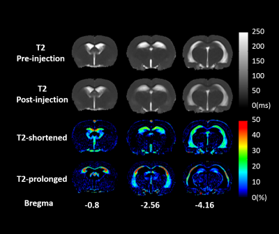 |
105 |
Transient hyperhydration modulates the brain fluid distribution of spontaneous hypertension rats: A T2 relaxometry study
Kun-I Chao, Xia-Yu Lyu, Pei-Lun Yu, Sheng-Min Huang, Shin-Lei Peng, Kung-Chu Ho, Ping-Huei Tsai, Fu-Nien Wang
After 2% of body weight fluid infusion, the homeostasis of fluid distribution was temporally modulated in spontaneously hypertensive rats, which is an animal model of neurodegenerative disease. After fluid infusion, shortened T2 showed in hippocampus and ventricles, and prolonged T2 was in corpus callosum and amygdala. The analysis of T2 distribution with rNNLS showed that a component with T2 around 100-200ms was increased in both T2-altered regions. We attributed this component as a restricted interstitial fluid, which could be correlated to the function of glymphatic system. Therefore, our experiment may shed a light on functional imaging of glymphatic system.
|
|
4971.
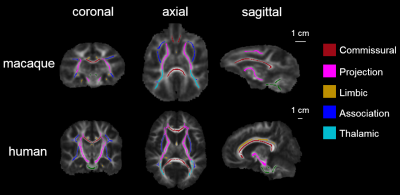 |
106 |
Comparison of white matter tracts between macaque and human brain
Qinlin Yu, Fang Fang, Hao Huang
White matter as the substrate of connectivity plays a critical role in brain evolution. However, the comprehensive tract-level comparison in morphology and microstructure between all common macaque and human white matter tracts has not been delineated. We aimed to qualitatively and quantitatively compare the tract-level microstructure and 3D pathways of corresponding white matter tracts between macaque and human brain using DTI measurements and tractography. Macaque white matter tract skeleton was obtained and comparison was conducted in five categorized tract groups. The differences are most prominent in association tracts with more extensive pathways and higher microstructural integrity in human brain association tracts.
|
|
4972.
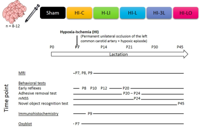 |
107 |
Lactate, the new hope against brain damages in neonatal hypoxia-ischemia
Hélène Roumes, Ursule Dumont, Leslie Mazuel, Stéphane Sanchez, Jordy Blanc, Véronique Bouchaud, Jean-François Chateil, Anne-Karine Bouzier-Sore
Hypoxia-ischemia remains a major perinatal mortality and chronic disability cause in newborns. Hypothermia is the only clinically therapeutic approach but new therapy must be developed. Our aim was to study the neuroprotective role of lactate in rat neonatal hypoxic-ischemic model and to determine the best pattern to counteract brain damages. Interestingly, lactate was much more neuroprotective when administered after hypoxia (curative) than before (preventive). Single versus 48h injection of lactate were compared. Lactate shows significant and important neuroprotection (ADC, FA, immunohistochemistry, behavioral studies).
|
|
4973.
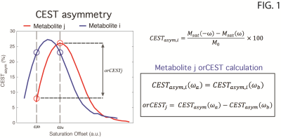 |
108 |
Mapping neurotransmitter variations upon water deprivation in rats using overlap-resolved CEST
Frederico Severo, Noam Shemesh
Thirst regulation involves several brain regions and Glutamatergic/GABAergic mechanisms. Imaging those noninvasively could make a big impact on understanding brain circuitry in-vivo. A recently proposed method termed overlap-resolved CEST (orCEST) was suggested for mapping both Glutamate and GABA in the brain with increased specificity. Here, orCEST is used to investigate changes in these neurotransmitters in the rat brain in vivo upon water deprivation. Decreases in GABA and Glutamate in hypothalamic preoptic areas and corpus callosum, respectively, were observed in the Water Deprived group. This bodes well for future applications of orCEST for studying neurotransmission in-vivo.
|
|
4974.
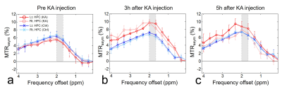 |
109 |
In Vivo Mapping and Quantification Approaches in Epileptic Seizure Rat Models Using Creatine Chemical Exchange Saturation Transfer (CrCEST) Imaging
Dong-Hoon Lee, Do-Wan Lee, Jae-Im Kwon, Chul-Woong Woo, Sang-Tae Kim, Jin Seong Lee, Choong Gon Choi, Kyung Won Kim, Jeong Kon Kim, Dong-Cheol Woo
CrCEST imaging is a novel imaging technique to provide in vivo image contrasts related with the changes of energy metabolism by creatine level changes. In this abstract, we attempted to evaluate signal changes in hippocampus at epileptic seizure rat models based on the quantified CrCEST signals. We also analyzed the correlations between CrCEST signals and PCr and PCr+Cr concentrations assessed by 1H-MRS spectra. Our results clearly showed that the CrCEST imaging can be a useful approach to evaluate the energy metabolism in hippocampus at epileptic seizure model, and to provide the quantitative results that related with creatine level changes.
|
|
4975.
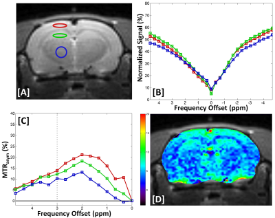 |
110 |
Chemical Exchange Saturation Transfer Imaging of Glutamate (GluCEST) in a Rat Model of Epilepsy using Proton Magnetic Resonance Spectroscopy (1H-MRS) at 7T
Do-Wan Lee, Jae-Im Kwon, Dong-Hoon Lee, Chul-Woong Woo, Sang-Tae Kim, Jin Seong Lee, Choong Gon Choi, Kyung Won Kim, Jeong Kon Kim, Dong-Cheol Woo
Chemical exchange saturation transfer imaging of glutamate (GluCEST) is a novel magnetic resonance imaging (MRI) method for measuring brain glutamate (Glu) in humans and animals. In vivo quantification of neurotransmitter signals can provide insight into functional roles of brain structures and knowledge about biochemical compounds. This study, based on quantified GluCEST contrast values and 1H-MR spectral concentrations, assessed signal changes in control rats and rats with kainic acid-induced epileptic seizures, and showed that in vivo GluCEST and 1H-MR spectral data can provide valuable information for interpreting changes in signals and concentrations of specific cerebral metabolites in kainic acid-induced rats.
|
|
4976.
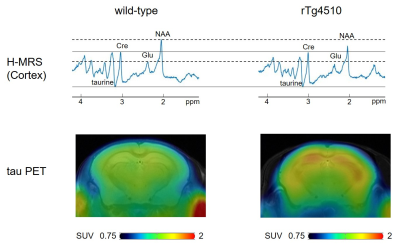 |
111 |
Correlations between brain metabolites and tau protein accumulation assessed by H-MRS and tau PET in Alzheimer’s disease model mice
Video Permission Withheld
Yuhei Takado, Hiroyuki Takuwa, Urushihata Takuya, Manami Takahashi, Maiko Ono, Jun Maeda, Masafumi Shimojo, Nobuhiro Nitta, Sayaka Shibata, Ichio Aoki, Naruhiko Sahara, Tetsuya Suhara, Makoto Higuchi
Amyloid beta protein and tau proteins are two major hallmarks of the pathology of Alzheimer’s disease. To elucidate the relationship between brain metabolites and tau protein, we performed H-MRS and tau PET imaging of tauopathy mouse model (rTg4510). Five rTg4510 mice and five wild-type mice were scanned at the age of 4 and 6 months by H-MRS and tau PET. We demonstrated that the amount of tau had negative correlations with the amount of taurine, tNAA, and glutamate, indicating that tau might reduce those metabolites selectively and caused neural impairment.
|
|
4977.
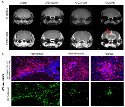 |
112 |
Longitudinal evaluation of lesion pathology in a novel murine model for Multiple Sclerosis
Caroline Guglielmetti, Christian Cordano, Ari Green, Myriam Chaumeil
We used conventional MRI to longitudinally assess lesion pathology in a combined cuprizone and experimental autoimmune encephalomyelitis (CPZ/EAE) model. The novelty of this model lies in the recruitment of cells from the innate and adaptive immune system into brain lesions. We used T2-weighted imaging to monitor white matter lesions. Interestingly, we showed a transient change in T2 contrast at the onset of clinical symptoms in the CPZ/EAE group. Using gadolinium-enhanced MRI, we showed transient opening of the blood-brain-barrier prior and/or following clinical symptoms. Altogether, these findings are of relevance to understand the dynamics of lesion formation in a novel MS model.
|
|
4978.
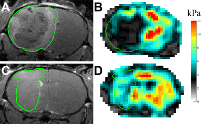 |
113 |
Anti-angiogenic treatment alters stiffness of glioblastoma in an orthotopic mouse model
Katharina Schregel, Michal Nowicki, Miklos Palotai, Navid Nazari, Rachel Zane, Ralph Sinkus, Sean Lawler, Samuel Patz
Glioblastoma (GBM) is the most common malignant brain tumor. As it is highly vascularized, anti-angiogenic treatment strategies have been tested. Such treatment hinders radiological tumor monitoring. Here, we probed the potential of magnetic resonance elastography (MRE) to evaluate GBM treated with anti-angiogenic therapy. GBM was orthotopically implanted in 10 nude mice, of which 5 were treated with B20 anti-VEGF antibody. MRI and MRE were performed repeatedly and brains were harvested for histology afterwards. Anti-angiogenic treatment slowed tumor growth, affected contrast-enhancement and slowed down tumor softening. The phase angle Y expressing the solid/liquid ratio appeared to indicate tumor progression.
|
|
4979.
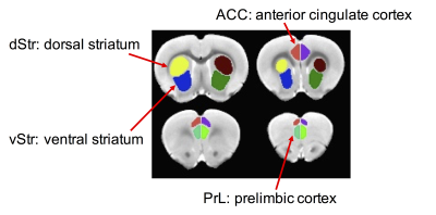 |
114 |
Fronto-striatal connectivity alterations in a prenatal ethanol exposure rat model: a resting-state functional MRI study
Shiyu Tang, Su Xu, Jaylyn Waddell, Marie Hanscom, Wenjun Zhu, Rao Gullapalli, Sandra Mooney
People with Fetal Alcohol Spectrum Disorders (FASDs) may present deficits in executive functions. The fronto-striatal circuit is a critical component of the fronto-basal ganglia pathway that plays an important role in executive processes. We used resting-state functional MRI to assess the effects of prenatal alcohol exposure on the functional interaction within the fronto-striatal circuit in a rat FASD model. Male alcohol-exposed rats, but not females, showed reduced fronto-striatal connectivity. Further exploratory analysis revealed a reduction in cortico-striatal connectivity in female alcohol-exposed rats, suggesting that prenatal alcohol exposure has sex dependent effects on executive and sensory processing functions.
|
|
4980.
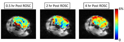 |
115 |
Longitudinal assessment of TAT-PHP treatment effect in improving brain function recovery from cardiac arrest resuscitation with endogenous perfusion using ASL and metabolic CEST MRI
Rong-Wen Tain, Chunpei Lee, Alessandro Scotti, Weiguo Li, Xiangdong Zhu, Terry L. Vanden Hoek, Jing Li, Kejia Cai
Sudden cardiac arrest is a leading cause of death in the US. A recently developed peptide-based (TAT-PHP) therapy has been shown to improve survival rate and heart metabolism in animal studies. There is a great need to evaluate the effect of the treatment in the brain as the brain is the most susceptible organ following cardiac arrest. We here demonstrated that the recovery of brain perfusion and metabolism is improved in mice with TAT-PHP treatment vs. controls with endogenous perfusion arterial spin labeling MRI and metabolic MRI based on chemical exchange saturation transfer.
|
|
4981.
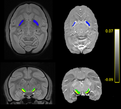 |
116 |
Longitudinal evaluation of iron load with quantitative suceptibility mapping (QSM) in a primate model of Parkinson's disease
Mathieu Santin, Maxime Verdier, Romain Valabregue, Elodie Laffrat, Lydia Cherif, Alexandra Petiet, Stéphane Lehéricy, Stéphane Hunot
In this study, we performed a longitudinal follow-up of iron load in a primate model of Parkinson's disease using both QSM and R2* mapping. ROI definition was automatically performed using an in-house template computed from all acquired data. Statistical analyses revealed that QSM is more sensitive to model-induced variation in iron-rich regions compared to R2* mapping.
|
|
4982.
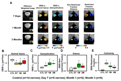 |
117 |
Optical Coherent Tomography (OCT) Detected Axonal Loss but Failed to Identify Acute Axonal Injury: A Diffusion Study
Tsen-Hsuan (Abby) Lin, Ying-Bo Shui, Ying Liu, Hsin-Chieh Yang, Michael Wallendorf, Carla Siegfriedand, Sheng-Kwei Song
Optical coherence tomography (OCT) is widely used to noninvasively assess retinal nerve fiber layer (RNFL) thickness to assess optic nerve damage. However, the impact of coexisting pathology on OCT-detected RNFL thickness remains unclear. We have previously developed a diffusion basis spectrum imaging (DBSI) to directly assess coexisting pathologies in mouse models of optic nerve crush (ONC) and optic neuritis. In the current study, we assessed retinal pathology and coexisting pathologies in optic nerve of ONC mice longitudinally, with OCT and DBSI, respectively. Our goal is to determine the relationship between OCT-detected RNFL change and DBSI-derived optic nerve pathologies.
|
|
4983.
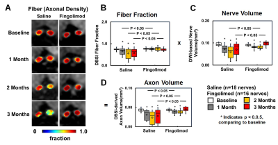 |
118 |
Diffusion Basis Spectrum Imaging (DBSI): Successful in vivo Use to Assess Axonal Protection in Mouse Optic Neuritis treated with Fingolimod
Tsen-Hsuan (Abby) Lin, Rui-Meng Yang, Jie Zhan , Chunyu Song, Peng Sun, Michael Wallendorf, Anne Cross, Sheng-Kwei Song
We previously introduced diffusion basis spectrum imaging (DBSI) to noninvasively assess coexisting pathologies in central nervous system (CNS) tissues.Previously, we showed in multiple sclerosis (MS) spinal cord specimens and mice with experimental autoimmune encephalomyelitis (EAE), the main animal model of MS, that DBSI reflected coexisting white matter pathologies in CNS. Fingolimod, a disease-modifying treatment approved for relapsing MS, is thought to preserve axons. In the current study, we employed longitudinal DBSI to noninvasively assess fingolimod treatment efficacy in EAE mouse optic nerve, followed by histological validation.
|
|
4984.
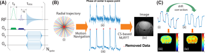 |
119 |
Protein Aggregation in Mouse Brain with Alzheimer's Disease Revealed by Saturation Transfer MRI
Lin Chen, Zhiliang Wei, Kannie Chan, Shuhui Cai, Guanshu Liu, Hanzhang Lu, Philip Wong, Peter van Zijl, Tong Li, Jiadi Xu
This study aims to detect the aggregated proteins involved in Alzheimer's Disease (AD) using chemical exchange saturation transfer (CEST) as a potential strategy for early diagnosis of the disease. A radial ultra-short echo time (UTE) image acquisition scheme was applied to map CEST signals with sufficient sensitivity and robustness to motion. Studies on cross-linked bovine serum albumin (BSA) and an AD transgenic model (mutant APP/PS1 mouse) demonstrated that the saturation transfer (ST) signal at -3.6 ppm, which is a combination of relayed nuclear Overhauser enhancement (rNOE)-CEST and a small portion of magnetization transfer contrast (MTC), can be used a potential biomarker for monitoring changes in brains of a mouse model of AD.
|
|
4985.
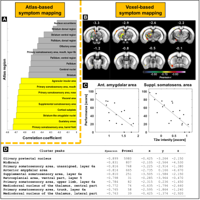 |
120 |
Atlas-based and voxel-based symptom mapping to explore anatomical substrates of functional recovery after stroke in the mouse
Stefan Koch, Felix Knab, Ulrike Grittner, René Bernard, Susanne Mueller, Marco Foddis, Janet Lips, Philipp Euskirchen, Norman Zerbe, Peter Hufnagl, André Rex, Ulrich Dirnagl, Tracy Farr, Christoph Harms, Philipp Boehm-Sturm
Post-stroke functional recovery remains a poorly understood process. We aimed to identify anatomical regions that statistically explain a deficit in the staircase test after stroke in the mouse using two animal models (MCAO, photothrombosis). MATLAB toolboxes are presented for co-registration of MRI and histology data to the Allen brain atlas and implementation of atlas region-wise or voxel-wise mapping of behavioral deficits. Automatic techniques are shown for validation of MRI results on stained tissue sections.
|
|
| Back |
| The International Society for Magnetic Resonance in Medicine is accredited by the Accreditation Council for Continuing Medical Education to provide continuing medical education for physicians. |
