Cervical & Intracranial Flow
Oral
Neuro
Thursday, 21 June 2018
| S03 |
15:30 - 17:30 |
Moderators: Huijun Chen, David Saloner |
15:30
|
1221.
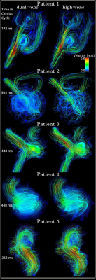 |
 Advantages of Dual-Venc 4D flow MRI in the evaluation of cerebral aneurysms Advantages of Dual-Venc 4D flow MRI in the evaluation of cerebral aneurysms
Susanne Schnell, Maria Aristova, Matthew Potts, Babak Jahromi, Liliana Ma, Alireza Vali, Amer Syed, Michael Hurley, Michael Markl, Sameer Ansari
We applied and investigated the benefits of using kt-GRAPPA accelerated dual-venc 4D flow MRI in five patients who presented with cerebral aneurysm between 4 and 12mm (smallest and largest dimension). Velocity values were systematically compared with the high-venc-only part of the same acquisition using correlation and histogram analysis. Blood flow was visualized with streamlines and quality was visually assessed. Results show that dual-venc 4D flow MRI provides refined information on slow flow and recirculating flow in cerebral aneurysms. Future studies will assess the feasibility of advanced hemodynamic measures obtained from dual-venc 4D flow MRI as predictive imaging biomarkers.
|
15:42
|
1222.
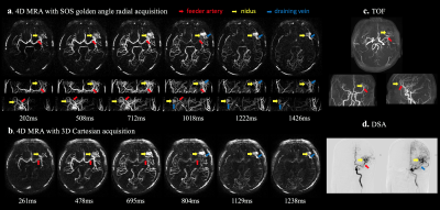 |
Evaluation of non-contrast enhanced 4-dimensional MRA with stack-of-stars golden angle radial trajectory in conjunction with KWIC reconstruction for the depiction of arteriovenous malformations
Lirong Yan, Songlin Yu, Samantha Ma, Jonathan Russin, Arun Amar, Yelong Shen, Qi Huang, Nader Pouratian, Hee Kwon Song, Danny Wang
This study evaluated a recently developed non-contrast enhanced 4-dimensional MRA (NCE 4D MRA) with stack-of-stars golden-angle radial trajectory and KWIC reconstruction for the detection of cerebral arteriovenous malformation (AVM) by comparison with DSA, TOF and standard Cartesian 4D MRA. The characterizations of AVM lesions using this radial 4D MRA are consistent with those of DSA. 4D MRA combined with TOF improves diagnostic confidence. Compared to Cartesian acquisition, radial acquisition shows improved delineation of dynamic flow with less motion artifacts and short scan time. Our findings indicate radial 4D MRA may be a promising alternative for the characterization of AVM features.
|
15:54
|
1223.
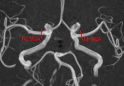 |
4D Flow assessment of arterial pulsation in the patients with internal carotid artery stenotic disease
Takahiro Ando, Tetsuro Sekine, Yasuo Murai, Ryo Takagi, Yasuo Amano, Erika Orita, Kotomi Iwata, Makoto Obara, Yoshio Matsumura, Shin-Ichiro Kumita
We performed 4D Flow MRI assessment of arterial pulsation in internal carotid artery stenotic disease (ICS) patients comparing to SPECT with acetazolamide challenge. Twelve patients with unilateral ICS were recruited. The blood flow volumes and the ratio of ΔF (rΔF) was calculated. In the affected-side MCA territory, the ratio of rest cerebral blood flow control (RCBFMCA) and cerebral vascular reserve (CVRMCA) were calculated from SPECT dataset. rΔF was significantly lower in the low CVR group than high group (P=0.008). The 6-min-standard 4D Flow MRI assessment of arterial pulsation in ICS patients can identify misery perfusion.
|
16:06
|
1224.
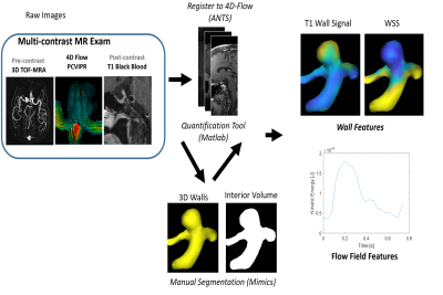 |
In-vivo correlations between hemodynamics and wall inflammation in patients with intracranial aneurysms: comparing 4D Flow MRI and vessel wall enhancement.
Myriam EDJLALI, Dahan Kim, Leonardo Rivera-Rivera, Pauline Roca, Catherine Oppenheim, Olivier Naggara, Patrick Turski, Oliver Wieben, Kevin Johnson
Recent studies point out hemodynamics and wall inflammation as individual risk factors for aneurysm evolution. We investigated on 28 aneurysms in-vivo correlations between hemodynamics and wall inflammation by comparing 4D Flow parameters and quantitative aneurysm wall enhancement. Viscous energy loss and kinetic energy were correlated to maximum values of enhancement with high correlation coefficients (p<0.0001; correlation coefficient 0.79 and p<0.0001; correlation coefficient 0.85, respectively). By showing that flow instability and spatial flow complexity are highly correlated with aneurysm wall enhancement, this study highlights the existence of a link between inflammation process depicted through vessel wall enhancement and hemodynamic patterns.
|
16:18
|
1225.
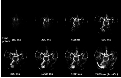 |
New 4D-MRA approach named 4D-APACK by integrating 4D-PACK and AccASL for visualization of long transit time artery: Investigating hemodynamics accuracy and AVM clinical case
Yuta Akamine, Makoto Obara, Osamu Togao, Shuhei Shibukawa, Masami Yoneyama, Tomoyuki Okuaki, Marc Van Cauteren
A new 4D-MRA approach named 4D-AccASL-PACK (4D-APACK) was implemented and images were acquired in six healthy volunteers and one AVM patient. 4D-APACK consists of 4D-PACK and acceleration-selective arterial spin labeling (AccASL) for obtaining efficiently both dynamic information and ATT independent visualization of very slow flow. 4D-APACK replaces last timepoint data of 4D-PACK with AccASL. To investigate hemodynamics accuracy for 4D-APACK, ATT correlation between 4D-pCASL and 4D-APACK and contamination of sinuses were measured. The vessel visualization of MCA M1 to M4 were compared with 4D-pCASL. We demonstrate that 4D-APACK brings reliable hemodynamic information comparable to 4D-pCASL, while retaining MCA visualization performance.
|
16:30
|
1226.
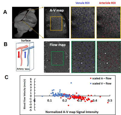 |
Sing-vessel cerebral blood flow (velocity) fMRI with phase-contrast imaging
Xuming Chen, Rolf Pohmann, Klaus Scheffler, Xin Yu
Single-vessel fMRI has been developed to map the BOLD signal from individual venules and the CBV signal from individual arterioles penetrating the cortex of anesthetized rats. Here, we applied phase-contrast (PC) imaging to measure the velocity of blood flow from individual penetrating arterioles and venules, which could be characterized as dark and bright dots in an arteriole-venule map with a multi gradient-echo sequence. The neuronal activity-coupled cerebral blood flow (CBF) changes can be directly measured with the PC-based velocity mapping from individual vessels. Thus, we have established single-vessel CBF fMRI mapping with phase-contrast imaging.
|
16:42
|
1227.
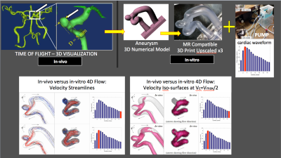 |
Sub-millimetric 4D Flow MR in small intracerebral aneurysms at 7 Tesla with experimental verification in upscaled 3D printed replica.
Pierre-Francois Van de Moortele, Mostafa Toloui, Omid Amili, Sean Moen, Sebastian Schmitter, Susanne Schnell, Michael Markl, Kamil Ugurbil, Filippo Coletti, Bharathi Jagadeesan
Asymptomatic intracerebral aneurysms of small size (<7mm) pose a difficult therapeutic challenge: left alone they may stay stable with no consequence or they may grow and/or rupture with devastating subarachnoid hemorrages. Pre-emptive treatment (surgical or endovascular) however carries non-negligible mortality and morbidity and there is no biomarker predicting these relative risk. Flow dynamics inside small aneurysms however could have a critical impact on their evolution. Here we investigate the use of 4D Flow MR to acquire submillimitric flow information in small aneurysms in vivo as well as in 3D printed up-scaled replica of actual aneurysms measured in patients.
|
16:54
|
1228.
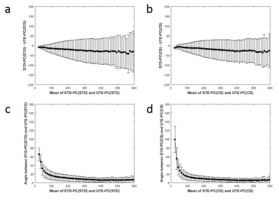 |
Characterization of Ultra-short Echo and Standard echo Phase-Contrast MRI for Neurovascular disease application
Dahan Kim, Katrina Ruedinger, David Rutkowski, Alejandro Roldán-Alzate, Patrick Turski, Kevin Johnson
We examined velocity measurements of standard (STD-PC) and ultra-short echo (UTE-PC) phase-contrast MRI in three different study cases, to characterize the effect of shortening echo time on artifacts from flow and metal. We found that UTE-PC measures higher velocity magnitudes not only in disturbed flow but also in normal vasculature, that UTE-PC results in smaller divergence of velocity field but no difference in erroneous flux through arterial wall within metal aneurysm stent, and that UTE-PC higher velocity magnitudes, less signal loss, and coherent flow directions in both untreated and un-treated aneurysm phantom.
|
17:06
|
1229.
 |
Intracranial flow measurements at 7T gated with Doppler Ultrasound
Karin Markenroth Bloch, Fabian Kording, Johannes Töger
Intracranial flow measurements at high field (7T) has the potential to give new information on hemodynamic properties. However, the strong field disturbs ECG gating, possibly compromising image quality. This work investigates possible benefits of using Doppler ultrasound (DUS) cardiac triggering compared to ECG–triggering in throughplane (2D) flow measurements at 7T. 2D flow was acquired using both ECG and DUS triggering in random order in healthy volunteers (n=8). The DUS was found to have a higher trigger sensitivity and fewer false negative and positive triggers. Flow and velocity results did not differ between ECG and DUS triggering.
|
17:18
|
1230.
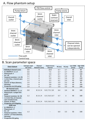 |
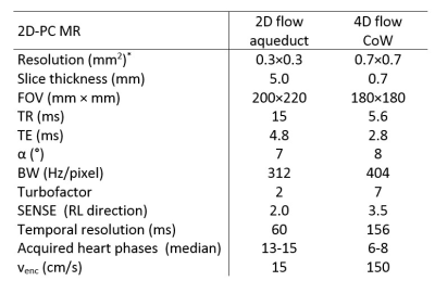
Scan parameter optimization of dual-venc 4D Flow MRI for the assessment of neurovascular flow networks in brain arteriovenous malformation
Maria Aristova, Alireza Vali, Alex Barker, Ali Shaibani, Sameer Ansari, Matthew Potts, Babak Jahromi, Michael Hurley, Susanne Schnell, Michael Markl
To optimize dual-venc 4D Flow MRI parameters for flow assessment in brain arteriovenous malformations, we conducted an in-vitro optimization analysis and compared it to in-vivo data from a patient with complex AVM. Using k-t acceleration factors of 2-5 and about 2-10 voxels across the imaged vessels, we quantified the agreement with the ground truth flow and geometry. We applied a flow distribution network graph concept to characterize flow conservation as an additional quality metric. Data indicated that approximately 5 voxels across imaged vessels are needed, consistent with results from previous publications.
|
|
