Perfusion & Permeability
Oral
Contrast Mechanisms
Tuesday, 19 June 2018
| S04 |
13:45 - 15:45 |
Moderators: Christopher Quarles, Peiying Liu |
13:45
 |
0493.
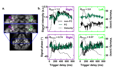 |
 Flow Compensation in Dynamic Susceptibility Contrast MRI for an Increase in Arterial Input Function Precision Flow Compensation in Dynamic Susceptibility Contrast MRI for an Increase in Arterial Input Function Precision
Benoît Bourassa-Moreau, Réjean Lebel, Guillaume Gilbert, David Mathieu, Martin Lepage
Arterial input functions (AIF) measured inside major arteries in perfusion MRI are derived from the contrast agent induced changes in the blood signal. Such signal measurements of flowing blood are prone to (in)flow induced effects. Here, the effects of pulsatile blood flow are studied for AIF measurement in dynamic susceptibility contrast (DSC) with dual-echo single-shot EPI. First moment flow compensation is shown to partially recover the noise attributed to flow effects. The resulting signal-to-noise ratio is increased by a factor of 14 (2) in the middle (internal) carotid artery providing a more precise AIF for cerebral blood flow calculation.
|
13:57
|
0494.
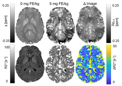 |
Comparison of Cerebral Blood Volume Estimates using Quantitative Susceptibility Mapping, and R2* relaxometry
Leonardo Rivera-Rivera, Ante Zhu, Timothy Colgan , Diego Hernando, Tilman Schubert, Patrick Turski, Kevin Johnson
Intracranial vascularity is modified in a wide array of diseases, including various forms of dementia, cancer, and stroke. When assessment of the vascular architecture is needed, one potential approach is steady state imaging with ferumoxytol to estimate cerebral blood volume (CBV). Building upon recent work, in this study we investigate the correlation between QSM and R2* based CBV estimates. Results from 19 healthy volunteers show that the QSM based CBV estimates demonstrated a high degree of correlation in gray and white matter, but larger variance than R2* based measures.
|
14:09
|
0495.
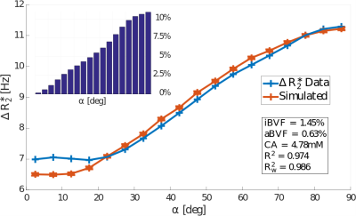 |
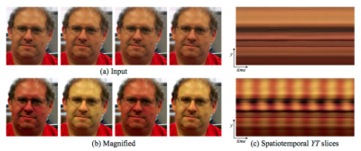
The effects of diffusion on the tissue orientation dependent gradient echo dynamic susceptibility contrast signal
Jonathan Doucette, Enedino Hernández-Torres, Christian Kames, Luxi Wei, Alexander Rauscher
Gradient echo dynamic susceptibility contrast (DSC) imaging of cerebral white matter tissue perfusion exhibits a strong dependency on the angle between white matter fibres and the main magnetic field. Here, we investigate the extent to which the diffusion of spins within the locally magnetically inhomogenous environment affects the orientation dependency of the DSC signal using a numerical model of the transverse magnetization within a voxel containing both isotropic and anisotropic vasculature. We found that diffusion increases the measured change in $$$R_2^*$$$ by up to an average of 3.2% for typical diffusion values, independent of fibre orientation, and further improves the fit between the simulated and measured $$$R_2^*$$$ values.
|
14:21
|
0496.
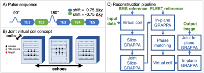 |
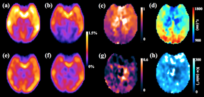
Accelerated dynamic quantitative perfusion imaging using an optimized simultaneous multi-slice (SMS) spin and gradient echo (SAGE) sequence with joint-virtual coil (JVC) reconstruction
Mary Kate Manhard, Berkin Bilgic, Congyu Liao, SoHyun Han, Thomas Witzel, Yi-Fen Yen, Kawin Setsompop
A 5-echo spin and gradient echo (SAGE) sequence was implemented with SMS to provide whole brain coverage at a high spatio-temporal resolution during DSC imaging. Complementary k-space sampling between echoes and joint-virtual coil (JVC) reconstruction were used to achieve high quality reconstruction at 9x acceleration, which enabled 1.9x1.9x5 mm whole-brain imaging at a TR of 1.6s. The multi-echo images from this sequence were fit to achieve quantitative R2 and R2* maps for each TR. This has been shown to limit T1 leakage effects and provide more accurate and detailed perfusion measures with the potential to better assess tumor diagnostics and progression.
|
14:33
|
0497.
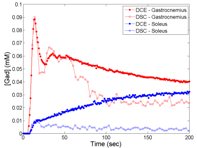 |
Quantitative assessment of exercise-stimulated muscle perfusion: a comparison between DCE and DSC imaging
Jeff Zhang, Christopher Conlin, Stephen Decker, Gwenael Layec, Jiawei Dong, Xiaowan Li, Nan Hu, Christopher Hanrahan, Lillian Khor, Michelle Mueller, Vivian Lee
For one group of healthy subjects, we measured exercise-stimulated perfusion in calf muscles using both DCE and DSC MRI, and found that the muscle perfusion estimates by the two methods were comparable, but the vascular-fraction estimates were not. Without confounding contribution from extravascular signals, DSC data has the potential of characterizing tissue vasculature more precisely. Acquisition of both DCE and DSC data in one exam can be achieved with either two injections of low contrast dose or acquisition techniques of interleaved T1and T2* imaging.
|
14:45
|
0498.
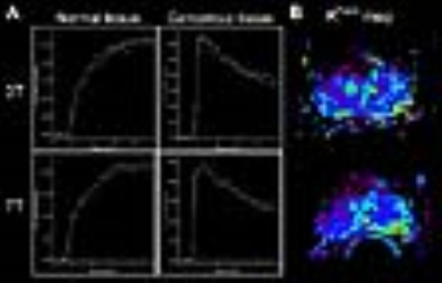 |
Evaluation of UTE for improved contrast enhanced DCE MRI at 7T
Naoharu Kobayashi, Patrick Bolan, Gregory Metzger
We investigated the feasibility of using an ultrashort echo time (UTE) technique for DCE MRI imaging of prostate cancer patients at 7T to minimize the impact of the increased R2* relaxivity on signal enhancement curves and pharmacokinetic parameter assessment. The Gd concentration-time curves and pharmacokinetic parameter maps were compared to clinical DCE MRI at 3T. The proposed method achieved DCE MRI at 7T with a signal time course comparable to 3T DCE MRI but with increased SNR for improved spatial resolution and no R2*effects, which are exacerbated with increasing field.
|
14:57
|
0499.
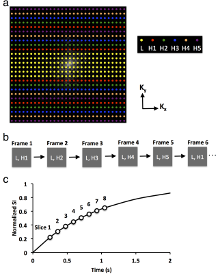 |
A Multislice TRICKS-Based DCE-MRI Method for Measurement of Murine Kidney Function
Kai Jiang, Prasanna Mishra, Slobodan Macura, Lilach Lerman
A multislice method based on time-resolved imaging of contrast kinetics was developed for measurement of murine kidney size and function. A total of eight slices covering both kidneys were imaged with a temporal resolution of 1.23 sec/scan. The estimated kidney volume showed a good agreement with a three-dimensional volume scan. By fitting contrast kinetics using a modified bi-compartment model, renal perfusion and normalized glomerular filtration rate (GFR) were quantified, and subsequently used to calculate renal blood flow and GFR. In conclusion, this method allows simultaneous and reliable measurement of mouse kidney volume, perfusion, blood flow, and GFR.
|
15:09
 |
0500.
 |
Improved reliability for mapping water exchange across blood-brain barrier by diffusion prepared three-dimensional pseudo-continuous arterial spin labeling
Xingfeng Shao, Danny J.J. Wang
A 3D diffusion-prepared gradient and spin echo (GRASE) pseudo-continuous ASL (pCASL) sequence was proposed to improve the reliability for mapping water exchange across the BBB non-invasively. The motion sensitive non-CPMG component signal was eliminated by gradients to spread the diffusion weighted signal across 4π before GRASE readout. Based on test-retest scans (~2 weeks apart) in aged subjects (67±8yrs), excellent reproducibility (ICC=0.87) was achieved for water exchange rate (Kw) measured by the proposed 3D sequence. Whole brain Kw was averaged to be 107.7±19.0 min-1 and 113.0±17.8 min-1 for the proposed 3D and 2D DW-pCASL method, respectively, well matching the literature results.
|
15:21
|
0501.
 |
Demonstrating a novel non-contrast MR perfusion technique: EVM-EPI MR perfusion
Steven Sherry, Emanuel Kanal, Gregory Walker, John Kirsch, Jonathan Polimeni, H. Harvey
EVM-EPI MR perfusion is a novel, non-contrast MR perfusion technique, which amplifies latent imperceptible contrast variation in perfused tissue of a T2*-weighted cine acquisition via Fourier methods at frequencies of cardiac perfusion.
|
15:33
|
0502.
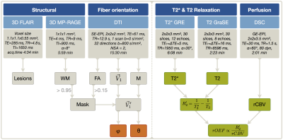 |
Assessment of white matter anisotropy effects in mq-BOLD based mapping of relative Oxygen Extraction Fraction
Stephan Kaczmarz, Jens Goettler, Andreas Hock, Dimitrios Karampinos, Claus Zimmer, Fahmeed Hyder, Christine Preibisch
Anisotropy effects were proven to strongly affect T2* and DSC-measurements, however influences on the relative Oxygen Extraction Fraction (rOEF) by multi-parametric quantitative BOLD (mq-BOLD) have not been investigated yet. Therefore, we present a methodolocigal study within healthy elderly. The major aim was to evaluate anisotropy effects on rOEF and all mq-BOLD related parameters at the same time. We found strong orientation dependencies of T2*, T2, R2' and rCBV on the main-nerve-fiber orientation towards B0 with variations up to 26.6%. However, our results show rather low angle dependency of rOEF with 13.6% as R2’ and rCBV effects show partially counteracting influences.
|
|
