Microstructure: A Stroll in the q-Space
Oral
Diffusion
Monday, 18 June 2018
| N04 |
16:15 - 18:15 |
Moderators: Noam Shemesh, Ileana Jelescu |
16:15
 |
0252.
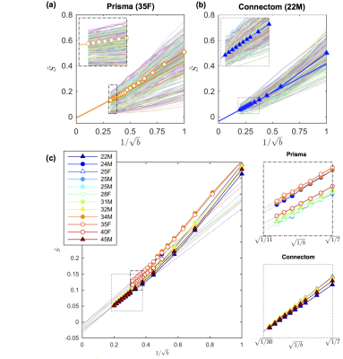 |
 Breaking the power law scaling of the dMRI signal on the Connectom scanner reveals its sensitivity to axon diameters Breaking the power law scaling of the dMRI signal on the Connectom scanner reveals its sensitivity to axon diameters
Jelle Veraart, Els Fieremans, Umesh Rudrapatna, Derek Jones, Dmitry Novikov
We demonstrate that in vivo diffusion MRI becomes sensitive to intra-axonal properties such as the inner axon diameters, when diffusion weightings in the range $$$b\sim7,000-25000\,\mathrm{s/mm^2}$$$ are achieved on the Human Connectom scanner (using gradients of 300mT/m). By analyzing the diffusion-weighted signal in the human white matter as a function of $$$b$$$, we observe significant deviations from the $$$b^{-1/2}$$$ scaling associated with the conventional “stick” model of infinitely-narrow axons. Our estimated effective MR diameters, heavily weighted by the tail of the axon diameter distribution, agree well with those calculated from histology histograms available from literature.
|
16:27
 |
0253.
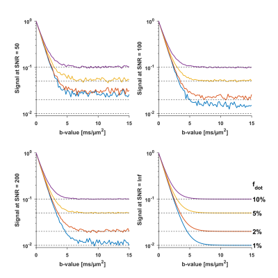 |
The Dot…wherefore art thou? Search for the isotropic restricted diffusion compartment in the brain with spherical tensor encoding and strong gradients
Chantal Tax, Filip Szczepankiewicz , Markus Nilsson, Derek Jones
The accuracy of biophysical models requires that all relevant tissue compartments are modelled. The so-called “dot compartment” is a conjectured compartment that represents small cells with apparent diffusivity approaching zero. We establish an upper limit of the “dot-fraction” across the whole brain in vivo, by using ultra-high gradients and optimized gradient waveforms for spherical tensor encoding. We report a notable signal above the noise floor in the cerebellar gray matter even for an extremely high b-value of 15000 s/mm2. For cerebral tissue, the dot-fraction seems negligible, and we consider how exchange may have affected this result.
|
16:39
|
0254.
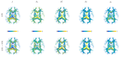 |
Effect of combining linear with spherical tensor encoding on estimating brain microstructural parameters
Els Fieremans, Jelle Veraart, Benjamin Ades-Aron, Filip Szczepankiewicz, Markus Nilsson, Dmitry Novikov
The diffusion MRI signal, as measured with conventional linear tensor encoding (LTE), has been shown to have not enough features to fully model the white matter microstructure. Here we investigate whether adding spherical encoding (STE) to LTE makes microstructural parameter estimation more robust. On signal simulations and in in vivo MRI data, we demonstrate that the intra-axonal diffusivity and axonal water fraction are estimated with higher precision, thereby enabling a 20 minute whole brain protocol to extract brain microstructural parameters without imposing constraints or priors.
|
16:51
|
0255.
 |
Unconstrained Estimation of Microstructure by the Combination of Single- and Double-planar diffusion encoding
Marco Reisert, Valerij G. Kiselev, Bibek Dhital
Modeling of white matter microstructure using standard diffusion MRI protocols is a poor-conditioned problem. With standard measurements there is a global degeneracy: There are two significantly different microstructural parameter sets explaining experimental data equally well. In this work we use an additional planar double diffusion encoding to resolve the degeneracy, and extend a Bayesian estimation framework to a combination of linear and planar diffusion encoding. This enables us to produce reliable estimates without applying any constraints to the microstructural diffusion model.
|
17:03
|
0256.
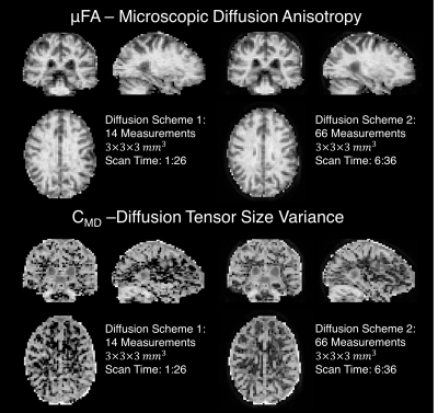 |
A Novel Method for Fast and Efficient Measurement of Diffusion Tensor Size and Shape Distributions
Grant Yang, Jennifer McNab
We demonstrate through simulations and empirical data that it is possible to simultaneously estimate the variance of the voxel-wise diffusion tensor shape and size distributions using efficient isotropic and linear diffusion encodings on a whole-body clinical MRI scanner with whole-brain coverage at 3mm isotropic resolution in under 2 minutes.
|
17:15
|
0257.
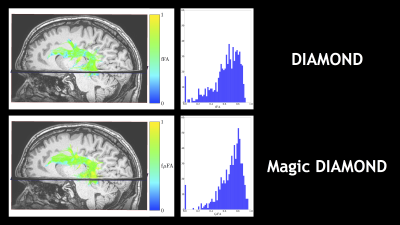 |
The "Magic DIAMOND" method: probing brain microstructure by combining b-tensor encoding and advanced diffusion compartment imaging
Alexis Reymbaut, Benoit Scherrer, Guillaume Gilbert, Filip Szczepankiewicz, Markus Nilsson, Maxime Descoteaux
Via q-trajectory encoding, b-tensors enable the disentanglement of isotropic and anisotropic diffusion components. Relevant metrics are usually extracted from data acquired with a combination of linear and spherical b-tensors with 1D parametric distributions of diffusivities. Independently, the DIAMOND model proposed an analytic result for a 6D parametric compartmental tensor distribution based on linearly acquired data. In this work, we extend DIAMOND’s analyticity to axisymmetric acquisitions. Evaluating this "Magic DIAMOND" approach on in vivo data, we show that it can tease apart isotropic diffusion and diffusivity compartments of crossing fascicles, hereby integrating specific compartments with intra-compartment diffusional variance.
|
17:27
|
0258.
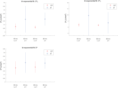 |
On the magnetic field and echo time dependence of the pseudo-diffusion coefficient
Andreas Riexinger, Andreas Wetscherek, Jan Martin, Tristan Kuder, Armin Nagel, Michael Uder, Bernhard Hensel, Lars Müller, Frederik Laun
It has been reported that a strong echo time dependence of the perfusion fraction f exists in liver and pancreas. The purpose of this work is to investigate whether a similar dependence exists for the pseudo-diffusion coefficient D*. Thereto, the livers of six healthy volunteers were examined at two echo times (TE = 40/80 ms) at two field strengths. In contrast to f, D* shows almost no echo time dependence in the healthy liver and the observed field strength dependence was smaller than the fit uncertainty.
|
17:39
|
0259.
 |
Cell specific anisotropy with double diffusion encoding spectroscopy in the human brain at 7T
Henrik Lundell, Andrew Webb, Itamar Ronen
The measurement of intracellular metabolite mobility with diffusion weighted spectroscopy (DWS) provides a cell-specific probe for microstructure. Measurements in animals and humans with conventional one dimensional diffusion encoding and model-guided analysis suggest that the main component of the intracellular space of both neurons and astrocytes comprises mainly anisotropic fibrous geometries. In this study we performed double diffusion encoded spectroscopy (DDES) in the human brain as a direct probe of anisotropic intracellular diffusion. As expected, our results support a main fibrous component but the results also suggest a more complex geometry of astrocytes that could include isotropic compartments.
|
17:51
|
0260.
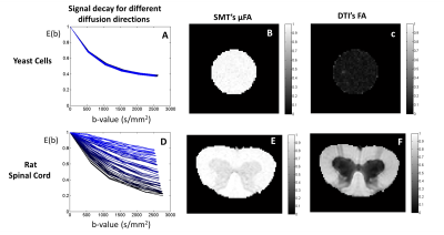 |
Validity Regimes of the Spherical Mean Technique
Rafael Neto Henriques, Noam Shemesh
The mean spherical technique (SMT) has been proposed as an attempt to disentangle microscopic diffusion properties from the mesoscopic tissue organization. However, it remains unclear under which conditions exactly the technique could still fruitfully deliver its metrics. In this study, SMT’s microscopic fractional anisotropy estimates (μFA) are investigated using synthetic and high-quality diffusion data. We find that any compartmental heterogeneity can crucially impact the µFA extracted from SMT. In addition, the b-value ranges where SMT delivers accurate information is specific to the microstructure itself. Our work suggests that SMT-driven µFA should be examined with care.
|
18:03
|
0261.
 |
Non-Invasive Determination of Sodium Pump Activity In Vivo with DWI
Charles Springer, Gregory Wilson, Brendan Moloney, Thomas Barbara, Xin Li, William Rooney, Jeffrey Maki
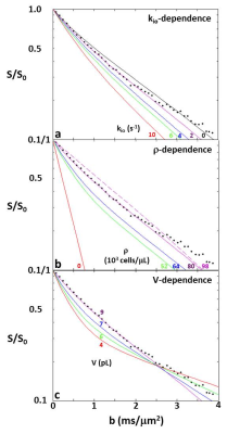
Using a very simple model, Monte Carlo random walk simulated DWI b-space decays exhibit sensitivity to parameters measuring membrane Na+,K+-ATPase activity, cell density, and voxel average cell volume. Furthermore, the simulation matching the literature experimental in vivo human cerebral cortex b-space decay has model parameters in near absolute agreement with the most pertinent literature values. The model parameters are: kio = 2 s-1, ρ = 80,400 cells/μL, and V = 9.2 pL. In addition, the ADC of this simulation agrees with published results.
|
|
