Poster: Body: Power Potpourri
Electronic Power Pitch Poster
Body: Breast, Chest, Abdomen, Pelvis
09:00 - 10:00
Thursday, 21 June 2018
Power Pitch Theater B - Exhibition Hall
| |
|
Plasma # |
|
0988.
 |
16 |
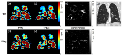
 Imaging collateral ventilation in patients with advanced Chronic Obstructive Pulmonary Disease – relative sensitivity of 3He and 129Xe MRI Imaging collateral ventilation in patients with advanced Chronic Obstructive Pulmonary Disease – relative sensitivity of 3He and 129Xe MRI
Helen Marshall, Guilhem Collier, Chris Johns, Ho-Fung Chan, Graham Norquay, Rod Lawson, Jim Wild
The success of lung volume reduction with endobronchial valves in patients with Chronic Obstructive Pulmonary Disease (COPD) depends on the absence of collateral ventilation into the target lung segment. Collateral ventilation can be assessed using time-resolved 3He MRI, but providing this clinical application with 129Xe MRI would be desirable. Two patients with severe COPD were scanned with time-resolved 3He and 129Xe MRI. In both patients, collateral ventilation visualised using 3He MRI was not observed with 129Xe MRI. Although the small sample size is a limitation, this suggests that 129Xe MRI may not be suitable for the assessment of collateral ventilation.
|
 |
0989.
 |
17 |
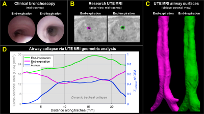
 Quantitative geometric assessment of regional airway collapse in neonates via retrospectively respiratory-gated 1H UTE MRI Quantitative geometric assessment of regional airway collapse in neonates via retrospectively respiratory-gated 1H UTE MRI
Nara Higano, Alister Bates, Erik Hysinger, Robert Fleck, Andrew Hahn, Sean Fain, Paul Kingma, Jason Woods
Neonatal airway malacia (dynamic larynx, trachea, and/or bronchi collapse) is a common airway complication often associated with preterm birth and congenital abnormalities but has not been extensively studied. This condition is currently diagnosed through visual bronchoscopy, which can be unreliable and poses increased risks to patients. We address these issues with an innovative technique using retrospectively respiratory-gated ultrashort echo time MRI and geometric analysis of moving airway anatomy for regional, quantitative evaluation of dynamic airway collapse in quiet-breathing, non-sedated neonates. This method has the potential to yield more accurate and objective assessment of neonatal airway collapse than current techniques allow.
|
|
0990.
 |
18 |
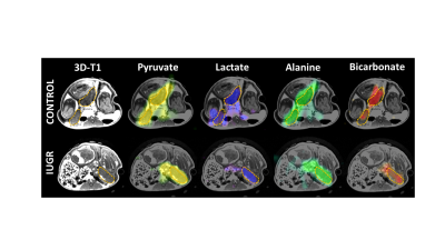
 Hyperpolarized [1-13C]Pyruvate Magnetic Resonance Imaging of Placentae Associated With Intrauterine Growth Restriction Hyperpolarized [1-13C]Pyruvate Magnetic Resonance Imaging of Placentae Associated With Intrauterine Growth Restriction
Lanette Friesen-Waldner, Conrad Rockel, Kevin Sinclair, Trevor Wade, Lauren Smith, Mohamed Moselhy, Cheryl Vander Tuin, Albert Chen, Barbra de Vrijer, Timothy Regnault, Charles McKenzie
Intrauterine growth restriction (IUGR) is associated with impaired placental metabolism and transport. Hyperpolarized carbon-13 (HP13C) MRI was used to detect metabolic differences between control and IUGR placentae in pregnant guinea pigs, and to determine the impact of maternal hyperoxygenation on placental metabolism. Area under the curve (AUC) was calculated for lactate, alanine, and bicarbonate and expressed as a ratio relative to pyruvate AUC. The ratio of alanine to pyruvate AUC decreased significantly in IUGR versus control placentae. Maternal hyperoxygenation resulted in significant increases in ratios of alanine and bicarbonate to pyruvate AUCs for both control and IUGR placentae.
|
|
0991.
 |
19 |
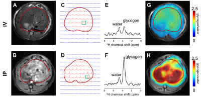
 Glycogen Synthesis Mapping Using In Vivo Deuterium Metabolic Imaging (DMI) Glycogen Synthesis Mapping Using In Vivo Deuterium Metabolic Imaging (DMI)
Henk De Feyter, Peter Brown, Kevin Behar, Douglas Rothman, Robin de Graaf
Deuterium Metabolic Imaging (DMI) is a novel approach providing high spatial resolution metabolic data from both animal models and human subjects. DMI relies on 2H MRSI in combination with administration of 2H-labeled substrates. We show how DMI combined with administration of [6,6’-2H2]-glucose can image liver glycogen synthesis in rats and human subjects. In rats at 11.7T, DMI revealed differences in liver glycogen synthesis between glucose administrations through an intravenous and intraperitoneal route. At 4T, we showed that DMI is feasible in humans and could detect labeling of liver glycogen after oral intake of [6,6’-2H2]-glucose.
|
|
0992.
 |
20 |
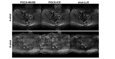
 High-Resolution Multishot Diffusion-Weighted Body and Breast MRI using Locally Low-rank regularization High-Resolution Multishot Diffusion-Weighted Body and Breast MRI using Locally Low-rank regularization
Yuxin Hu, Evan Levine, Catherine Moran, Valentina Taviani, Shreyas Vasanawala, Bruce Daniel, Brian Hargreaves
Multishot imaging has been shown to provide high resolution diffusion-weighted images (DWIs) with reduced distortion, however, significant aliasing artifacts and signal cancellation still occur due to the mismatch of the motion-induced phase between different shots. Several proposed SENSE-based methods without using navigators work well for brain DWI. However, these methods may fail when phase variations are more complex, in body DWI. In this work, we circumvent the challenging phase estimation step and efficiently solve this problem by using a locally low-rank reconstruction approach. The proposed reconstruction enables high resolution imaging with reduced geometric distortion compared with other approaches.
|
 |
0993.
 |
21 |
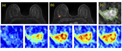
 Prediction of Breast Cancer Molecular Subtypes Using Conventional Feature Extraction and Two Machine Learning Architectures Based on DCE-MRI Prediction of Breast Cancer Molecular Subtypes Using Conventional Feature Extraction and Two Machine Learning Architectures Based on DCE-MRI
Yang Zhang, Siwa Chan, Jeon-Hor Chen, Daniel Chow, Peter Chang, Melissa Khy, Dah-Cherng Yeh, Xinxin Wang, Min-Ying Su
Two different convolutional neural network architectures were applied to differentiate subtype breast cancer based on 5 DCE-MRI time frame images: (1) a conventional serial convolutional neural network; (2) a convolutional long short term memory (CLSTM) Network. In addition, a logistic classifier was trained using morphology and texture features, selected using a random forest algorithm. For CNN, a bounding box based on the automated tumor segmentation was used to create a cropped image of the tumor as network input. A total of 94 cancers were analyzed, including 14 triple negative, 29 HER2-positive, and 51 Hormonal-positive, HER2-negative. Upon 10-fold validation, the differentiation accuracy is 0.81-0.86 using serial CNN, and 0.88-0.95 using the CLSTM.
|
|
0994.
 |
22 |
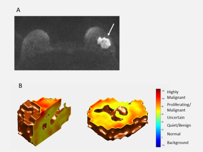
A diffusion MRI based computer-guided assistance approach for the diagnosis of breast lesions with high accuracy and without the need for contrast agents.
Video Permission Withheld
Mariko Goto, Denis Le Bihan, Koji Sakai, Kei Yamada
This prospective study included 37 patients with 39 breast lesions. DWI images were acquired at 2 b values (200, 1500s/mm²) on a 3T-MRI scanner with a dedicated 16-channel breast coil. From the images a non-Gaussian diffusion based absolute quantitative biomarker, so-called Signature Index (S-index), was calculated by comparing tissue signals to a library of tissue reference signals to provide a classification of tumor types. The median S-index for malignant lesions was significantly higher (p < 0.0001) than for benign lesions, and the overall S-index diagnostic performance was significantly higher than BI-RADS (AUC=0.96 and 0.83, respectively), without contrast agents.
|
|
0995.
 |
23 |
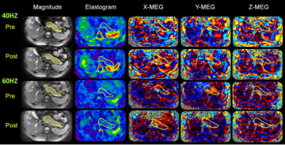
 Changes in Pancreatic Stiffness in Obese Adults Receiving an Oral Glucose Load, as Measured by Magnetic Resonance Elastography Changes in Pancreatic Stiffness in Obese Adults Receiving an Oral Glucose Load, as Measured by Magnetic Resonance Elastography
Ruoyun Ji, Yu Shi, Yanqing Liu, Lizhuo Cang, Min Wang, Qiyong Guo
When applying MR elastography (MRE) in pancreas, the waves propagated in individuals with high Body Mass Index (BMI) tend to travel longer distances with more challenges. In this study, we performed MRE in 18 obese subjects before and after an administration of a 75 g oral glucose load (OGL), and we found that 40Hz had better wave image quality than 60Hz and was more sensitive to detect the stiffness decrease after OGL.
|
 |
0996.
 |
24 |
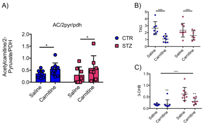
 L-Carnitine Shows Beneficial Effects on Cardiac Metabolism and Function: A Hyperpolarized MRS and Langendorff Perfusion Study L-Carnitine Shows Beneficial Effects on Cardiac Metabolism and Function: A Hyperpolarized MRS and Langendorff Perfusion Study
Dragana Savic, Vicky Ball, Kerstin Timm, Lisa Heather, Damian Tyler
L-carnitine acts as a buffer of acetyl-CoA units in the mitochondria, as well as facilitating transport of fatty acids. In addition, L-carnitine levels are decreased in the diabetic heart. The purpose of this study was to investigate the effect of L-carnitine supplementation on cardiac function and metabolism in the diabetic rat heart. We show that daily injections of L-carnitine can alter cardiac metabolism in the in-vivo diabetic rat heart, and can improve functional recovery as well as fatty acid oxidation rates post ischemia. Such studies allow a better understanding of the interactions between metabolism and function in the diabetic heart and may provide new insight into novel therapeutics.
|
 |
0997.
 |
25 |
 MRI Cine-Tagging of Cardiac-Induced Motion: Diagnostic Performance for Noninvasive Staging of Liver Fibrosis MRI Cine-Tagging of Cardiac-Induced Motion: Diagnostic Performance for Noninvasive Staging of Liver Fibrosis
Thierry Lefebvre, Léonie Petitclerc, Laurent Bilodeau, Giada Sebastiani, Hélène Castel, Claire Wartelle-Bladou, Bich Nguyen, Guillaume Gilbert, An Tang
Elastography techniques for staging liver fibrosis assess the right liver and require additional hardware. MRI cine-tagging evaluates the strain of liver tissue and shows promise for staging liver fibrosis without additional hardware. It can be performed routinely during MRI examinations. Strain showed high correlation with fibrosis stages (ρ = -0.60, P < 0.001). AUC was 0.78 to distinguish fibrosis stages F0 vs. ≥ F1, 0.78 for ≤ F1 vs. ≥ F2, 0.87 for ≤ F2 vs. ≥ F3, and 0.87 for ≤ F3 vs. F4. Larger studies in cohorts with specific liver disease are required to validate this technique.
|
 |
0998.
 |
26 |
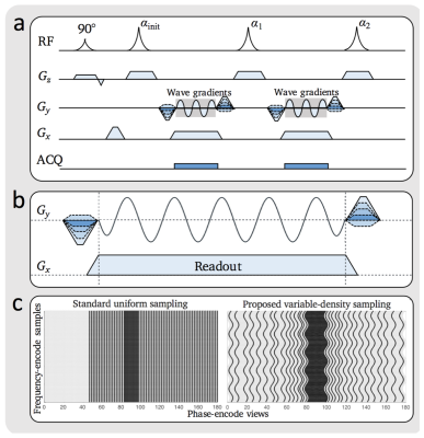
 Improved Speed and Image Quality for Imaging of Liver Lesions with Auto-calibrated Wave Encoded Variable Density Single-Shot Fast Spin Echo. Improved Speed and Image Quality for Imaging of Liver Lesions with Auto-calibrated Wave Encoded Variable Density Single-Shot Fast Spin Echo.
Jamil Shaikh, Feiyu Chen, Valentina Taviani, Kim Vu, Shreyas Vasanawala
Abdominal T2-weighted imaging is conventionally lengthy, but single shot approaches significantly improve current acquisition times. For single shot fast spin echo (SSFSE), axial imaging speed and sharpness are constrained by limited parallel imaging acceleration. Here, SSFSE technique with wave encoding and variable-density sampling (wSSFSE) was developed to enable higher accelerations and improve overall image quality. The purpose of this study is to assess image quality, delineation of anatomical structures, lesion conspicuity, and speed improvements with wSSFSE.
|
|
0999.
 |
27 |
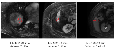
 A Fully Convolutional Neural Network for 3D Volumetric Liver Lesion Segmentation A Fully Convolutional Neural Network for 3D Volumetric Liver Lesion Segmentation
Sean Sall, Anitha Krishnan, Jesse Lieman-Sifry, Felix Lau, Matthieu Le, Matt DiDonato, Albert Hsiao, Claude Sirlin, John Axerio-Cilies, Daniel Golden
We present an automated approach to liver lesion segmentation in abdominal MRI scans. We use a 3D fully convolutional neural network to segment liver lesions; segmentations are then used to estimate longest linear diameter (LLD) and volume. We show that the median LLD error is 2.01 mm and that these estimates are within limits of clinically usability as part of a semi or fully-automated workflow. Automating lesion segmentation may pave the way for tracking lesion volume and tumor burden as well as treatment response.
|
|
1000.
 |
28 |
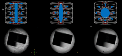
High spatial and temporal free breathing T1 contrast enhanced imaging using a novel 4D variable density, elliptical centric radial stack-of-stars sharing approach
Video Permission Withheld
Gabriele Beck, Suthambhara Nagaraj, Joao Silva Canaveira Tourais, Jan Hendrik Wuelbern, Johannes Peeters
Non-Cartesian acquisitions combined with parallel imaging and compressed sensing have been proposed for high spatial and temporal resolution free breathing motion robust 4D contrast enhanced applications. While these approaches provide substantial reduction of contrast blurring and motion robustness, the sampling in itself may still provide limitations to capture the temporal image content. Our approach focuses on the optimization of the 4D sampling strategy with a unique 4D variable density, elliptical-centric radial stack of stars sharing approach. This approach reduces not only the minimum scan time of the 4D scan but also provide possibilties to optimally adjust and capture temporal information like motion and contrast enhancement.
|
|
1001.
 |
29 |
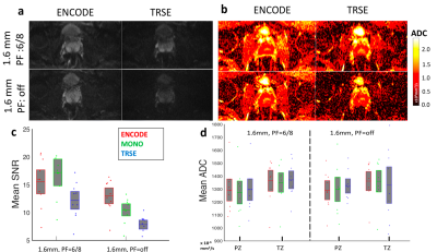
 Higher-Resolution Prostate Diffusion MRI with Minimized Echo Time using Eddy Current Nulled Convex Optimized Diffusion Encoding (ENCODE) Higher-Resolution Prostate Diffusion MRI with Minimized Echo Time using Eddy Current Nulled Convex Optimized Diffusion Encoding (ENCODE)
Zhaohuan Zhang, Kevin Moulin, Eric Aliotta, Sepideh Shakeri, Sohrab Mirak, Daniel Ennis, Holden Wu
Higher-resolution prostate DWI has the potential to improve prostate cancer diagnosis, but suffers from additional SNR reduction due to increased TE at longer EPI readouts for current diffusion encoding schemes. In this work, we evaluated the newly proposed Eddy Current Nulled Convex Optimized Diffusion Encoding to achieve eddy-current-free diffusion encoding with minimized TE for prostate diffusion MRI using standard and higher-resolution protocols. The ENCODE higher-resolution protocol achieves higher scores for image sharpness and contrast compared to standard-resolution prostate DWI.
|
|
1002.
 |
30 |
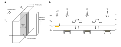
 Evaluating the Accuracy of Multi-component T2 and Fractions for Luminal Water Imaging of the Prostate using 3D GRASE with Inner Volume Selection Evaluating the Accuracy of Multi-component T2 and Fractions for Luminal Water Imaging of the Prostate using 3D GRASE with Inner Volume Selection
Rachel Chan, Angus Lau, Garry Detzler, Vivekanandan Thayalasuthan, Robert Nam, Masoom Haider
Prostate cancer can be detected using a multi-component T2 mapping technique termed luminal water imaging (LWI). The purpose of our study is: i) to accelerate the LWI acquisition by using inner volume selection (IVS) in GRASE and ii) to assess the accuracy of estimated T2 values and fractions. Simulations, phantom and in-vivo prostate experiments were performed. Results show that the estimated parameters should be interpreted with caution in noisy scenarios and at low fractions of the long T2 component. Results demonstrate that GRASE with IVS is effective for accelerating prostate LWI by at least a factor of three.
|
|

 Watch the full Pitch Session Here


