Poster: Musculoskeletal Madness
Electronic Power Pitch Poster
Musculoskeletal
09:15 - 10:15
Tuesday, 19 June 2018
Power Pitch Theater B - Exhibition Hall
| |
|
Plasma # |
|
0327.
 |
16 |
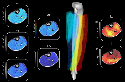
Intramuscular variability and sex difference in diffusion properties and 3D architecture of human lower leg muscles assessed with ultra-high-field diffusion tensor imaging and tractography
Video Permission Withheld
Alexandre Fouré, Augustin Ogier, Christophe Vilmen, Arnaud Le Troter, Thorsten Feiweier, Maxime Guye, Julien Gondin, Pierre Besson, David Bendahan
Skeletal muscle function impairment can be associated to changes in both muscle volume and structural arrangement of fascicles/fibers within the muscle thereby leading to the reduction of muscle force production. Diffusion properties and 3D structural organization of muscle fibers were quantified from high-resolution diffusion tensor images recorded at ultra-high-field MRI (UHF-7T). These parameters were assessed regarding their intramuscular variability and sensitivity to sex difference. This application of UHF-7T might be of high interest for the assessment of muscular injuries in both athletes and patients with muscular disorders.
|
|
0328.
 |
17 |
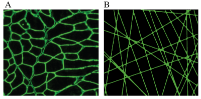
 Time-dependent diffusion and the random permeable barrier model predict muscles fiber dimensions in Duchenne muscular dystrophy mice Time-dependent diffusion and the random permeable barrier model predict muscles fiber dimensions in Duchenne muscular dystrophy mice
Bauke Kogelman, Kevin Adamzek, Ernst Suidgeest, Gregory Lemberskiy, Dmitry Novikov, Els Fieremans, Maaike Putten, Louise Weerd
We used time-dependent diffusion, with diffusion times up to 500ms to assess the muscle tissue microstructure in two different mouse models of Duchenne muscular dystrophy (DMD). We found significant differences in diffusivities between DMD models and the genetic background matched wild types in diffusion times up to 100ms. We modeled diffusion time-dependence with the random permeable barrier model (RPBM), which yielded surface-to-volume ratios indicative of fiber size and compared these values to gold-standard histology of the same muscles. The RPBM predicted relative differences in fiber dimensions between mdx and wild type within each genetic background.
|
|
0329.
 |
18 |
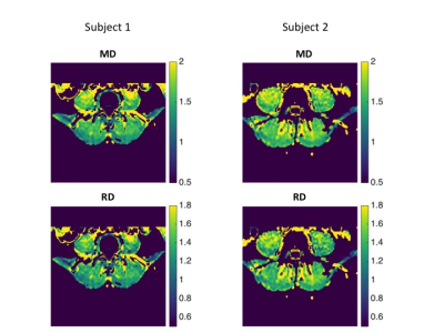
Relationship of paraspinal muscle DTI metrics to isometric strength measurements
Elisabeth Klupp, Barbara Cervantes, Sarah Schlaeger, Stephanie Inhuber, Florian Kreuzpointer, Michael Dieckmeyer, Friedemann Freitag, Ernst Rummeny, Claus Zimmer, Jan Kirschke, Dimitrios Karampinos, Thomas Baum
Diffusion Tensor Imaging (DTI) enables the microstructural examination of muscle tissue as well as its pathological and stress-dependent changes. Little is known about the associations between muscular DTI parameters and corresponding muscle strength measurements. The present study investigated the correlations of DTI parameters of paraspinal muscles with isometric strength measurements in healthy subjects. The results indicate that DTI may potentially track slight changes of back muscle tissue microstructure that relate to muscle strength and may be useful in the early diagnosis of back muscle diseases and back pain.
|
|
0330.
 |
19 |
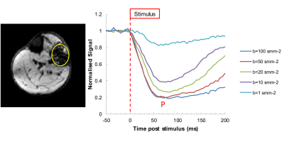
Imaging human motor unit activity using MRI.
Video Permission Withheld
Paola Porcari, Ian Schofield, Roger Whittaker, Andrew Blamire
Neuromuscular diseases can lead to characteristic changes in the anatomy of motor units (MU). In this study we developed an MR approach to image motor unit activity. In-scanner electrical stimulation of MU was synchronized to a pulsed-gradient spin-echo imaging sequence sensitive to microscopic contraction of muscle fibers. Experimental data was compared to a simple theoretical model and scan parameters evaluated. The spatial pattern of muscle fibers forming different MU was imaged for the first time in healthy controls.
|
|
0331.
 |
20 |
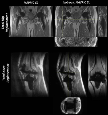
Clinical Feasibility of Isotropic MAVRIC SL Imaging of Total Joint Arthroplasties
Matthew Koff, Suryanarayanan Kaushik, Parina Shah, Erin Argentieri, Hollis Potter
Multi-spectral MRI reduces susceptibility when imaging near total joint arthroplasty (TJA). MAVRIC SL acquires 24 bins of off-resonance data to generate images, but most implants require fewer bins. This study uses a calibration scan, to determine an adequate number of bins, coupled with long echo trains and variable flip angles to permit an isotropic MAVRIC SL acquisition in clinically feasible scan times. The isotropic MAVRIC SL acquisition showed an improved detection of the implant-bone interface surrounding TJA, while retaining image contrast and overall SNR. An isotropic acquisition may improve the diagnostic capability of MAVRIC SL images.
|
|
0332.
 |
21 |
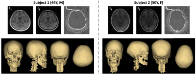
Solid-State MRI as a noninvasive alternative to computed tomography for craniofacial imaging
Hyunyeol Lee, Xia Zhao, Hee Kwon Song, Rosaline Zhang, Scott Bartlett, Felix Wehrli
Computed tomography (CT) imaging is the imaging modality of choice for 3D visualization of bone. However, there is growing concern about repeated exposure to ionizing radiation, in particular during infancy, for instance, in patients with craniosynostosis pre- and post-surgery. Here, we developed a dual-RF, dual-echo, 3D UTE sequence using view-sharing to minimize scan time. Images are reconstructed by combining long- and short-RF, first and second echoes, yielding soft-tissue suppressed skull images at 1.1 mm isotropic resolution in 6 minutes scan time in a human skull ex vivo and test subjects in vivo. 3D renderings display the relevant craniofacial skeleton similar to CT.
|
|
0333.
 |
22 |
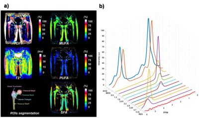
Subregional bone marrow adipose tissue composition in the proximal femur: Comparison of 3T Chemical Shift Encoded-MRI and Magnetic Resonance Spectroscopy
Dimitri MARTEL, Benjamin LEPORQ, Mary BRUNO, Stephen HONIG, Amit SAXENA, H.Michael BELMONT, Gabrielle TURYAN, Ravinder R. REGATTE, Gregory CHANG
Studies of bone marrow adipose tissue (bMAT) composition in osteoporosis has revealed increased [a1] in fat content and decreased unsaturated fatty acid in the proximal femur in osteoporosis (OP) patients compared to controls. Advancements in MR image-based methods for fat assessment have allowed quantification of fat content though chemical-shift encoded MRI (CSE-MRI). CSE-MRI methods have not been compared to MRS for bMAT composition assessment. The aim of this study was to quantify, within subregions of the proximal femur, bone marrow PDFF as well as saturated and unsaturated fat using both CSE-MRI and MRS.
|
|
0334.
 |
23 |
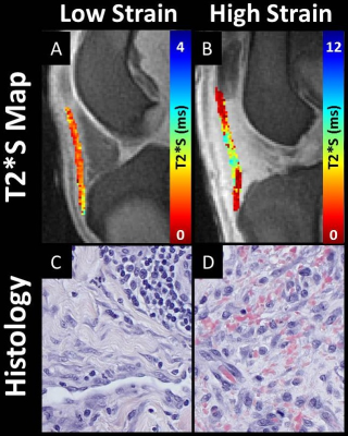
Temporal Changes of a Canine Model of Patellar Tendinopathy Using UTE MRI T2* Assessment: A Pilot Study
Sarah Pownder, Kei Hayashi, Brian Caserto, Bin Lin, Hollis Potter, Matthew Koff
Tendon damage has been traditionally evaluated invasively with biopsy. This pilot study evaluated the effects of an induced mechanical strain of the patellar tendon on UTE-MRI metrics and corresponding histology. The induced strain had a greater effect on the quantitative MRI metrics over time, by percentage change, than by the level of induced strain. A corresponding histologic analysis had similar findings. The results indicate the capability of utilizing MRI to evaluate differences in the quantitative T2* metrics of the patellar tendon. Time dependent changes in quantitative UTE-MRI metrics may be detected using a model of induced subclinical tendon damage.
|
|
0335.
 |
24 |
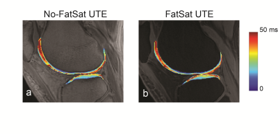
Knee Cartilage UTE T2* Quantification with Water-Fat Decomposition
Misung Han, Peng Cao, Michael Carl, Thomas Link , Peder Larson, Roland Krug
Recently, there has been a growing interest in assessing lesions in the deep layers of knee cartilage as a pathogenesis of osteoarthritis. Ultrashort echo-time (UTE) T2* mapping in the deep layers has demonstrated great potential to detect early degenerative changes. Fat suppression is normally accompanied to reduce off-resonance artifacts from adjacent fatty tissues; however, it can also suppress significant amount of short T2 tissues and affect quantification. In this work, we quantified cartilage T2* by decomposing fat and water components using no fat-suppressed 3D multi-echo UTE images and demonstrated shorter T2* compared to that from fat-suppressed 3D UTE images.
|
|
0336.
 |
25 |
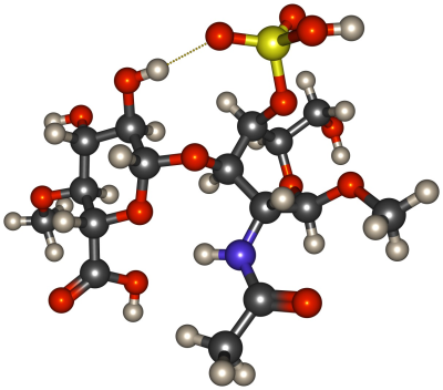
Strategies for Obtaining Relaxation Rates due to Chemical Exchange from Parameter Free Atomistic Simulations
Henning Henschel, Matti Hanni, Miika Nieminen
Chemical exchange has likely a significant impact on the dispersion of T1ρ in cartilaginous tissue. In order to quantify this effect, we are developing atomistic simulation strategies for direct modelling of proton transfer reactions between biomacromolecules – first and foremost chondroitin sulfate – and water. Here we present benchmarking results from a number of semi-empirical quantum mechanics methods for several key system properties. Results suggest feasibility of the chosen approach.
|
|
0337.
 |
26 |
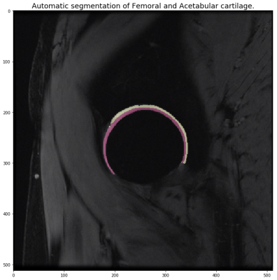
Automatic Segmentation of Hip Cartilage with Deep Convolutional Neural Nets for the evaluation of Acetabulum and Femoral T1? and T2 relaxation times.
Michael Girard, Valentina Pedoia, Berk Norman, Jasmine Rossi-Devries, Sharmila Majumdar
In this study we utilize a deep learning approach to automatically segment the femoral and acetabular cartilages in the hip. From these segmentations we also calculate T1ρ and T2 relaxation times then compare to manual segmentations and their T1ρ and T2 values. We show the T1ρ and T2 relaxation times, calculated using manual and automatic segmentations, are very correlated, R values above .94, and comparable.
|
|
0338.
 |
27 |
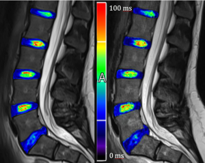
Assessment of the clinical feasibility of routine T2 mapping of the intervertebral disc using highly undersampled k-space data
Marcus Raudner, Markus Schreiner, Tom Hilbert, Tobias Kober, Vladimir Juras, Claudia Kronnerwetter, David Stelzeneder, Siegfried Trattnig
Even though the measurement of T2 relaxation time is one of the most widespread available assets of quantitative MRI in clinical routine, it is left with untapped potential as the required scan time is too long. GRAPPATINI is a model-based iterative algorithm reconstructing T2 maps and T2-weighted images from highly-undersampled k-space data. The aim of this study was to compare the quantitative results of an established standard method with this novel technique in the intervertebral disc with the additional benefit of GRAPPATINI to simulate T2w contrasts at arbitrarily chosen echo times.
|
|
0339.
 |
28 |
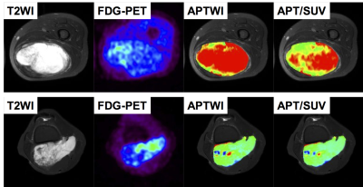
Voxel-wise ratios of amide proton transfer (APT) signals and standardized uptake values (SUVs) of fluorodeoxyglucose (FDG) in the differentiation of myxoid-rich soft-tissue tumors with FDG-PET/MR imaging
Koji Sagiyama, Yuji Watanabe, Keisuke Ishimatsu, Takeshi Kamitani, Yuzo Yamasaki, Takuya Hino, Sungtak Hong, Jochen Keupp, Yoshihiro Matsumoto, Hiroshi Honda
It is often difficult to differentiate myxoid-rich soft-tissue tumors on conventional imaging. In this study, we performed a direct voxel-wise comparison of amide proton transfer (APT) signals and standardized uptake values (SUVs) obtained on FDG-PET/MR imaging. Among myxoid-rich tumors including myxoid liposarcomas, myxofibrosarcomas, myxoid chondrosarcomas, and schwannomas, the mean APT/SUV was significantly higher in liposarcomas than in the other myxoid-rich tumors while a single parameter such as APT, SUV or apparent diffusion coefficient (ADC) did not show any significant differences between the two groups. The APT/SUV could be a reliable bio-imaging marker for differentiating soft-tissue tumors.
|
|
0340.
 |
29 |
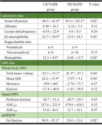
Assessment of early treatment response by multiparametric whole-body MRI as a 1-step approach to prediction of overall response rate in patients with multiple myeloma
Miyuki Takasu, Takayuki Tamura, Yuji Akiyama, Yoko Kaichi, Shota Kondo, Chihiro Tani, Kazuo Awai
We compared remission status at completion of chemotherapy with changes in MRI biomarkers obtained by advanced MRI techniques, including total tumor volume calculated from whole-body diffusion-weighted imaging and fat fraction by mDIXON soon after induction of chemotherapy for patients with multiple myeloma. The early change in fat fraction of lumbar bone marrow and serum monoclonal protein after 2 cycles of chemotherapy contributed significantly to the prediction of CR/VGPR status. Results of this study may indicate that prediction of remission status can be achieved by assessing bone marrow on lumbar spinal MRI with mDIXON.
|
 |
0341.
 |
30 |
Simultaneous Bilateral Knee MR Imaging
Feliks Kogan, Evan Levine, Akshay Chaudhari, Uchechukwuka Monu, Kevin Epperson, Edwin Oei, Garry Gold, Brian Hargreaves
Osteoarthritis (OA) is commonly a bilateral disease. While long scan time and costs have precluded separate scanning of both knees in clinical MRI, there is evidence that bilateral examinations are beneficial for evaluation of OA changes, especially for longitudinal studies. In this study, we demonstrate that a bilateral coil-array setup can image both knees simultaneously in similar scan times as conventional unilateral knee scans with comparable image quality and quantitative accuracy. This has the potential to improve the value of MRI knee evaluations.
|
|
