Joint Annual Meeting ISMRM-ESMRMB • 16-21 June 2018 • Paris, France

Weekend Educational Course
Advanced Clinical MR Imaging in MSK
Advanced Clinical MR Imaging in MSK: Part 1
Weekend Course

ORGANIZERS: Eric Chang, Garry Gold, Emily McWalter, Edwin Oei, Philip Robinson

Saturday, 16 June 2018
S01  08:00 - 09:30 Moderators:  Paolo Felisaz, Matthew Koff

Skill Level: Intermediate

Session Number: WE-05A

Overview
This course will provide an in-depth discussion on current and next-generation MR imaging techniques for use in the musculoskeletal system. Clinical applicability for imaging normal and abnormal tissues will be featured. An interactive expert panel discussion on overcoming challenges for implementation will conclude the session.

Target Audience
Technologists, radiologists, and clinical practitioners who perform MSK imaging.

Educational Objectives
As a result of attending this course, participants should be able to:
-Describe the most appropriate uses of new MR imaging techniques;
-Explain the impact of MR imaging in clinical decision making for commonly imaged joints; and
-Describe the MR imaging appearances of common internal derangements.


08:00
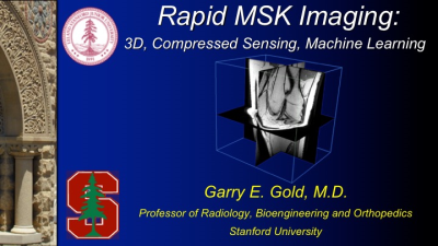
Rapid MR (3D, CS, Machine Learning)
Garry Gold
This presentation will cover new methods for rapid three-dimensional musculoskeletal imaging, including compressed sensing and machine learning methods.
08:25   SAMs: Rapid MR (3D, CS, Machine Learning)
08:30
Shoulder
Luis Beltran
This presentation will discuss imaging of the shoulder with an emphasis on operative management of anterior shoulder instability and rotator cuff tears.  The learning objectives are to a) understand the mechanisms of injury and associated pathologies in anterior shoulder instability and rotator cuff tears, b) be familiar with current treatment guidelines for management of anterior shoulder instability and rotator cuff disease, c) recognize the normal and abnormal appearances of postoperative MRI studies for anterior shoulder instability surgery and rotator cuff repairs.   I give permission to record this presentation.

09:00
  Advanced Imaging in Hand & Wrist in Common Rheumatologically & Traumatic Conditions
Mikael Boesen
The lecture will show the conventional ways of imaging most common rheumatically and traumatic conditions in the hand and wrist and show the potential added value of new 3-dimensional and multi-parametric imaging technologies like dynamic contrast enhanced MRI (DCE-MRI), PET-CT and PET MRI, ultrasound, dual energy- and conebeam CT of the hand and wrist as promising methods to complement the imaging portfolio that adds further to the understanding and quantification of the pathophysiology, inflammatory load and morphological disease patterns in patients with rheumatoid arthritis (RA), psoriasis arthritis (PsA), crystal deposition diseases (Urate and CPPD), osteoarthritis (OA) and trauma.

09:30
  Break & Meet the Teachers
 
Advanced Clinical MR Imaging in MSK: Part 2
Weekend Course

ORGANIZERS: Jenny Bencardino, Eric Chang, Garry Gold, Emily McWalter, Edwin Oei, Philip Robinson

Saturday, 16 June 2018
S01  10:00 - 11:30 Moderators:  Paolo Felisaz, Matthew Koff

Skill Level: Intermediate

Session Number: WE-05B

Overview
This course will provide an in-depth discussion on current and next-generation MR imaging techniques for use in the musculoskeletal system. Clinical applicability for imaging normal and abnormal tissues will be featured. An interactive expert panel discussion on overcoming challenges for implementation will conclude the session.

Target Audience
Technologists, radiologists, and clinical practitioners who perform MSK imaging.

Educational Objectives
As a result of attending this course, participants should be able to:
-Describe the most appropriate uses of new MR imaging techniques;
-Explain the impact of MR imaging in clinical decision making for commonly imaged joints; and 
-Describe the MR imaging appearances of common internal derangements.


10:00
Peripheral Nerves
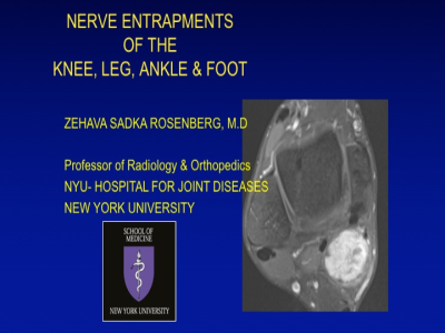
Zehava Rosenberg
This talk will focus on MR imaging of peripheral nerve of the knee, ankle and foot.    The talk will be subdivided into two parts.  A.  A discussion of general MRI features of entrapment neuropathies in the lower extremity.  The listener will be provided with distinctive direct and indirect features of neuropathy.  B.  Focused discussion of specific nerves commonly susceptible to neuropathy, with a brief initial review of normal anatomy followed by MRI examples of common neuropathies.   

10:30
  Imaging Around Metal (Physics)
Kevin Koch
11:00
  Hip
Andoni Toms
11:30
  Lunch & Meet the Teachers
Back
The International Society for Magnetic Resonance in Medicine is accredited by the Accreditation Council for Continuing Medical Education to provide continuing medical education for physicians.