|
Weekend Educational Course
Molecular Imaging for Beginners |
|
Molecular Imaging for Beginners: Part 1
Weekend Course
ORGANIZERS: Ichio Aoki, Arvind Pathak
Saturday, 16 June 2018
| S04 |
08:00 - 09:15 |
Moderators: Christin Sander, Arvind Pathak |
Skill Level: Basic
Session Number: WE-06A
Overview
This educational session is designed to provide an introduction to the principles of molecular imaging. This course begins with an overview of MR molecular imaging, followed by an overview of complementary molecular imaging methods. Next, the beginner is introduced to the principles of relaxation mechanisms, spectroscopy, probe design, and cellular molecular imaging. The session culminates in a lecture describing how to design molecular imaging experiments.
Target Audience
Students, fellows and researchers who need a “gentle" introduction to molecular imaging concepts and applications.
Educational Objectives
As a result of attending this course, participants should be able to:
-Describe the fundamental concepts and methods in the diverse field of molecular imaging;
-Discuss how to evaluate, design, and implement molecular imaging studies and protocols; and
-Summarize "good" practices of molecular imaging and experimental design.
08:00
|
|
 Introduction to MR Molecular Imaging Introduction to MR Molecular Imaging
Peter Caravan
|
08:25
|
|
 Multimodality Molecular Imaging for Beginners Multimodality Molecular Imaging for Beginners
Kristine Glunde
Multimodality molecular imaging applies imaging modalities beyond visualizing anatomy and morphology to include the ability of imaging disease-specific biomolecules and pathways in cancer, cardiovascular disease, and inflammation, among others. Imaging modalities used in molecular imaging are computed tomography (CT), magnetic resonance imaging (MRI), magnetic resonance spectroscopic imaging (MRSI), optical imaging, positron emission tomography (PET), single-photon-emission computerized tomography (SPECT), and ultrasound (US). This lecture will give an overview of the most important concepts and applications in multimodality molecular imaging for beginners.
|
08:50
|
|
 Fundamentals of MR Relaxation for Molecular Imagers Fundamentals of MR Relaxation for Molecular Imagers
Silvio Aime
|
09:15
|
|
Break & Meet the Teachers |
|
| |
|
Molecular Imaging for Beginners: Part 2
Weekend Course
ORGANIZERS: Ichio Aoki, Arvind Pathak
Saturday, 16 June 2018
| S04 |
10:00 - 11:40 |
Moderators: Christin Sander, Arvind Pathak |
Skill Level: Basic
Session Number: WE-06B
Overview
This educational session is designed to provide an introduction to the principles of molecular imaging. This course begins with an overview of MR molecular imaging, followed by an overview of complementary molecular imaging methods. Next, the beginner is introduced to the principles of relaxation mechanisms, spectroscopy, probe design, and cellular molecular imaging. The session culminates in a lecture describing how to design molecular imaging experiments.
Target Audience
Students, fellows and researchers who need a “gentle" introduction to molecular imaging concepts and applications.
Educational Objectives
As a result of attending this course, participants should be able to:
-Describe the fundamental concepts and methods in the diverse field of molecular imaging;
-Discuss how to evaluate, design, and implement molecular imaging studies and protocols; and
-Summarize "good" practices of molecular imaging and experimental design.
10:00
|
 |
 MR Spectroscopy - The Forgotten Molecular Imaging Modality MR Spectroscopy - The Forgotten Molecular Imaging Modality
Robin de Graaf
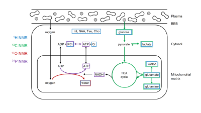
MR-based molecular imaging typically relies on the high detection sensitivity of water to indirectly observe the underlying molecular and cellular processes. The ubiquitous presence of water demands that image contrast and specificity is obtained through molecular imaging probe design. MR spectroscopy uses the intrinsic specificity provided by the chemical shift to allow detection of a wide range of metabolites and metabolic pathways. When combined with spatial imaging gradients, MR-based metabolic imaging can provide a unique and complementary addition to the arsenal of molecular imaging techniques.
|
10:25
|
 |
 Principles of Probe Design for Molecular Imagers Principles of Probe Design for Molecular Imagers
Horacio Cabral, Ichio Aoki
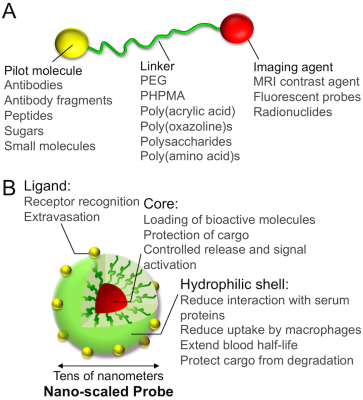
Molecular imaging allows the visualization of biological events in real-time at tissue, cellular and subcellular levels in living systems by merging conventional imaging techniques with probes designed to report the expression of biomarkers or variations in physiological factors. Successful molecular imaging agents should provide high contrast intensity with low noise-to-signal ratio at the target in vivo for sufficient time. Herein, I recapitulate the principles for designing effective molecular imaging probes for various imaging modalities, and provide fundamental strategies for the development of these probes and their application in biology, diagnosis and therapy.
|
10:50
|
|
 Introduction to "Cellular" Molecular Imaging Introduction to "Cellular" Molecular Imaging
Ben Bartelle
|
11:15
|
|
 How to Design a Molecular Imaging Experiment How to Design a Molecular Imaging Experiment
Emmanuel Barbier
This course will address the steps that a beginner should follow to develop a molecular imaging experiment in small animals: after setting the target and choosing the appropriate animal model, we will evaluate the pros and cons of existing imaging modalities, with a focus on MRI data acquisition and integration during post-processing. Examples will be taken from oncology and neuroscience.
|
11:40
|
|
Adjournment & Meet the Teachers |
|
| Back |
| The International Society for Magnetic Resonance in Medicine is accredited by the Accreditation Council for Continuing Medical Education to provide continuing medical education for physicians. |


