Female Pelvis
Oral
Body: Breast, Chest, Abdomen, Pelvis
Monday, 13 May 2019
| Room 518A-C |
08:15 - 10:15 |
Moderators: Johannes Heverhagen, Rebecca Rakow-Penner |
08:15
|
0096.
 |
Technical Feasibility of Three-Dimensional Magnetic Resonance Elastography for Assessing Endometrial Carcinoma

Xi Long, Tianhui Zhang, Sichi Kuang, Ying Deng, Bingjun He, Jingbiao Chen, Phillip Rossman, Kevin J Glaser, Sudhakar K Venkatesh, Bing Wu, Richard L Ehman, Jin Wang
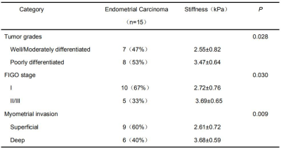
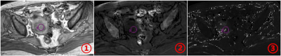
Endometrial carcinoma (EC) is one of the most common primary malignant tumors in women worldwide. Tumor grades, International Federation of Gynecology and Obstetrics (FIGO) stage and myometrial invasion of EC are important factors for treatment planning and prognosis. We explored the potential value of MR elastography (MRE) for the prediction of tumor grades, FIGO stage and myometrial invasion of EC. Our study showed that mean tumor stiffness may be a useful metric for differentiating well or moderately differentiated EC from poorly differentiated EC and for differentiating superficial invasion from deep myometrial invasion in EC.
|
08:27
|
0097
 |
Value of R2* and T2* in differential diagnosis of uterine sarcoma and degenerated hysteromyoma
Video Permission Withheld
Miao Niu, Ailian Liu, Lizhi Xie
To investigate the clinical value of enhanced T2 star weighted angiography (ESWAN) quantitative parameters in differential diagnosis of uterine sarcoma and degenerated hysteromyoma.
|
08:39
|
0098.
 |
High b-value diffusion-weighted MRI in cervical cancer detection: Preliminary results
Kaibao Sun, Qi Zhang, Zheng Zhong, Muge Karaman, Xiaoduo Yu, Han Ouyang, Xiaohong Zhou
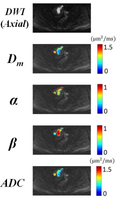
Cervical cancer remains one of the leading causes of cancer-related deaths in women. High b-value DWI with non-Gaussian modeling has made it possible to probe tumor tissue complexity, microstructures, and heterogeneity. We employed a non-Gaussian diffusion model based on continuous-time random walk (CTRW) theory to differentiate normal from cancerous cervical tissues. The CTRW parameters (Dm and β) exhibited a statistically significant difference between cancerous cervical tissue and normal. Our preliminary results illustrate the added value of high b-value DWI for cervical cancer detection, and point to a possible direction of diagnosing or staging cervical cancer using non-Gaussian diffusion models.
|
08:51
|
0099.
 |
Three-Dimensional Amide Proton Transfer MR Imaging for Cervical Cancer: Initial Experience
Yong-Lan He, Cheng-Yu Lin, Ya-Fei Qi, Xiaoqi Wang, Hai-Long Zhou, Hua-Dan Xue, Zheng-Yu Jin
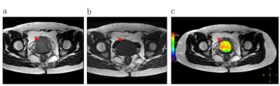
The present study demonstrates the feasibility of 3D APT MR imaging for uterine cervix on the largest sample size to our knowledge. As the clinical robustness of APT imaging in the pelvis is what researchers concerned, our study investigated and revealed the excellent agreement in both image quality assessment and APT values measurement, even on small cervical lesions with maximum diameter less than 2cm. Our studies demonstrated that APT imaging could differentiate cervical cancer from normal cervix. Cervical cancer showed significant higher APT values than that of normal cervix.
|
09:03
|
0100.
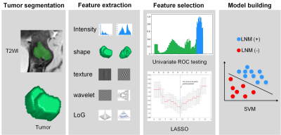 |
Radiomics Analysis of tumor and peri-tumor tissue on T2-Weighted Imaging Improves Diagnostic Performance of Lymph Node Metastasis in Patients with Cervical Cancer
Qingxia Wu, Shuo Wang, Xi Chen, Yan Wang, Yusong Lin, Meiyun Wang
The tumor margin and peritumoral tissue play an important role in the process of LN metastasis. The aim of this study was to utilize radiomics analysis of tumor and peri-tumor tissue on T2 weighted image (T2WI) to improve LNM prediction ablility in cervical cancer patients. We found that peritumoral tissue of cervical cancer on T2WI showed favorable value in predicting LNM. The decision tree we proposed which incorporates the radiomics features of intratumoral and peritumoral tissue on T2WI and c-LN status can be potentially used for personalized preoperative evaluation of LNM and optimal treatment regimen selection in cervical cancer patients.
|
09:15
|
0101.
 |
Prediction of Neoadjuvant Chemoradiation Therapy Response in Rectal Cancer Using Radiomics Compared to Deep Learning Based on Pre-Treatment and mid-RT MRI
Yang Zhang, Liming Shi, Ke Nie, Xiaonan Sun, Tianye Niu, Ning Yue, Tiffany Kwong, Peter Chang, Daniel Chow, Jeon-Hor Chen, Min-Ying Su
The capability to predict patients’ response to neoadjuvant chemoradiation therapy is important for improving their management. The multi-parametric MRI (T2, DWI, DCE) performed before treatment and after 3-4 weeks of radiation were analyzed to predict final pathological response. Quantitative radiomics was performed using GLCM texture and histogram parameters, and also ROI and deep learning using convolutional neural network (CNN) were performed. Combining quantitative radiomics features with tumor volume and diffusion coefficient could achieve accuracy of 0.86 for pCR vs. non-pCR and 0.93 for GR vs. non-GR, and adding follow-up to pre-treatment MRI could improve accuracy, especially for CNN analysis.
|
09:27
 |
0102.
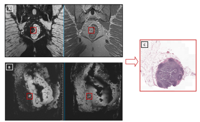 |
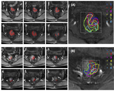
The feasibility of matching lymph nodes detected on USPIO-enhanced MRI with histopathology in rectal cancer
Rutger Stijns, Bart Philips, Iris Nagtegaal, Fatih Polat, Johannes de Wilt, Carla Wauters, Patrik Zamecnik, Jurgen Futterer, Tom Scheenen
Lymph node staging in rectal cancer based on imaging is a major challenge. Node-to-node matching is crucial to determine the histopathology of lymph nodes that are detected on in-vivo MRI. A workflow of in-vivo MRI, ex-vivo MRI and MR-guided pathology was set up for lymph nodes that were characterized on USPIO-enhanced MRI. Difficulties were seen in the node-to-node matching, in despite of the use of high-resolution 3D ex-vivo MRI to link in-vivo detected nodes to final pathology.
|
09:39
|
0103.
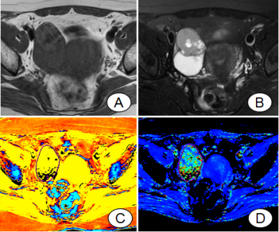 |
Enhanced T2 Star-Weighted Angiography (ESWAN) for Differentiating Borderline From Malignant Epithelial Ovarian Tumors
Xu Han, Meiyu Sun, Lizhi Xie, Kaiyu Wang, Mengyao Wang, Rui Fan
The aim of this study is to assess the fitted parameters of ESWAN in ovarian tumors and to investigate their potential in distinguishing borderline from malignant epithelial ovarian tumors, which can provide detailed information for clinical treatment. R2* and T2* in ESWAN were the key parameters for distinguishing borderline from malignant epithelial ovarian tumors. MR-ESWAN sequence can be used as non-enhancement quantitative indexes, which has a good application prospect.
|
09:51
|
0104.
 |
Ex-vivo MR-guided pathology to improve lymph node staging in rectal cancer
Rutger Stijns, Bart Philips, Carla Wauters, Johannes de Wilt, Iris Nagtegaal, Tom Scheenen
Pathological lymph node yield can be influenced by multiple factors. The use of ex-vivo MR guided pathology of rectal specimens could provide insight in the number and size of lymph nodes present in a rectal specimen and aid in an increased pathological lymph node yield. Therefore two series of rectal specimens were examined, one control group and one MR-guided group. Ex-vivo MRI revealed significantly more and significantly smaller lymph nodes without increasing the pathological yield. Small nodes appear to be difficult to harvest, presumably requiring a 3D approach for further improvement of pathological evaluation.
|
10:03
|
0105.
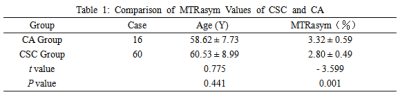 |
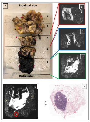
Using Amide Proton Transferto indentify Cervical Squamous Carcinoma/Adenocarcinoma and Evaluate its Differentiation Grade
Nan Meng, Jing Wang, Wenling Liu, Xuejia Wang, Dandan Zheng, Huijia Yin, Hongxia Wang, Dongming Han*
Amide proton transfer (APT) weighted imaging provides information about concentration of proteins/peptides with amide backbones. At present, there is no report on whether APT can be applied to cervical cancer. Our results show that APT can be used to preliminarily identify cervical squamous carcinoma, cervical adenocarcinoma and evaluate its differentiation grade.
|
|
