Advanced Imaging of Cartilage-Bone Interactions
Advanced Imaging of Cartilage-Bone Interactions
Weekday Course
Weekday Course
ORGANIZERS: Jung-Ah Choi, Riccardo Lattanzi, Miika Nieminen, Jan Fritz, Edwin Oei
Tuesday, 14 May 2019
| Room 518A-C | 15:45 - 17:45 | Moderators: Neal Bangerter, Ashley Williams |
Skill Level: Intermediate to Advanced
Session Number: TU-08
Overview
Changes occur to both the deep cartilage and subchondral bone in musculoskeletal diseases such as osteoarthritis, and interactions between these adjacent tissues are important. Advanced MRI techniques are required to obtain signal from these regions of tissue due to their short T2 relaxation times. This session explores the biology of interactions at the cartilage-bone interface, the advanced MRI techniques required to study this region, clinical studies of this interaction, and the use of PET/MR systems to study metabolic changes.
Target Audience
Trainees, researchers, and clinicians with an interest in changes at the bone-cartilage interface in musculoskeletal disease such as osteoarthritis.
Educational Objectives
As a result of attending this course, participants should be able to:
- Describe the biologic interactions at the cartilage-bone interface;
- List and summarize MRI techniques used to image the cartilage-bone interface;
- Describe how the cartilage-bone interface has been studied clinically; and
- Explain how PET/MR can be used to study metabolic changes at the cartilage-bone interface.
| 15:45 |
 |
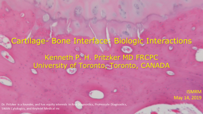
Biology of the Interactions at the Cartilage-Bone Interface
Kenneth Pritzker
The Cartilage-Bone interface acts as a viscoelastic material that can actively adapt to mechanical loads on both an instantaneous and long term basis. Loading this structure produces shear stresses on tissue components which, if excessive, lead to injury and its reaction. Reaction to injury has an early phase consisting of edema in subjacent bone marrow and overlying articular cartilage. Reaction to injury may extend as microfractures through the calcified cartilage,subchondral bone and/or trabecular bone. This is followed by repair which has the capacity to functionally regenerate the cartilage and bone tissues.
|
| 16:15 |
Imaging Early: Advanced Imaging of Short T2 Species
Mikko Nissi
This part of the educational course covers MRI techniques used to image the short/ultra-short T2 spins, abundant for example in the cartilage-bone interface and discuss the potential of the methods in the assessment of the short T2 species. The lectures will cover the main imaging methods, namely the UTE, ZTE, and SWIFT imaging sequences and their applications in the musculoskeletal system, especially in cartilage, bone, and the interface in between.
|
|
| 16:45 |
Clinical Applications of Imaging Cartilage-Bone Interactions
Christine Chung
Cartilage-bone interactions are structurally and functionally complex. Understanding this complexity is crucial with regard to non-invasive diagnosis and characterization of disease, as well as for development of treatment methods.
|
|
| 17:15 |
Metabolic Changes at the Cartilage-Bone Interface with PET/MR
Sharmila Majumdar
Osteoarthritis (OA) is a degenerative whole joint disease. Simultaneous PET-MR imaging provides a unique opportunity to study bone-cartilage interactions in joint degeneration, and provides a key to the pathophysiology of OA.
|
|
| 17:45 |
Adjournment |
 Back to Program-at-a-Glance |
  Back to Top