
Combined Educational & Scientific Session
MRI of Knee Stabilizers
Session Topic: Muscle, Cartilage, Knee stabilizers
Session Sub-Topic: MRI of Knee Stabilizers
Combined Educational & Scientific Session
ORGANIZERS: Jan Fritz, Riccardo Lattanzi, Kimberly Amrami, Jung-Ah Choi, Miika Nieminen, Hiroshi Yoshioka
| Monday Parallel 4 Live Q&A | Monday, 10 August 2020, 15:15 - 16:00 UTC | Moderators: Bragi Sveinsson & Anup Singh |
Skill Level: Intermediate to Advanced
Session Number: C-M-03
Overview
Instability and laxity are important effects of knee injuries. An accurate diagnosis forms the foundation for conservative versus surgical management, the type of operative management, and prognosis. In a multifaceted workup consisting of history and skilled physical examination, MRI plays a crucial role in the assessment of the integrity of primary and secondary knee stabilizers, including ligaments and muscle-tendon units, respectively. Structural and morphological MRI allows for the detection and grading of injured structures, injury patterns, and progression of healing. Functional and quantitative MRI techniques are advanced MRI tools that promise to provide additional information about joint mobility, stability, injury healing, and graft integration.
Target Audience
Researchers, clinicians, and technologists interested in MRI of knee tissues.
Educational Objectives
As a result of attending this course, participants should be able to:
- List the primary and secondary stabilizers of the knee;
- Apply current clinical MRI techniques for the assessment of the structural integrity of the knee stabilizers;
- Diagnose and grade structural injuries of the knee stabilizers;
- Define current and future roles of functional MRI of the knee stabilizers; and
- Use quantitative MRI techniques to diagnose morphologically occult injuries, as well as monitor healing and graft integration.
| Structural MRI of the Knee Stabilizers
Darryl Sneag
|
||
| Functional & Quantitative MRI of the Knee Stabilizers
James Griffith
|
||
0358.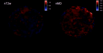 |
Feasibility of dynamic DTI exercise response and resistance dependence in quadriceps muscle
Eric E. Sigmund1, Steven H. Baete1, and Danielle Costanzo1
1Radiology, NYU Langone Health, New York, NY, United States
We describe measurement of resistance dependence diffusion metric exercise response in thigh muscle with a multiple echo diffusion tensor imaging (MEDITI) on a clinical 3 T scanner. With radial imaging, accelerated diffusion encoding, and compressed sensing reconstruction, spatial resolution of 3.4 mm and temporal resolution of 16 s was achieved. Using an MR-compatible ergometer with pneumatic resistance and force/displacement monitoring, post-exercise recovery of DTI metrics in the rectus femoris following quadriceps extension was monitored as a function of resistance. Significant dependences of response on resistance were observed.
|
|
 |
0359.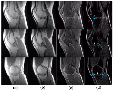 |
3D UTE Cones Double Echo Steady State Imaging – a New Approach for High Resolution Morphological and Quantitative Evaluation of Short T2 Tissues
Hyungseok Jang1, Michael Carl2, Yajun Ma1, Mei Wu1, Zhao Wei1, Saeed Jerban1, Eric Chang1,3, and Jiang Du1
1University of California, San Diego, San Diego, CA, United States, 2GE Healthcare, La Jolla, CA, United States, 3VA San Diego Healthcare System, San Diego, CA, United States
Double echo steady state (DESS) imaging allows acquisition of two MR images with different contrasts from FID and echo images. In this study, we explored the feasibility and efficacy of 3D UTE Cones-based DESS (3D UTE-Cones-DESS) imaging of short T2 tissues in the knee joint. In ex vivo study of four cadaveric knees and in vivo study of three healthy volunteers, the UTE-Cones-DESS sequence provided high contrast imaging of the osteochondral junction (OCJ), the menisci, and other short T2 tissues, as well as T2 maps, under a total scan time of three minutes.
|
0360.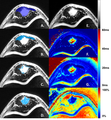 |
Sub-regional Quantification of Tissue-Specific Hydration State in Patellar Tendinopathy with 3D Ultrashort Echo Time MRI
Stephan Breda1, Dirk Poot1, Dorottya Papp1, Gabriel Krestin1, Robert-Jan de Vos2, and Edwin Oei1
1Radiology & Nuclear Medicine, Erasmus University Medical Center, Rotterdam, Netherlands, 2Orthopedics & Sports Medicine, Erasmus University Medical Center, Rotterdam, Netherlands
Patellar tendinopathy (PT) is an overuse injury of the patellar tendon in athletes, often resulting from jumping activities such as playing basketball or volleyball. MR imaging with ultrashort echo times (3D-UTE MRI) is used to image the typical degenerative process of the proximal patellar tendon in PT. However, image analysis can be challenging within the heterogeneous patellar tendon affected by tendinopathy. Therefore, we propose a novel method for image analysis, in which voxels are sub-selected based on a parameter from bi-exponential fitting. This resulted in the identification of T2* biomarkers, specific for distinct tissue-compartments within the patellar tendon.
|
|
 |
0361.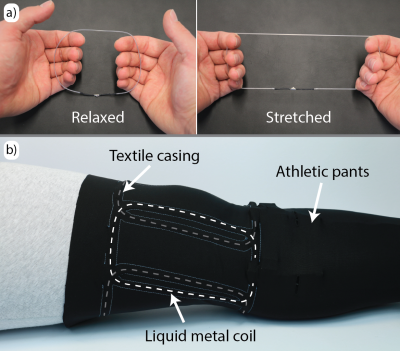 |
Towards kinematic knee imaging with a liquid metal array
Andreas Port1, Roger Luechinger1, David Otto Brunner1, and Klaas Paul Pruessmann1
1Institute for Biomedical Engineering, ETH Zurich and University of Zurich, Zurich, Switzerland
Kinematic MR studies provide functional insights that corresponding static methods may not be able to provide. However, MR signal reception from body parts with large flexion ranges, such as the knee, can be challenging. Wearable RF coils that adapt well to a specific anatomy would offer good sensitivity and patient comfort at the same time. In the present work, we explore the practical utility of a wearable liquid metal coil. For this purpose a MR compatible knee bending setup is used. Static and kinematic imaging of a volunteer’s knee confirm sensitivity and coverage over the whole range of flexion.
|
|
0362.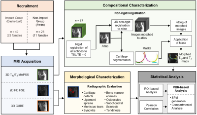 |
Compositional and Morphological Characterization of Knee Articular Cartilage in Collegiate Basketball Players using Multiparametric MRI
Kenneth T. Gao1, Valentina Pedoia1, Radhika Tibrewala1, Katherine A. Young2, Feliks Kogan2, Matthew F. Koff3, Garry E. Gold2, Hollis Potter3, and Sharmila Majumdar1
1Department of Radiology and Biomedical Imaging, University of California, San Francisco, San Francisco, CA, United States, 2Department of Radiology, Stanford University, Stanford, CA, United States, 3Department of Radiology and Imaging, Hospital for Special Surgery, New York, NY, United States
Magnetic resonance imaging (MRI) is commonly used to evaluate the morphology of athletes with high knee impact; however, the biochemical composition of their cartilage is not as well understood. In this study, we utilized voxel-based relaxometry (VBR), a fully automatic registration technique, to compare local distribution of knee articular cartilage T1ρ and T2 relaxation times between high knee impact athletes (basketball players) and non-knee impact athletes (swimmers). Statistical analysis revealed laminar differences near the patella, with basketball players having prolonged values in the deep layer. These findings, amongst others, related well to morphological evaluation of the image set.
|
 Watch the Video