
Combined Educational & Scientific Session
From Microvasculature Dynamics to Functional Signals
| Thursday Parallel 4 Live Q&A | Thursday, 13 August 2020, 14:20 - 15:05 UTC | Moderators: Xin Yu & Luca Vizioli |
Session Number: C-Th-02
Overview
It has been 30 years since the development of functional MRI (fMRI) method in the 1990s. The hemodynamic basis of the fMRI signal has been extensively studied to better interpret the broad basic and clinical applications of the modern fMRI methods. This course will provide an overview and sampling of current work in this area, including the blood flow, blood volume, and oxygen saturation issues related to the neurovascular coupling studies of both animal and human models. In particular, studies on the vessel-specific contribution to the fMRI signal across different scales are covered using methodology with multiple modalities.
Target Audience
Anyone interested in research into the neurovascular origins of the fMRI signal, its dependence on field strength and its applications.
Educational Objectives
As a result of attending this course, participants should be able to:
-Identify the cerebrovascular hemodynamic features related to the fMRI signals; and
-Describe how multimodal experimental methods and modeling schemes are combined to interpret the fMRI signal as an indirect measure of brain function.
1311.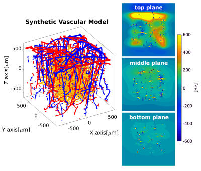 |
Investigation of the dynamic fingerprint of the BOLD fMRI signal based on a novel statistical 3D cortical vascular network of the human brain
Mario G. Báez-Yáñez1, Jeroen Siero1,2, and Natalia Petridou1
1Department of Radiology, Center for Image Sciences, UMC Utrecht, Utrecht, Netherlands, 2Spinoza Centre for Neuroimaging Amsterdam, Royal Netherlands Academy of Arts and Sciences, Amsterdam, Netherlands
In order to quantify the hemodynamic contributions to the BOLD fMRI signal in humans, it is necessary to adopt a computational model that resembles the cortical vasculature and mimics hemodynamic changes triggered by neurovascular coupling. Moreover, simulation of the local magnetic disturbance induced by the geometry, hemodynamic changes, and the biophysical properties of the tissues can provide accurate insights on the physiological fingerprint of the BOLD fMRI signal. In this work, based on a realistic 3D computational approach of the human cortical vasculature, we simulate the biophysical effects produced by hemodynamic changes to compute a dynamic BOLD fMRI signal response.
|
|
| Contrast Mechanisms & Field Strength Dependence
Klaus Scheffler
|
||
 |
Vascular Network & Signal Origins
James Mester, Paolo Bazzigaluppi, Matthew Rozak, Bojana Stefanovic
This lecture will review recent work characterizing neurovascular coupling on the microscopic level and underscore the significance of studying network-level behaviour.
|
|
1101. |
The role of rapid capillary resistance decreases in the BOLD response assessed through simulations in a realistic vascular network
Joerg Peter Pfannmoeller1, Louis Gagnon2, Avery Berman1, and Jonathan Polimeni1
1Imaging, Athinoula A. Martinos Center for Biomedical Imaging, Massachusetts General Hospital, Boston, MA, United States, 2Physics, Engineering Physics and Optics, Laval University, Quebec, QC, Canada The brain’s physiology may fundamentally limit the achievable spatial and temporal specificity of gradient-echo fMRI. Even if the physiology does not pose such a limitation a better understanding would allow for data analysis techniques that improve the spatial specificity. Microscopy allows for highly detailed investigations of local physiological mechanism and provides a growing knowledge from which fMRI may benefit profoundly. A current challenge is the transition from focal mechanisms to their consequence on the mesoscopic scale of BOLD examinations. In this abstract we present our recent work on this transition using simulations of the BOLD effect.
|
|
 |
1102. |
On the relation between positive and negative functional changes of cerebral blood flow and T2* in the human visual cortex.
Ratnamanjuri Devi1, Toralf Mildner1, Torsten Schlumm1, Jöran Lepsien 1, and Harald E. Möller 1
1Max Planck Institute for Human Cognitive and Brain Sciences, Leipzig, Germany Measurement of functional CBF is challenging due to inherently low SNR and signal amplitudes, especially in regions of negative BOLD response. Here, multi-echo center-out EPI is introduced which allows for simultaneous measurement of functional changes in CBF and T2* with improved sensitivity. Using a visual stimulus inducing positive and negative BOLD responses, a linear relationship between absolute changes in CBF and T2* along both positive and negative directions was found with similar coupling ratios. Negative absolute functional CBF changes were found to be almost independent of the baseline CBF, in agreement with previous work on the positive BOLD response. |
1103.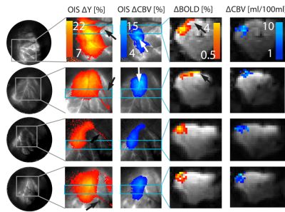 |
Cross species validation of the layer-fMRI VASO contrast mechanism: data comparison against pre-clinical 2D-OIS and CBV-MRI gold standards.
Aneurin J Kennerley1, Benedikt A Poser2, Frida H Torkelsen1, Rainer Goebel2, Amanda Kaas2, and Laurentius Huber2
1Chemistry, University of York, York, United Kingdom, 2Maastricht Brain Imaging Centre, Maastricht University, Maastricht, Netherlands
With recent advances in ultra-high-field MRI hardware and sequence mechanisms, it has become possible to capture CBV-weighted fMRI signal across cortical layers. However, the exact contrast mechanisms of layer-dependent VASO has not been fully validated with gold-standard pre-clinical methods.
|
|
|
1104.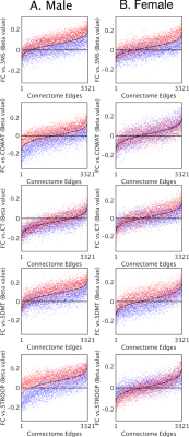 |
Individual differences in haemoglobin concentration influence BOLD fMRI functional connectivity and its correlations with behaviour
Phillip G D Ward1,2,3, Edwina R Orchard1,2,3, Stuart Oldham3, Aurina Arnatkevičiūtė3, Francesco Sforazzini1, Alex Fornito3, Gary F Egan1,2,3, and Sharna D Jamadar1,2,3
1Monash Biomedical Imaging, Monash University, Melbourne, Australia, 2Australian Research Council Centre of Excellence for Integrative Brain Function, Melbourne, Australia, 3Turner Institute for Brain and Mental Health, Monash University, Melbourne, Australia
The BOLD signal detects changes in relative concentrations of oxy/deoxy-haemoglobin. Thus, individual blood haemoglobin levels may influence the BOLD signal-to-noise ratio in a manner independent of neural activity. In this study, we emulate group-differences in haemoglobin by performing a median split on 524 healthy elderly individuals based on individual measurements of haemoglobin. When compared, the two haemoglobin subgroups showed no differences in cognitive measures, however, significant differences in linear relationships between cognitive performance and functional connectivity were observed in four cognitive tests. Our findings confirm that haemoglobin levels are an important confounding variable in BOLD-fMRI-based studies in the elderly.
|
 Watch the Video