
Digital Poster Session
Neuro: Brain tumors
Neuro
1691 -1702 Brain tumors - Brain Tumour
1703 -1716 Brain tumors - Brain Tumour: Perfusion & Permeability Imaging
1717 -1731 Brain tumors - Brain Tumour: Radiomics
1732 -1746 Brain tumors - Brain Tumour: Diffusion Imaging
1691.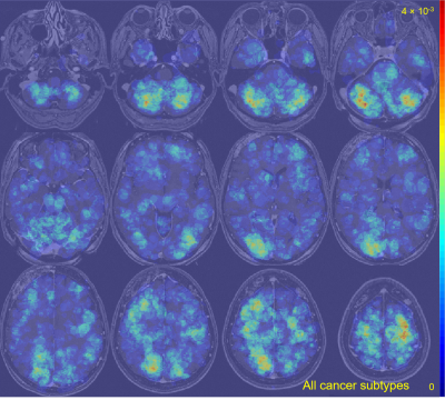 |
An anatomic atlas of brain metastases: probability maps based on 13 primary cancer subtypes from post-contrast T1-weighted MRI of 955 patients
Jeremiah W Sanders1, Jason M Johnson2, Henry Szu-Meng Chen1, Melissa Chen2, Maria Gule-Monroe2, Zijian Zhou1, Tina M Briere3, Jing Li4, Jingfei Ma1, and Ho-Ling Liu1
1Imaging Physics, University of Texas MD Anderson Cancer Center, Houston, TX, United States, 2Neuroradiology, University of Texas MD Anderson Cancer Center, Houston, TX, United States, 3Radiation Physics, University of Texas MD Anderson Cancer Center, Houston, TX, United States, 4Radiation Oncology, University of Texas MD Anderson Cancer Center, Houston, TX, United States Poster Permission Withheld
Metastasis is regarded as a highly inefficient process in that less than 0.01% of circulating tumor cells eventually succeeds in forming secondary tumor growths. The seed and soil theory suggests that certain tumor cells have a specific affinity for the milieu of certain organs. Numerous studies have attempted to better understand the biology behind the distribution of brain metastasis and theories include both variables involving the metastasis and the target brain parenchyma. This work establishes probabilistic maps of locations where 13 primary cancer subtypes metastasize to the brain from T1-weighted MRIs of 955 patients undergoing stereotactic radiosurgery.
|
|
1692.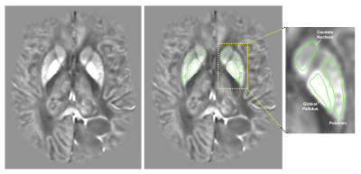 |
Correlation of glioma grade with basal ganglia iron content using QSMnet+
Thomas Reith1, Eun-Jung Choi2, Jongho Lee2, Melissa Prah1, Robert Wujek1,3, Mona Al-Gizawiy1, Christopher Chitambar4, and Kathleen Schmainda1
1Biophysics, Medical College of Wisconsin, Milwaukee, WI, United States, 2Electrical and Computer Engineering, Seoul National University, Seoul, Korea, Republic of, 3Biomedical Engineering, Medical College of Wisconsin, Milwaukee, WI, United States, 4Hematology & Oncology, Medical College of Wisconsin, Milwaukee, WI, United States
Basal ganglia iron levels were assessed in 55 patients with gliomas by using a deep neural network-powered quantitative susceptibility mapping method (QSMnet+). Basal ganglia QSM values increased with higher tumor grade, suggesting a correlation between BG iron content and glioma severity.
|
|
1693.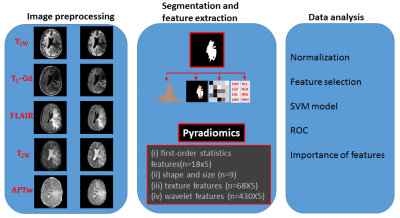 |
Radiomics features from APTw MRI improve the diagnostic performance of structural MRI in early response assessment of malignant gliomas
Shanshan Jiang1, Pengfei Guo2, Hye-Young Heo1, John Laterra3, Charles Eberhart4, Michael Lim5, Peter C.M. van Zijl1,6, and Jinyuan Zhou1
1Radiology, Johns Hopkins University, Baltimore, MD, United States, 2Computer Science, Johns Hopkins University, Baltimore, MD, United States, 3Neurology, Johns Hopkins University, Baltimore, MD, United States, 4Pathology, Johns Hopkins University, Baltimore, MD, United States, 5Neurosurgery, Johns Hopkins University, Baltimore, MD, United States, 6F.M. Kirby Research Center, Kennedy Krieger Institute, Baltimore, MD, United States
Assessment of glioma treatment is based on pathological evaluation via biopsies or radiological criteria using follow-up MRI, which is either invasive or time consuming. Amide protein transfer weighted (APTw) MRI has been validated to accurately detect recurrent malignant gliomas by multiple studies. The cutting edge methodology of radiomics provides quantitative measurements for imaging diagnosis. Here, we develop an automated framework that integrates APTw MRI radiomic features with a machine learning model to evaluate treatment response for gliomas. Our results suggest that the use of APTw features enabled the radiomic model to reach a more accurate assessment of the treatment effect.
|
|
1694.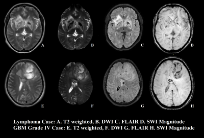 |
Differentiating lymphoma from grade-IV glioma using susceptibility weighted imaging based texture feature at 3T MRI
Rupsa Bhattacharjee1,2, Rakesh Kumar Gupta3, Rana Patir4, Sandeep Vaishya4, Sunita Ahlawat4, and Anup Singh1,5
1Center for Biomedical Engineering, Indian Institute of Technology Delhi, New Delhi, India, 2Philips Health Systems, Philips India Limited, Gurugram, India, 3Department of Radiology, Fortis Memorial Research Institute, Gurugram, India, 4Department of Neurosurgery, Fortis Memorial research Institute, Gurugram, India, 5Department of Biomedical Engineering, All India Institute of Medical Sciences, New Delhi, India
No standalone method is yet reported to differentiate PCNSL and grade-IV glioma with highest accuracy and diagnostic confidence. Imaging based differentiation between these two types has been a challenging problem. Objective of this study is to evaluate performance of texture-based features of SWI in improved differentiation of PCNSL from grade-IV gliomas. Proposed approach based upon texture feature-extraction from segmented SWI lesion, enabled automatic classification of tumors into primary central nervous system lymphoma and grade-IV glioma cases. One of the texture feature, Contrast, provided highest AUC along with high sensitivity and specificity. This classification might improve diagnosis and grading of tumors.
|
|
1695.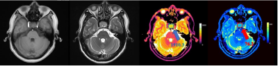 |
MRF for preoperative differentiation of non-functioning gonadotroph adenoma and non-functioning non-gonadotropin adenoma
Rushi Chen1, Yan Bai2, Mathias Nittka3, Gregor Koerzdoerfer3, Xianchang Zhang4, and Meiyun Wang2
1Henan provincial people's hospital& Zhengzhou University People’s Hospital, Zhengzhou, China, 2Henan provincial people's hospital, Zhengzhou, China, 3MR Pre-development, Siemens Healthcare, Erlangen, Germany, 4MR Collaboration, Siemens Healthcare Ltd, Beijing, China
Currently, there is no effective pharmacological treatment for non-functioning pituitary adenoma, including gonadotroph adenoma (GPA). Recent findings showed that an alternative pharmacological treatment targeted at somatostatin receptor 3 may be promising, which required accurate diagnosis of GPA before surgery. This study aimed to evaluate the utility of magnetic resonance fingerprinting (MRF) in the pre-surgical differentiation of GPA from non-functioning, non-gonadotropin adenoma (NGPA). The results showed that GPAs have significantly higher T1 and T2 values in the solid tumor than NGPA, suggesting that MRF may have potential for diagnosing GPA and benefit its treatment plan.
|
|
1696.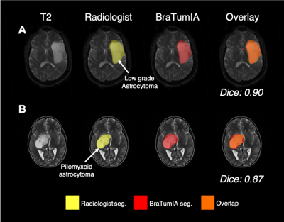 |
Assessment of Automated Brain Tumour Segmentation Tools for Clinical Data
Reneira Seeamber1, Katherine L Ordidge2,3, Felice d’Arco4, Kshitij Mankad4, Tara D Barwick2,3, Adam D Waldman5,6, Patrick W Hales7, and Matthew Grech-Sollars2,3
1Department of Computing, Imperial College London, London, United Kingdom, 2Department of Surgery and Cancer, Imperial College London, London, United Kingdom, 3Department of Imaging, Imperial College Healthcare NHS Trust UK, London, United Kingdom, 4Great Ormond Street Children’s Hospital, London, United Kingdom, 5Department of Medicine, Imperial College London, London, United Kingdom, 6Centre for Clinical Brain Sciences, The University of Edinburgh, Edinburgh, United Kingdom, 7Department of Developmental Neurosciences, UCL Great Ormond Street Institute of Child Health, London, United Kingdom
Radiological diagnosis of certain brain tumours remains poor. The development of automated tools that can extract valuable, yet currently underused, information from routine MRI acquisitions have been explored. The present study aimed to evaluate the accuracy of three automatic tumour segmentation programmes using the BRATS validation 2018 glioma dataset (n=66). The performance of BraTumIA was assessed across an adult glioma dataset from Imperial College Healthcare NHS Trust (n=13) and a paediatric brain tumour dataset from Great Ormond Street Hospital NHS Foundation Trust. DeepMedic segments adult gliomas more accurately than BraTumIA and ONCOhabitats. However, BraTumIA provides a more user-friendly segmentation tool.
|
|
1697.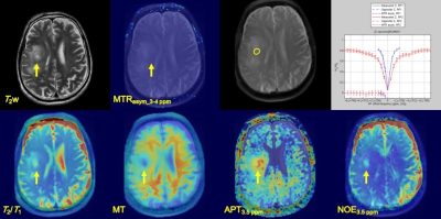 |
Characterization of Brain Tumors using Amide Proton and Nuclear Overhauser Effect at 3 Tesla MR Scanner.
Yuki Kanazawa1, Masafumi Harada1, Mitsuharu Miyoshi2, Takashi Abe1, Yuki Matsumoto1, and Yasushi Takagi3
1Tokushima University, Tokushima, Japan, 2Global MR Applications and Workflow, GE Healthcare Japan, Hino, Japan, 3Department of Neurosurgery, Tokushima University, Tokushima, Japan
To achieve detailed characterization of brain tumors, we demonstrated CEST imaging with multi-pool model [bulk water, magnetization transfer (MT), amide proton transfer (APT), and nuclear Overhauser effect (NOE)] derive from the Bloch equation. This study was performed using five patients with brain tumors. As a result, there was high linearity between MTRasym and T2/T1 in each tumor (R2 = 0.91, P<0.05). Moreover, there were significantly different NOE signals for tumors and normal white matter (P<0.05). Detection using APT and NOE make it possible to provide more detailed information of brain tumors than MTRasym analysis.
|
|
1698.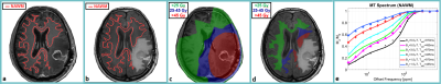 |
Effects of Chemo-Radiation on Normal Appearing White Matter in GBM patients
Hatef Mehrabian1, Mark Ruschin2, Sten Myrehaug3,4, Hany Soliman1,3,4, Arjun Sahgal1,3,4, and Greg J Stanisz1,5
1Physical Sciences, Sunnybrook Research Institute, Toronto, ON, Canada, 2Medical Physics, Sunnybrook Health Sciences Centre, Toronto, ON, Canada, 3Radiation Oncology, Sunnybrook Health Sciences Centre, Toronto, ON, Canada, 4Radiation Oncology, University of Toronto, Toronto, ON, Canada, 5Medical Biophysics, University of Toronto, Toronto, ON, Canada
Chemo-radiation treatment for GBM damages normal brain tissues and leads to significant deterioration in patients’ quality of life months or years after treatment. Magnetization transfer (MT) is sensitive to white matter damage and may detect it early. We observed significant reduction in MT exchange rate (RM0B) that quantifies white matters integrity as early as one month after 6-week chemo-radiation. The treatment impact on normal brain tissue was observed even in regions that received low radiation dose. More importantly, not all patients experienced MT reduction showing the heterogeneity of response to treatment and the potential of MT in identifying vulnerable patients.
|
|
1699.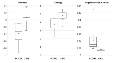 |
Differential diagnosis of primary CNS lymphoma and glioblastoma using statistics derived from commercial software
Tatsuya Yamamoto1, Kuniyoshi Hayashi2, and Hirohiko Kimura3
1Department of Diagnostic Radiology, The Cancer Institute Hospital of the Japanese Foundation for Cancer Research, Tokyo, Japan, 2Division of Biostatistics and Bioinformatics, Graduate School of Public Health, St. Luke’s International University, Tokyo, Japan, 3Department of Radiology, Faculty of Medical Sciences, University of Fukui, Fukui, Japan Poster Permission Withheld
Primary central nervous system lymphomas (PCNSLs) are sometimes difficult to distinguish from glioblastomas (GBMs) based on routine magnetic resonance examination. This study assessed the utility of histogram analysis of intratumoral contrast-enhanced region using contrast-enhanced T1WI (CET1WI) with a 3D-spoiled gradient recalled acquisition in the steady state sequence for PCNSLs and GBMs to determine whether histogram statistics differed between the two tumors using the commercial software. There were significant differences in skewness, entropy, and angular second moment between PCNSLs and GBMs. This suggests the possibility of the differential diagnosis of PCNSLs and GBMs using histogram analysis of CET1WI.
|
|
1700.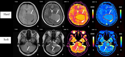 |
Magnetic Resonance Fingerprinting for Preoperative Differentiation of Soft and Hard Meningiomas
Rui Zhang1, Yan Bai1, Xianchang Zhang2, Qin Feng1, Mengke Wang1, Menghuan Zhang1, Mathias Nittka3, Gregor Koerzdoerfer3, Yusong Lin4, and Meiyun Wang1
1Department of Medical Imaging, Henan Provincial People’s Hospital & Zhengzhou University People’s Hospital;Henan Key Laboratory of Neurological Imaging, Zhengzhou, China, 2MR Collaboration, Siemens Healthcare Ltd., Beijing, China, 3MR Pre-development, Siemens Healthcare, Erlangen, Germany, 4Cooperative Innovation Center of Internet Healthcare & School of Software and Applied Technology, Zhengzhou University, Zhengzhou, China
Tumor consistency is an important factor in determining the resectability and surgical outcome of meningiomas. This study investigated the use of magnetic resonance fingerprinting (MRF) to distinguish between meningiomas characterized as soft or hard based on intraoperative patient records. Mann-Whitney U tests were used to compare longitudinal relaxation time (T1) and transverse relaxation time (T2) values derived from MRF. Soft meningiomas had significantly higher T1 and T2 values than hard meningiomas. Both T1 and T2 showed good diagnostic performance, suggesting that MRF may provide valuable information for preoperative differentiation of meningioma consistency and help guide the treatment plan.
|
|
1701. |
Application of Half-Dose Contrast-Enhanced T2-Fluid-Attenuated Inversion Recovery (T2 FLAIR) Sequence in Brain Metastasis
Teng Jin1, Jing Wang1, and Zhenwei Yao2
1Radiology, Union Hospital, Tongji Medical College, Huazhong University of Science and Technology, Wuhan, China, 2Department of Radiology, Huashan Hospital, Fudan University, Shanghai, China
For reducing the gadolinium dose of patients with metastasis, we seek a valuable tool to lower contrast agent doses but keep MR image quality, which is important for patients’ healthcare. We analyzed the optimum scanning time for 1/2 dose CE-T2FLAIR, and compared the CR value of metastases and enhancement degree among the 1/2 dose CE-T2 FLAIR, 1/2 dose CE-T1WI and full dose CE-BRAVO sequence. Our team further analyzed the relationship between enhancement pattern, lesion size and image quality. Our study not only reduced the harm from GBCA administrations but also improved detection rate of brain metastases.
|
|
1702.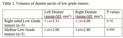 |
SWI changes in the dentate nucleus in high and low-grade brain stem neoplasms
Kamlesh Jobanputra1, Bhaswati Roy2, Karuna Raj1, Amit Agarwal1, and Frank Yu1
1Radiology, University of Texas Southwestern Medical Center, Dallas, TX, United States, 2Anesthesiology, University of California at Los Angeles, Los Angeles, CA, United States
An investigation that stemmed from observation that there were dentate volume changes on SWI in low grade tumor of the brainstem showed significant decrease in signal intensity in the ipsilateral dentate nucleus in high grade neoplasms and decrease in ipsilateral dentate volumes in low grade neoplasms. We think that dentate iron deposition and its imaging is yet an unexplored field, especially in brain tumor imaging. Further evaluation with QSM and STI with histopathological correlation may also help explore pathways of iron homeostasis.
|
 Watch the Video
 View the Poster
1703.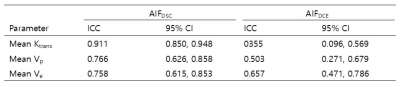 |
Differentiation of glioblastoma and lymphoma: Improvement in Accuracy and Reliability of DCE Parameters by Using AIF Obtained from DSC-MRI
Koung Mi Kang1, Seung Hong Choi1, Inpyeong Hwang1, Roh-Eul Yoo1, Tae Jin Yun1, Ji-hoon Kim1, and Chul-Ho Sohn1
1Seoul National University Hospital, Seoul, Republic of Korea
We investigated that whether arterial input functions (AIFs) obtained from Dynamic susceptibility-contrast (DSC)-MRI, or AIFDSC values, improved reliability and diagnostic accuracy of DCE parameters, for differentiating glioblastoma (GBM) from primary CNS Lymphoma (PCNSL), compared with AIFs derived from DCE-MRI (AIFDCE). This study demonstrated that AIFDSC –driven DCE parameters had better diagnostic accuracy and reliability for the differential diagnosis than those from AIFDCE. In addition, the AIFDSC-driven mean Ktrans showed better accuracy than the other DCE parameters and rCBV in the discrimination of GBM without marked vascularity from PCNSL.
|
|
1704. |
Quantitative analysis of MR perfusion imaging in diagnosis and differential diagnosis of alveolar echinococcosis and brain metastases
Jian Wang1, Xin Gao1, Chunhui Jiang1, and Jundi Liu1
1The First Affiliated Hospital of Xinjiang Medical University, Urumqi, China, Xinjiang, China
Due to similar appearance in conventional MR images, it is often difficult to differentiate cerebral vesicular echinococcosis (CAE) from brain metastases (BM). In this study, user perfusion weighted imaging technique, perfusion characteristics of these 2 diseases were systematically investigated. Our results indicated that significant difference of semi-quantitative parameters relative cerebral blood flow (rCBF) and relative cerebral blood volume (rCBV) from PWI between CAE and BM. Hence PWI can provide valuable additional information in differential disagnosis of alveolar echinococcosis and brain metastases.
|
|
1705. |
Response prediction of the vestibular schwannoma after gamma-knife radiosurgery using DCE-MRI: preliminary results after 1-year follow-up.
Inpyeong Hwang1, Seung Hong Choi1, Jin Wook Kim2, Roh-Eul Yoo1, Koung Mi Kang1, Tae Jin Yun1, Ji-hoon Kim1, and Chul-ho Sohn1
1Department of Radiology, Seoul National University Hospital, Seoul, Korea, Republic of, 2Department of Neurosurgery, Seoul National University Hospital, Seoul, Korea, Republic of
There are few known predictive factors for response to gamma-knife radiosurgery (GKRS) in vestibular schwannoma (VS). This study investigated the relationship between DCE-MR derived pharmacokinetic parameters and tumor volume change at one year after GKRS follow-up in VS. This prospective study performed a volumetric measurement of DCE-MR pharmacokinetic parameters before GKRS. The patients underwent followed-up MR at post one year, tumor volume was measured, and pharmacokinetic parameters were compared between tumor growth group and stable group. Only the Ktrans value showed statistical differences. Our results suggest that Ktrans value has the potential to predict tumor response in VS after GKRS.
|
|
1706. |
Pixel-wise and regional comparison of a low flip angle, single-dose versus moderate flip angle, double-dose DSC-MRI protocol
Laura C. Bell1, Leland S. Hu2, Yuxiang Zhou2, Kathleen M. Schmainda3, Jerrold L. Boxerman4, and C. Chad Quarles1
1Barrow Neurological Institute, Phoenix, AZ, United States, 2Mayo Clinic, Scottsdale, AZ, United States, 3Medical College of Wisconsin53226, Milwaukee, WI, United States, 4Rhode Island Hospital and Alpert Medical School of Brown University, Providence, RI, United States
Current recommendations for brain tumor dynamic susceptibility contrast MRI include the use of a moderate flip angle (MFA) and a full standard preload dose.1 Recently, simulations2 identified an equally accurate protocol that requires no preload and uses a low flip angle (LFA) and optimized echo time. This was validated in vivo3 by comparing LFA- and MFA-based mean tumor cerebral blood volume. Here we aim to systematically characterize agreement by evaluating the pixel-wise agreement in enhancing tumor, edema, white matter, and gray matter regions. We found high agreement in all regions further strengthening the clinical utility of the LFA protocol.
|
|
1707.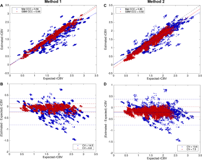 |
Analysis of Accuracy and Precision of Recommended Protocols for Dynamic Susceptibility Contrast MRI for Brain Metastases
Natenael B Semmineh1, Usha Bhalla2, Laura C Bell1, Ashley M Stokes1, Matthew D Lee3, Leland Hu4, Jerrold L Boxerman3,5, and C Chad Quarles1
1Division of Neuroimaging Research, Barrow Neurological Institute, Phoenix, AZ, United States, 2Brown University, Providence, RI, United States, 3Alpert Medical School - Brown University, Providence, RI, United States, 4Department of Radiology, Mayo Clinic Arizona, phoenix, AZ, United States, 5Department of Diagnostic Imaging, Rhode Island Hospital, Providence, RI, United States
Numerous methods have been proposed to overcome the confounding influence of contrast agent leakage on DSC-MRI derived CBV maps in brain tumors. Recently, we leveraged a digital reference object to identify the optimal acquisition and analysis methods for GBM patients, a protocol that is now being recommended as the standardized approach for all clinical scans. It is unknown if the biological differences between GBM and metastases would confound the use of this protocol in patients with brain metastases. In this study we characterize the influence of this GBM-centric protocol resulted in lower metastases CBV accuracy and precision.
|
|
1708.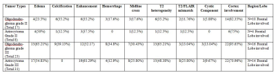 |
Comparative evaluation of Oligodendroglioma and Astrocytoma subtypes with similar grade using Conventional MRI and T1 perfusion MRI imaging
Mamta Gupta1, Abhinav Gupta1, Anup Singh2, Jitender Saini3, Rana Patir4, Sunita Ahlawat5, Vani Santosh3, Neha Vats2, Manish Awasthi2, Suhail Parvaze6, and Rakesh Kumar Gupta1
1Department of Radiology, Fortis Memorial Research Institute, Gurgaon, India, 2Centre for Biomedical Engineering, Indian Institute of Technology, New Delhi, India, 3National Institute of Mental, Health and Neurosciences, Bangalore, India, 4Department of Neurosurgery, Fortis Memorial Research Institute, Gurgaon, India, 5SRL Diagnostics, Fortis Remorial Research Institute, Gurgaon, India, 6Philips Innovation Campus, Bangalore, India
The differentiation of oligodendroglioma and astrocytoma has become increasingly important due to the distinct sensitivity of oligodendroglioma (OD) to chemotherapy and prolonged survival compared to astrocytoma (AT) of similar grades. The purpose of the present study was to determine whether unique characteristics of OD and AT are visible on MR imaging and to assess the added value of perfusion imaging in differentiating OD from AT tumors across similar grades in large study population. Our results demonstrated no characteristic/specific conventional MRI features and perfusion parameters those could clearly differentiate similar grades of OD’s and AT tumors.
|
|
1709.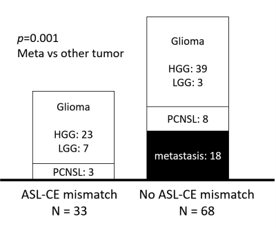 |
"ASL-CE mismatch": An Imaging Finding for Diagnosis of Brain Tumor, Multicenter Research
Takashi Abe1, Maki Otomo1, Mihoko Kondo1, Yuta Arai1, Yoichi Otomi1, Yuki Matsumoto2, Yuki Kanazawa2, Kohei Nakajima3, Yoshifumi Mizobuchi3, Yasushi Takagi3, and Masafumi Harada2
1Radiology and Radiation Oncology, Tokushima University Hospital, Tokushima, Japan, 2Tokushima University, Tokushima, Japan, 3Neurosergery, Tokushima University Hospital, Tokushima, Japan
One hundred one cases with brain tumor were included (62: HGG, 10: LGG, 18: metastasis, 11: lymphoma), and divided into two groups by the presence or absence of ASL hyperintensity outside of CE area (ASL-CE mismatch). Only the cases with glioma or lymphoma (23 HGG, 7 LGG, 3 lymphoma) revealed ASL-CE mismatch, and the cases with metastasis didn't (p = 0.001). Similar results were obtained in a sub-analysis where data were divided by MRI vendor. This novel finding may reflect tumor invasion and/or vascular growth in the surrounding brain parenchyma, and is useful for identifying metastasis from the other tumors.
|
|
1710.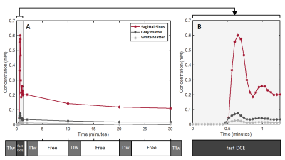 |
Time-efficient method for detecting subtle blood-brain barrier leakage – a clinical feasibility study
Paulien H.M. Voorter1,2, Marieke van den Kerkhof2,3, Daniëlle B.P. Eekers4, Richard A.M. Canters4, Wouter van Elmpt4, Joost J. de Jong2,3, Lisanne P.W. Canjels2,3,5, Jacobus F.A. Jansen2,3,5, Alida A. Postma2, and Walter H. Backes2,3
1Department of Biomedical Engineering, Eindhoven University of Technology, Eindhoven, Netherlands, 2Department of Radiology and Nuclear Medicine, Maastricht University Medical Center, Maastricht, Netherlands, 3School for Mental Health and Neuroscience, Maastricht University, Maastricht, Netherlands, 4Department of Radiation Oncology (MAASTRO), GROW – School for Oncology and Developmental Biology, Maastricht University Medical Center, Maastricht, Netherlands, 5Department of Electrical Engineering, Eindhoven University of Technology, Eindhoven, Netherlands
Blood-brain barrier (BBB) disruption underlies the origin of many brain disorders. Measuring subtle BBB leakage of gadolinium-based contrast agents is commonly performed by a continuous and time-consuming dynamic MRI protocol. However, BBB leakage is subtle, for which discrete sampling at strategic time-points might be sufficient. This study explores the feasibility of the time-efficient interleaved protocol by applying it to brain tumor patients. We were able to measure high permeability in a craniopharyngioma and low permeability in a low-grade tumor and in healthy tissue. The time-efficient protocol is promising and will be further evaluated in more patients.
|
|
1711.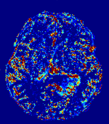 |
Simultaneous Fast and Slow DCE: temporal multiresolution and spatial high resolution T1 dynamics of the whole brain from a single continuous scan
Ingomar Gutmann1, Julia Furtner2, Gregor Kasprian2, Gerhard Krexner3, Daniela Prayer2, and Patrick Thurner2
1University of Vienna, Vienna, Austria, 2Medical University Vienna, Vienna, Austria, 3University of Vienna, Wien, Austria
We present DCE acquisition with high spatial resolution that excels the guidelines of the ACR for spatial resolution, maintaining whole brain coverage at all times, while at the same time providing a temporal sampling rate of fast enough to make accurate flow and permeability measurements from T1-weighted DCE MRI in a clinical neuro-oncology setting possible.Furthermore we demonstrate the use of radial spoke aggregation in GRASP reconstruction for DCE perfusion imaging of the brain and show preliminary results on the impact of variable temporal resolution on estimating pharmacokinetic models.
|
|
1712.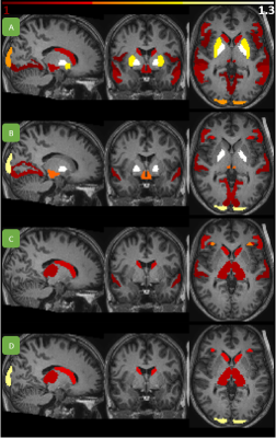 |
Increased cerebral blood volume in cortical and subcortical brain regions in Glioblastoma patients after EPI distortion correction
Ivar Thokle Hovden1, Oliver M. Geier1, Ingrid Digernes1, Elies Fuster-Garcia1, Grethe Løvland2, Einar Vik-Mo3, Torstein R. Meling3,4, and Kyrre E. Emblem1
1Department of Diagnostic Physics, Oslo University Hospital, Oslo, Norway, 2The Intervention Centre, Oslo University Hospital, Oslo, Norway, 3Department of Neurosurgery, Oslo University Hospital, Oslo, Norway, 4Department of Neurosurgery, Geneva University Hospitals, Geneva, Switzerland
We studied the changes in rCBV introduced by echo-planar imaging (EPI) distortion correction methods when applied to EPI dynamic susceptibility contrast (DSC)-MRI. The results obtained using FSL TOPUP and EPIC distortion correction methods indicate that both subcortical and cortical regions are affected and returning an overall increase in rCBV. In the context of longitudinal EPI-based analysis in glioblastoma patients, EPI distortions and subsequent corrections are important determiners for assessing robust responses of rCBV change.
|
|
1713.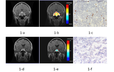 |
Preoperative Proliferation Prediction of Pituitary Macroadenoma Based on Radiomics Analysis on DCE-MRI
Yangyingqiu Liu1, Yanwei Miao1, Ailian Liu1, Qingwei Song1, and Lizhi Xie2
1Department of Radiology, First Affiliated Hospital of Dalian Medical University, Dalian, China, 2GE healthcare, Beijing, China
Proliferative states of pituitary macroadenomas is assessed by radiomics analysis on Dynamic Contrast Enhanced MRI(DCE-MRI). The results show that DCE-MRI could be used as a new noninvasive method for identification the proliferative states of pituitary macroadenomas.
|
|
1714.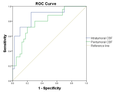 |
3D arterial spin labeling (3D-ASL) MR perfusion imaging application in differentiation primary CNS lymphoma from metastatic brain tumors
zongqiong sun1, weiqiang dou2, and shudong hu1
1MR, Affiliated hospital of Jiangnan university, Wuxi, China, 2MR research, GE healthcare,MR research China, Beijing, China
CBF values of PCNSL are significant lower than those of MBT, and this is helpful to differentiate the two tumors by using 3D ASL MR perfusion imaging from quantitative parameters analyses.
|
|
1715.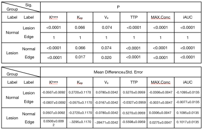 |
Value of quantitative dynamic contrast-enhanced MR imaging in analyzing microvascular permeability
Li Xiang1, Lihua Sun1, Longsheng Wang1, and Yaqiong Ge2
1Radiology, The second affiliated hospital of Anhui Medical University, Hefei, China, 2GE Healthcare, Shanghai, China
DCE-MRI was used to conduct quantitative analysis of microvascular permeability in three different areas of meningioma: Lesion, Edge and Normal. 37 patients with pathologically confirmed meningioma were enrolled, while quantitative parameters were measured. The parameters of ktrans, TTP, MAX. Conc, iAUC in Lesion group and Edge group were statistically significant, and the parameters of Ktrans, kep, Ve, TTP, Max.Conc as well as AUC in Lesion group and Normal group, were statistically significance. The ktrans parameter has the highest AUC value, which means the better performance for permeability study of WHO with different grades of meningiomas and invasive meningiomas.
|
|
1716.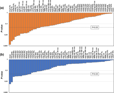 |
Histogram-based CBF quantification allows prediction of histopathologic grade and molecular markers in de novo brain gliomas
David Lu1, Jay J. Pillai1,2, Hanzhang Lu1, and Yang Li1
1Department of Radiology, Johns Hopkins University School of Medicine, Baltimore, MD, United States, 2Department of Neurosurgery, Johns Hopkins University School of Medicine, Baltimore, MD, United States
We developed a histogram-based CBF analysis method for the prediction of tumor histopathologic grade and molecular marker (including IDH1 mutation, ATRX loss, p53 mutation, MGMT methylation, and 1p/19q co-deletion) in a group of de novo brain glioma patients. ASL MRI may be a non-invasive and cost-effective approach to assist in brain tumor diagnosis and prognosis.
|
| 1717. | Correlation of MRI signal characteristics of intracranial melanoma metastases with BRAF mutation status
Arian Lasocki1,2 and Grant McArthur2,3
1Cancer Imaging, Peter MacCallum Cancer Centre, Melbourne, Australia, 2The Sir Peter MacCallum Department of Oncology, The University of Melbourne, Parkville, Australia, 3Medical Oncology, Peter MacCallum Cancer Centre, Melbourne, Australia
In small intracranial melanoma metastases, MRI evidence of melanin was more common in BRAFV600-mutant patients than BRAF-wildtype, supporting that melanin content is greater in not just the primary, but also in the distant metastases. Haemorrhage and central necrosis were more common in larger metastases. Central necrosis was also more common in BRAF-mutant patients who had not previously received anti-BRAF therapy, suggesting that metastases in BRAF-mutant patients have a tendency to central necrosis, though this is modified by anti-BRAF therapy, thus resulting in more solid metastases.
|
|
1718.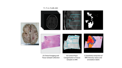 |
MRI signatures associated with pathologically relevant histological features of brain cancer at autopsy
Samuel Bobholz1, Allison Lowman2, Alexander Barrington3, Michael Brehler2, Sean McGarry1, Jennifer Connelly4, Elizabeth Cochran5, Anjishnu Banerjee6, and Peter LaViolette2,3
1Biophysics, Medical College of Wisconsin, Wauwatosa, WI, United States, 2Radiology, Medical College of Wisconsin, Wauwatosa, WI, United States, 3Biomedical Engineering, Medical College of Wisconsin, Wauwatosa, WI, United States, 4Neurology, Medical College of Wisconsin, Wauwatosa, WI, United States, 5Pathology, Medical College of Wisconsin, Wauwatosa, WI, United States, 6Biostatistics, Medical College of Wisconsin, Wauwatosa, WI, United States
This study sought to assess the ability for voxel-wise MRI intensity values to distinguish between co-registered pathological annotations of autopsy tissue samples from brain cancer patients. Though single image and pairwise image assessments did not reveal separable intensity distributions for the pathological annotation classes, ensemble-based predictive modelling using multiparametric MRI intensities proved able to predict pathological annotations with modest accuracy. These results suggest a complex relationship between MRI values and pathological features that are most accurately assessed in terms of multiple MR imaging modalities.
|
|
1719.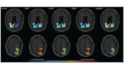 |
Comparison of radiomics-based and deep learning techniques for predicting nuclei density in brain cancer patients from MRI
Samuel Bobholz1, Allison Lowman2, Alexander Barrington3, Michael Brehler2, Jennifer Connelly4, Elizabeth Cochran5, Anjishnu Banerjee6, and Peter LaViolette2,3
1Biophysics, Medical College of Wisconsin, Wauwatosa, WI, United States, 2Radiology, Medical College of Wisconsin, Wauwatosa, WI, United States, 3Biomedical Engineering, Medical College of Wisconsin, Wauwatosa, WI, United States, 4Neurology, Medical College of Wisconsin, Wauwatosa, WI, United States, 5Pathology, Medical College of Wisconsin, Wauwatosa, WI, United States, 6Biostatistics, Medical College of Wisconsin, Wauwatosa, WI, United States
This study sought to compare localized predictions of cellular density via radiomics-based and neural network-based modeling, using co-registered autopsy tissue samples from 16 brain cancer patients as ground truth. We found that radiomics models tended to slightly outperform the neural networks, despite evidence of overfitting in all radiomics-based models and an Alexnet-based transfer learning model. These results suggest that radiomics models tend to perform at least as well as neural network when applied to this dataset, but the propensity of these models for overfitting highlights further needs to be addressed with modelling on larger data sets.
|
|
1720.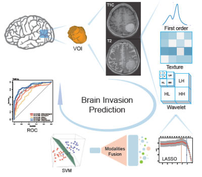 |
A radiomics model for preoperative prediction of brain invasion in meningioma based on MRI
jing Zhang1, kuan Yao2, Zhenyu Liu3, Junlin Zhou1, Guojing Zhang4, and Yuntai Cao1
1Radiology, Lanzhou University Second Hospital, Lanzhou, China, 2School of Biomedical Engineering, Shanghai Jiao Tong University, Shanghai, Shanghai, China, 3CAS Key Laboratory of Molecular Imaging, Institute of Automation, Beijing, Beijing, China, 4Lanzhou University Second Hospital, Lanzhou, China
Objectives Using a radiomics method to predict brain invasion by meningioma. Methods 1595 quantitative imaging features were extracted. LASSO was performed to select features. SVM was used to fit the predictive model. Furthermore, a nomogram was constructed, and validated using decision curve analysis (DCA). Results 8 features were significantly correlated with brain invasion. The radiomics model derived from the fusing MRI sequences resulted in the best discrimination ability, with AUC of0.855(95%CI, 0.829-0.882), sensitivity of 80.32% (95%CI, 75.56%-85.25%). Conclusions The radiomics model developed in this study provided a new non-invasive way to facilitate the preoperative prediction of brain invasion in meningioma.
|
|
1721.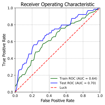 |
Radiomics features of magnetic resonance images as novel predictive factors of bone invasion in meningiomas
Jing Zhang1, Jianqing Sun2, Guojing Zhang1, Yuntai Cao1, and Junlin Zhou1
1Radiology, Lanzhou University Second Hospital, Lanzhou, China, 2Philips Healthcare, Shanghai, Shanghai, China
Objectives Radiomics method was used to predict bone invasion. Methods 1227 quantitative imaging features were extracted. Recursive Feature Elimination (RFE) was performed to select the most informative features. Ridge Classifier was chosen to predict model. Results The AUCof the radiomics model derived from CET1WI and T2WI sequence were0.72,0.72and 0.72,0.64 in the training and test datasets , respectively, and combined CET1WI and T2WI sequences were 0.73and 0.72 when predict bone invasion. Conclusions The radiomics model developed in this study may aid neurosurgeons in the pre-operative prediction of bone invasion by meningiomas ,which can contribute to make clinical strategies and predict prognosis.
|
|
1722.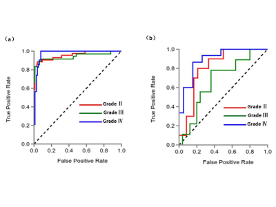 |
MRI-based radiomics analysis for differentiating subtypes of glioma
Jing Zhang1, Yanghong Ou1, and Shuangfeng Dai2
1Lanzhou University Second Hospital, Lanzhou, China, 2Huiying Medical Technology Co., Ltd., Beijing, China
Radiomics provides a tool for comprehensive quantification and visualization of intra-tumor heterogeneity at the radiological level. Several radiomics studies have been reported in prediction of the survival and treatment response of glioma. And most researches had focused on binary classification of the Low-grade glioma (grade II) and high-grade glioma (grade III and grade IV). However, the influence of different MRI scan plane on the radiomic features of glioma has not been investigated. The purpose of this retrospective study was to demonstrate the feasibility of radiomics methods to determine the three subtypes (grade II, III, and IV) of glioma based on multi-sequences and different scan plane of magnetic resonance imaging (MRI).
|
|
1723.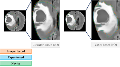 |
Experience counts! Comparison of ROI placement strategies for radiomics analysis of gliomas
Bárbara Schmitz Abecassis1,2, Koji Sakai2, Tomonori Toyotsuji2, Yoshiaki Ota2,3, and Kei Yamada2
1Department of Radiology, Leiden University Medical Center, Leiden, Netherlands, 2Department of Radiology, Kyoto Prefectural University of Medicine, Kyoto, Japan, 3Department of Radiology, University of Michigan, Ann Arbor, MI, United States
Glioma delineation is complex and time-consuming. We evaluated whether a technically less challenging method could potentially bypass this tedious process, when using a simple manual circular-ROI by less trained personnel. We assessed whether applying a circular-ROI to clinical 3T-MRIs from 12 glioma patients, would extract significantly different GLI features. We compared it to the gold-standard manual delineation approach, having 3 observers with different levels of expertise placing the ROIs. Experience matters to extract consistent GLI features across delineation runs. The different ROI methods extracted significantly different GLI features, emphasizing the importance of delineation strategies when analyzing heterogeneous tumors like gliomas.
|
|
1724.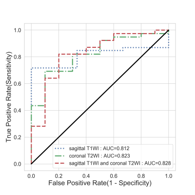 |
Radiomics approach in preoperative differentiation of prolactinoma and non-prolactinoma pituitary adenomas
Rushi Chen1, Yan Bai2, Mengke Wang2, Feng Qin2, Menghuan Zhang2, Xueping Zhang2, Shang Wang2, Yongchao Zhao2, Ting Fang2, Xinhui Wang2, Huan Zhao2, Li Mao3, Xiuli Li3, and Meiyun Wang2
1Henan provincial people's hospital& Zhengzhou University People’s Hospital, Zhengzhou, China, 2Henan provincial people's hospital, Zhengzhou, China, 3Deepwise AI Lab, Beijing, China
The primary treatment for prolactinomas is dopamine agonist, while transsphenoidal surgery is recommended as primary therapy for the non-prolactinomas. This study aimed to evaluate the radiomic model in the differentiation of prolactinoma from non-prolactinomas before surgery. Our results demonstrated that the eXtreme Gradient Boosting model based on 2264 radiomic features extracted from the original and preprocessed sagittal contrast-enhanced T1WI and coronal T2WI yielded the AUC value of 1.000 and 0.828 on the train set and test set, respectively. The radiomic model may have potential for differentiating the prolactinomas from non-prolactinomas, which may be beneficial to guide the treatment plan.
|
|
1725.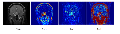 |
A preliminary study on predicting the ACTH&GH hormone immunohistochemistry typing of pituitary macroadenoma based on Radiomics DCE-MRI
Yangyingqiu Liu1, Yanwei Miao1, Ailian Liu1, Qingwei Song1, and Bing Wu2
1Department of Radiology, First Affiliated Hospital of Dalian Medical University, Dalian, China, 2GE healthcare, Beijing, China
Hormone immunohistochemistry typing of pituitary macroadenomas is assessed by radiomics analysis on Dynamic Contrast Enhanced MRI(DCE-MRI). The results show that DCE-MRI could be used as a new noninvasive method to predictive the hormone immunohistochemistry typingof pituitary macroadenomas.
|
|
1726.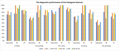 |
The Diagnostic Performance of Multiparametric MRI Radiomics for Classification of Untreated Adult Gliomas
Amirah Faisal Alsaedi1,2, Jasmina Panovska-Griffiths3, Xavier Golay2, and Sotirios Bisdas2,4
1Department of Radiology Technology, Taibah University, Medina, Saudi Arabia, 2Department of Brain Repair & Rehabilitation, UCL, Queen Square, Institute of Neurology, London, United Kingdom, 3Department of Applied Health Research, UCL, London, United Kingdom, 4Lysholm Department of Neuroradiology, The National Hospital for Neurology and Neurosurgery, University College Hospitals NHS Trust, London, United Kingdom
This study aimed to assess the diagnostic performance of multiparametric MRI radiomics for glioma class prediction according to the WHO 2016 classification. Histogram features were extracted from prospectively acquired multiparametric MRI (pCASL, DSC-MRI, DCE-MRI, and DWI) in 32 patients with primary gliomas. The uncombined significant features of ASL, ADC, DSC, and DCE, revealed diagnostic performances varying from low (44% ) to fair (86%) and unable to predict all the histomolecular classes. However, combining them for each MRI method, independently, enhanced the diagnostic accuracy up to 100% and predict all the classes. This alludes the use of multimodal radiomics for glioma classification.
|
|
1727.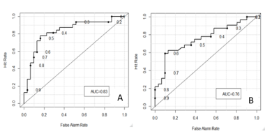 |
Addition of peritumoral area improves T1-weighted texture-based prediction of glioblastoma multiforme progression
George Zenzerovich1 and Tim Q Duong2
1Radiology, Stony Brook University, Stony Brook, NY, United States, 2Stony Brook University, Stony Brook, NY, United States
Texture features obtained from peritumoral area of diagnostic MR image of Glioblastoma Multiforme were used to improve texture based prediction of tumor progression. The peritumoral area was drawn and examined then derived texture features were added to conventional model to improve performance.
|
|
1728.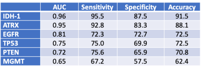 |
Multiparametric MRI Texture Analysis in Prediction of Genetic Biomarkers in Patients with Brain Glioma
Shingo Kihira1, Nadejda Tsankova2, Adam Bauer3, Yu Sakai1, Nicole Zubizarreta4, Jane Houldsworth2, Fahad Khan2, Adilia Hormigo5, Constantinos Hadjipanayis6, and Kambiz Nael1
1Radiology, Icahn School of Medicine at Mount Sinai, New York, NY, United States, 2Pathology, Icahn School of Medicine at Mount Sinai, New York, NY, United States, 3Radiology, Kaiser Permanente Fontana Medical Center, Fontana, CA, United States, 4Institute for Health Care Delivery Sciences, Icahn School of Medicine at Mount Sinai, New York, NY, United States, 5Neurology, Icahn School of Medicine at Mount Sinai, New York, NY, United States, 6Neurosurgery, Icahn School of Medicine at Mount Sinai, New York, NY, United States
In this retrospective study, we used a commercially available texture analysis software (Olea Medical) to construct a multiparametric MRI radiomic model that can be used to predict several important prognostic biomarkers in a cohort of patients with brain glioma. A total of 92 texture features were calculated from both FLAIR and T1C+ images using a volume-of-interest analysis encompassing the entire FLAIR hyperintense tumor. Radiomic features obtained from our multiparametric MR texture model were able to predict genetic biomarkers of brain glioma with predictive accuracies ranging from modest (62.4%) for MGMT to nearing 90% for IDH-1 and ARTX.
|
|
1729.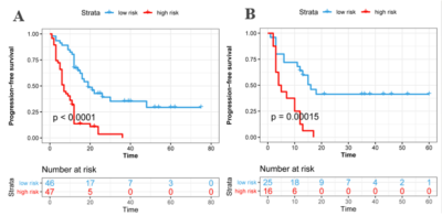 |
Prediction of high-grade glioma prognosis based on radiomics features of tumor and peritumoral edema areas from magnetic resonance imaging
YiXin HU1, Lan Li1, JiuQuan Zhang1, and Jianqing SUN2
1Chongqing University Cancer Hospital & Chongqing Cancer Institute & Chongqing Cancer Hospital, ChongQing, China, 2Philips Healthcare, Shanghai, China
To build a radiomics signature from MR images to predict high grade glioma (HGG, WHO grade III and IV) patients with different prognosis. Then, to explore the predictive value based on the edge of the tumor area and PBZ, respectively.
|
|
1730. |
Differential diagnosis of solitary fibrous tumor/hemangiopericytoma and angiomatous meningioma using 3D-MRI texture feature model
Junyi Dong1, Yangyingqiu Liu2, Yanwei Miao1, Huicong Shen3, and Yan Guo4
1First Affiliated Hospital of Dalian Medical University, Dalian,116011,China, Dalian, China, 2Department of Radiology, First Affiliated Hospital of Dalian Medical University, Dalian,116011,China, Dalian, China, 3Beijing Tiantan hospital, Beijing, China, 4Life science, Shenyang, China
Intracranial solitary fibrous tumor(SFT)/hemangiopericytoma (HPC) is a rare malignant tumor originating from the intracranial vasculature, while Angiomatous meningioma (AM) is also a rare benign one as a histological subtype of meningioma with World Health Origination (WHO) grade I. The two tumors have similar location and conventional MRI features, but the treatment and prognosis are quite different. Texture analysis can get more information that can't be seen in conventional MRI images. It is very necessary to establish the differentiation model of texture analysis between the two kinds of tumors.
|
|
| 1731. | WITHDRAWN |
1732.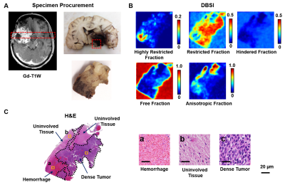 |
Diffusion Histology Imaging Characterizes and Differentiates Various Tumor Histological Features in High-Grade Brain Tumors
Zezhong Ye1, Sam E. Gary2, Jeffrey D. Viox3, Anthony T. Wu4, Joshua Lin5, Peng Sun1, Joshua B. Rubin6, Sonika Dahiya7, and Sheng-Kwei Song1
1Radiology, Washington University School of Medicine, St. Louis, MO, United States, 2Medical Scientist Training Program, The University of Alabama at Birmingham, Birmingham, AL, United States, 3School of Medicine, University of Missouri - Kansas City, Kansas City, MO, United States, 4Biomedical Engineering, Washington University, St. Louis, MO, United States, 5Keck School of Medicine, The University of Southern California, Los Angeles, CA, United States, 6Pediatrics, Washington University School of Medicine, St. Louis, MO, United States, 7Pathology and Immunology, Washington University School of Medicine, St. Louis, MO, United States
Current clinical diagnosis, surgical resection, and assessment of treatment response for high-grade brain tumor patients relies heavily on gadolinium-enhanced T1-weighted MRI, although such imaging is non-specific for tumor and merely reflects a disrupted blood-brain barrier. The complex tumor microenvironment and spatial heterogeneity make high-grade brain tumor very difficult to characterize using current clinical imaging modalities. We developed a novel imaging strategy to characterize key tumor histological features and demonstrated its capability to accurately predict these histology. Extending this approach to larger cohorts of both tumor specimens and patients could provide further validation and facilitate its clinical translation for patient management.
|
|
1733.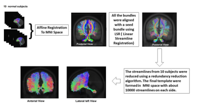 |
Assessment of Cerebello-Thalamo-Cortical (CTC) Fiber Pathways in Post-Surgical Medulloblastoma Patients Using a CTC Template
Qing Ji1, Matthew Scoggins1, Angela Edwards1, John O. Glass1, Tara Brinkman2,3, Zoltan Patay1, and Wilburn E. Reddick1
1Diagnostic Imaging, St.Jude Children's Research Hospital, Memphis, TN, United States, 2Psychology, St.Jude Children's Research Hospital, Memphis, TN, United States, 3Epidemiology and Cancer Control, St.Jude Children's Research Hospital, Memphis, TN, United States A CTC fiber pathway template was built in MNI space from the CTC data of 10 healthy controls. The template was aligned with the principle diffusion directions of each individual DTI data of additional 10 healthy controls and 11 post-surgical medulloblastoma patients using a linear algorithm developed in this study. The aligned template can accurately mimic the real CTC pathways in an individual subject in both healthy and post-surgical subjects. The DTI parameter values in post-surgical medulloblastoma patients can be accessed using the transformed CTC pathway. |
|
1734.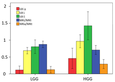 |
Non-invasive separation of low and high grade gliomas with diffusion kurtosis decomposition (DKD)
Sirui Li1, Wenbo Sun1, Yuan Zheng2, Qing Wei3, Samo Lasic4, Shihong Han3, Shuheng Zhang3, Danielle van Westen5, Karin Bryskhe4, Daniel Topgaard4,5, and Haibo Xu1
1Zhongnan Hospital of Wuhan University, Wuhan, China, 2UIH America, Inc., Houston, TX, United States, 3United Imaging Healthcare, Shanghai, China, 4Random Walk Imaging, Lund, Sweden, 5Lund University, Lund, Sweden
Diffusion kurtosis decomposition (DKD) is a novel advanced diffusion MRI modality relying on customized pulse sequences and high-performance hardware to assess cell shapes and density heterogeneity via the anisotropic and isotropic mean kurtosis parameters, MKa and MKi, which are fundamentally different microstructural properties that are inextricably entangled in conventional diffusion kurtosis imaging (DKI). We have investigated DKD imaging of gliomas in a clinical setting, and for the first time established the correlations between MKa, MKi, and tumor grade. In comparison to conventional diffusion methods, DKD more accurately describes the microstructural changes and provides a useful tool for glioma diagnosis.
|
|
1735.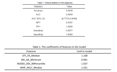 |
Tumor Grading of Glioma by Histogram Analysis Based on Multiple Advanced Diffusion Models, including DTI, DKI, MAP-MRI and NODDI
Gao Ankang1, Zhang Huiting2, Yang Guang3, Wang Shaoyu2, Yan Xu2, Bai Jie1, and Cheng Jingliang1
1MRI, Dept. of MRI, The First Affiliated Hospital of Zhengzhou University, Zhengzhou, China, 2MR Scientific Marketing, Siemens Healthcare, Shanghai, China, 3Shanghai Key Laboratory of Magnetic Resonance, East China Normal University, Shanghai, China
Diffusion imaging is widely used to noninvasively detect the microscopic diffusion properties of biological tissues in vivo. Advanced diffusion models were recently proposed to provide additional microstructure information. In present work, we applied four diffusion models in glioma grading, including DTI, DKI, MAP-MRI and NODDI model, which could be acquired within a single scan.
|
|
1736.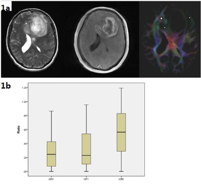 |
Track-density imaging (TDI): Evaluating the peritumoral white matter areas of glioblastoma
Gao Ankang1, Chen Qianqian1, Zhu Jinxia2, Stefan Huwer3, Bai Jie1, and Cheng Jingliang1
1MRI, Dept. of MRI, The First Affiliated Hospital of Zhengzhou University, Zhengzhou, China, 2MR Collaboration, Siemens Healthcare Ltd., Beijing, China, Beijing, China, 3Siemens Healthcare GmbH, Erlangen, Germany, Erlangen, Germany
By observing the gradually developing characteristics of track density in the area of peritumoral edema, the extent of possible tumor invasion was judged. This may contribute to neurosurgical planning, the postoperative quality of life, and longer survival expectancy. Simultaneously, TDI was able to show the crossing fibers of the tumor part but could not resolve the disturbed diffusion of severe peritumoral edema’s influence on the fibers.
|
|
1737.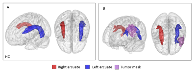 |
Along-tract quantitative analysis of arcuate fasciculus for awake glioma surgery
Lia Talozzi1, Matteo Martinoni2, Micaela Mitolo 3, Filippo Badaloni2, Sofia Asioli1,4, Claudia Testa5, Raffaele Lodi1,6, and Caterina Tonon1,3
1Biomedical and Neuromotor Sciences, University of Bologna, Bologna, Italy, 2IRCCS Istituto delle Scienze Neurologiche di Bologna, Neurosurgery Unit, Bologna, Italy, 3IRCCS Istituto delle Scienze Neurologiche di Bologna, Neuroradiology Functional Diagnostic Unit, Bologna, Italy, 4Section of Anatomic Pathology 'M. Malpighi', Bellaria Hospital, Bologna, Italy, 5Physics and Astronomy, University of Bologna, Bologna, Italy, 6, IRCCS Istituto delle Scienze Neurologiche di Bologna, Bologna, Italy
Probabilistic arcuate fasciculus (AF) tractography was computed in healthy controls (HC) and for a patient with a diffuse glioma, IDH 1 wild type left frontal lobe. Bilateral curvature mapping and along-tract DTI measures were evaluated, modelling surface geometry. In HC, hemispheric asymmetries in tract curvature showed a more lateral and inferior trajectory on the left, not measured for the patient’s left AF, medially dislocated. In HC, a left-lateralized asymmetries (higher FA and volume) was measured, and it was preserved for the patient. During pre-surgical evaluation, the proposed developed tractography analyses allowed a quantitative investigation of AF dislocation and microstructural integrity.
|
|
1738.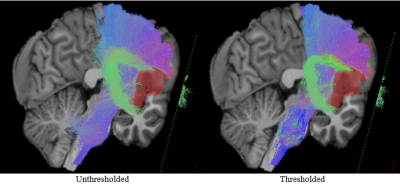 |
Minimizing false streamlines in anatomically constrained tractography for neurosurgery guidance in patients with brain neoplasms
Daniel Krahulec1, Ahmed Radwan2, Stefan Sunaert2, Maarten Versluis1, Kim van de Ven1, and Marcel Breeuwer1,3
1MR R&D Clinical Science, Philips Healthcare, Best, Netherlands, 2Department of Imaging & Pathology, Translational MRI, KU Leuven, Leuven, Belgium, 3Department of Biomedical Engineering – Medical Image Analysis, Eindhoven University of Technology, Eindhoven, Netherlands
A data analysis pipeline comprising KUL neuroimaging tools was applied to analyze diffusion and anatomical MRI datasets with neoplastic lesions and perform diffusion tractography with anatomical priors. We utilized a novel framework for reducing the amount of false positive streamlines to allow for improved visualization of different fiber bundles. Results illustrate the meaningfulness of this approach in neurosurgery workflow through offering clinicians more information on the quality of perilesional fiber bundles as well as where to resect with care to preserve functionally eloquent areas.
|
|
1739.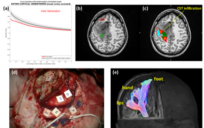 |
Do advanced fMRI and tractography preoperative evaluations correspond to intraoperative findings? A pilot evaluation in brain tumour patients.
Marco Borri1, Jose Pedro Lavrador2, Irene Brumer1,3, Enrico De Vita4, Jonathan Ashmore1,5, Francesco Vergani2, Ranjeev Bhangoo2, Keyoumars Ashkan2, and Jozef Jarosz1
1Neuroradiology, King's College Hospital, London, United Kingdom, 2Neurosurgery, King's College Hospital, London, United Kingdom, 3Computer Assisted Clinical Medicine, Medical Faculty Mannheim, Heidelberg University, Mannheim, Germany, 4Department of Biomedical Engineering, School of Biomedical Engineering and Imaging Sciences, London, United Kingdom, 5Department of Medical Physics and Bioengineering, NHS Highland, Inverness, United Kingdom
In this work we have evaluated the feasibility of incorporating advanced fMRI and tractography evaluations into the real-life presurgical management of patients undergoing brain lesion resections. In this pilot cohort of patients, all major preoperative imaging findings were validated by intraoperative measurements. With the inclusion of fMRI end regions, probabilistic tractography allowed a better reconstruction of the corticospinal tract and its branches in regions adjacent or within the tumour with altered or damaged fibre architecture. Robust fMRI-based language lateralization was able to describe likely dominance, in agreement with intraoperative findings and initial postoperative deficit.
|
|
1740.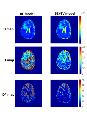 |
IVIM analysis using Total Variation Penalty Regularization Based Model for Brain Tumor Analysis
Mridula Vij1, Archana Vadiraj Malagi1, Esha Badiya Kayal1, Jitender Saini2, and Amit Mehndiratta1
1Centre for Biomedical Engineering, Indian Institute of Technology Delhi, New Delhi, India, 2Department of Neuroimaging & Interventional Radiology, National Institute of Mental Health and Neuro Sciences, Bengaluru, India
The occurrence of brain tumor is increasing; it is believed that early detection will prove to be beneficial for its treatment. IVIM has proved to provide valuable information and has been explored for brain tumor in prior studies. In the following study, IVIM analysis for brain tumor detection was performed using a novel Biexponential model with Total Variation regularization function (BE+TV model), and a comparison with established Biexponential model was performed. Improvement in coefficient of variation by 16.5 to 43.4% was observed across IVIM parameter. Perfusion faction was estimated to be reliably with this novel methodology.
|
|
1741.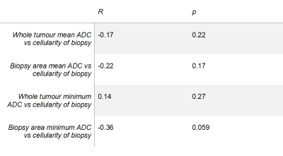 |
Do targeted biopsies improve the correlation between ADC and cellularity in patients with glioma?
Simran Kukran1,2, Marianna Inglese2, Katherine L Ordidge2,3, Claire Davies 2, Lesley Honeyfield3, Babar L Vaqas4, Sophie Camp4, David Peterson4, Kevin O'Neill4, Clara Limback-Stanic5, Tara Barwick2,3, Adam Waldman6,7, and Matthew Grech-Sollars2,3
1Department of Radiotherapy and Imaging, Institute of Cancer Research, Sutton, United Kingdom, 2Department of Surgery and Cancer, Imperial College London, London, United Kingdom, 3Department of Imaging, Imperial College London, London, United Kingdom, 4Department of Neurosurgery, Imperial College London, London, United Kingdom, 5Department of Histopathology, Imperial College London, London, United Kingdom, 6Department of Medicine, Imperial College London, London, United Kingdom, 7Centre for Clinical Brain Sciences, The University of Edinburgh, Edinburgh, United Kingdom
It is thought that hypercellular regions of glioma have lower ADC values via a restriction in flow path, but there is no consensus regarding the correlation between ADC values and the cellularity of histological biopsies. In this study a slightly stronger negative correlation was observed between sample cellularity and the average ADC across the biopsy region as compared to the average ADC across the whole tumour in patients with glioma. However, neither correlation was found to be significant, which could be due to a small cohort size and the variation in tumour biological factors other than cellularity affecting ADC.
|
|
| 1742. | Histogram analysis in predicting the grade and histological subtype of meningiomas based on diffusion kurtosis imaging
Xiaodan Chen1, Ying Chen2, Lin Lin3, Jie Wu4, and Guang Yang4
1Radiology, Fujian Cancer Hospital, Fuzhou, China, 2Fujian Cancer Hospital, Fuzhou, China, 3Fujian Medical University Union Hospital, Fuzhou, China, 4Shanghai Key Laboratory of Magnetic Resonance, East China Normal University, Shanghai, China, Shanghai, China
Objectives Presurgical grading is particularly important for selecting the best therapeutic strategy for meningioma patients. Therefore, our study is to investigate the value of histogram analysis of diffusion kurtosis imaging (DKI) maps in the differentiation of grades and histological subtypes of meningiomas.Methods A total of 172 patients with histopathologically proven meningiomas underwent preoperative magnetic resonance imaging (MRI) and were classified into low-grade and high-grade groups. Mean Kurtosis (MK), fractional anisotropy (FA), mean diffusivity (MD) histograms were generated based on solid components of the whole tumour. The following parameters of each histogram were obtained:10th, 25th, 75th, and 90th percentiles, mean,median,maximum,minimum,and kurtosis, skewness, and variance. Comparisons of different grades and subtypes were made by Mann-Whitney U test,Kruskal-Wallis tests, ROC curves analysis, and multiple logistic regression.Results Significantly higher maximum, Skewness, and variance of MD, mean, median, maximum, variance, 10th, 25th, 75th, and 90th percentile of MK were found in high-grade than low-grade meningiomas (all P<0.05). DKI histogram parameters differentiated 7 of 10 pairs of subtype pairs (atypical versus meningothelial/ fibrous/transitional/angiomatous meningiomas; angiomatous versus fibrous/transitional meningioma; fibrous versus meningothelial meningiomas). The 90th percentile of MK yielded the highest AUC of 0.870 and was the only independent indicators for grading meningiomas.Conclusion The histogram analysis of DKI is useful for differentiating meningioma grades and subtypes. The 90th percentile of MK may serve as an optimal parameter for predicting the grade of meningiomas.
|
|
1743.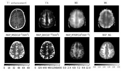 |
Evaluation of glioma grade and proliferation activity by different diffusion-weighted-imaging models: including DKI and MAP-MRI
shenghui Xie1, shaoyu Wang2, xu Yan2, huapeng Zhang2, guang Yang3, and yang Gao1
1Department of Radiology, Affiliated Hospital of Inner Mongolia Medical University, Hohhot, China, 2Siemens Healthineers, Shanghai, China, 3East China Normal University, Shanghai, China
The purpose of this study was to explore the diagnostic value of multimode diffusion imaging in the evaluation of glioma tumor grade and proliferation activity. (DKI) and MAP-MRImodel). The results showed that there were significant differences in MK, RTOP and QIV between high and low grade glioma, and the values of MK and QIV of the tumor parenchyma were positively correlated with ki-67. MK and QIV have great potential in predicting tumor proliferation activity. Multimode diffusion-weighted imaging is valuable for the evaluation of preoperative glioma grade and tumor proliferation activity.
|
|
1744.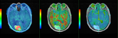 |
The value of 3D ASL and DTI combined with conventional MRI features in differential diagnosis of hemangiopericytoma and angiomatous meningioma
rui hu1, yi jun wu1, wen chen1, ke hua zheng1, dai zhong wang1, and lin xu1
1taihe hospital, shiyan, China
To investigate the value of MRI characteristics in the differential diagnosis of hemangiopericytoma and angiomatous meningioma.15 cases of AM and 11 cases of the HPC which were confirmed by pathology. MRI sequences were routinely available. The mean ADC values of HPC was significantly higher than that of AM (t = 2.613, p =0.02). The mean CBF values (t= -8.99, P<0.01) and FA values (t= -3.66, P<0.01) were lower for HPC compared with AM. MRI imaging characteristics of HPC and AM have significant difference. Combining structural MRI comparative analysis and functional MRI was useful for distinguishing HPC from MA.
|
|
1745.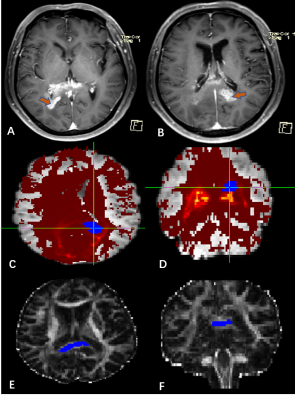 |
The difference between multicentric gliomas and diffuse gliomas: evidence from the probabilistic fiber tracking by diffusion tensor imaging
Simin Zhang1, Xiaorui Su1, Weina Wang1, Qiang Yue1, and Qiyong Gong1
1Huaxi MR Research Center,Department of Radiology, West China Hospital of Sichuan University, Chengdu, China
Multicentric gliomas are rare lesions of central nervous system. Due to the invasive nature of glioma, differentiating multicentric gliomas from diffuse gliomas is difficult. As yet there remains lack of methods to elucidate lesions are totally separated unless surgical biopsy. We use probabilistic fiber tracking to identify the white matter dissemination routes. Our results showed the connectivity value and probabilistic value of the lesions in multicentric gliomas were significantly lower than the lesions in diffuse gliomas. The features provide new insight into multicentric gliomas and may aid in accurate classification of multicentric glioma and diffuse glioma in vivo.
|
|
1746.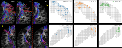 |
Multi-level fiber tracking: evaluation on clinical data
Andrey Zhylka1, Nico Sollmann2,3, Alberto De Luca4, Daniel Krahulec5, Marcel Breeuwer1,5, Alexander Leemans4, and Josien Pluim1
1Biomedical Engineering department, Eindhoven University of Technology, Eindhoven, Netherlands, 2Department of Diagnostic and Interventional Neuroradiology, , Klinikum rechts der Isar, Technische Universität München, Munich, Germany, 3TUM-Neuroimaging Center, Klinikum rechts der Isar, Technische Universität München, Munich, Germany, 4University Medical Center Utrecht, Utrecht, Netherlands, 5MR R&D Clinical Science, Philips Healthcare, Best, Netherlands
Conventional deterministic fiber tractography approaches commonly used in clinical applications are prone to generating false-negative reconstructions, which might influence further decision-making related to treatment and repeated surgery in patients with brain tumors. Surgery-related effects, such as blood inflow into white matter and edema, further distort the diffusion signal, complicating the task of tractography. We evaluated a novel multi-level fiber tractography approach on data of subjects who had undergone tumor resection. A comparison with conventional deterministic approaches is performed. The results were correlated with the reported motor-function deficit grades.
|