Digital Poster Session
Cancer: Preclinical & Miscellaneous
Cancer
4720 -4732 Cancer Imaging: Preclinical & Miscellaneous - Cancer Imaging: Preclinical Models
4733 -4747 Cancer Imaging: Preclinical & Miscellaneous - Cancer Imaging: Quantitative
4748 -4763 Cancer Imaging: Preclinical & Miscellaneous - Cancer Imaging: Miscellaneous 1
Session Topic: Cancer Imaging: Preclinical & Miscellaneous
Session Sub-Topic: Cancer Imaging: Preclinical Models
Digital Poster
Cancer
| 4720. | Monitoring Tumor Vascular Microenvironment Response by Multiparametric MRI after Dual Inhibition of PFKFB3 and VEGF in Glioblastoma
Junfeng Zhang1 and Weiguo Zhang1
1Radiology, Daping Hospital, Army Medical University, Chongqing, China
Tumor vascular normalization induced by antiangiogenic therapy such as bevacizumab (BEV) is a promising strategy to remodel tumor microenvironment. However, this effect is transient and finally vanished because of inevitable adaptive therapy resistance. In this study, we found that targeting tumor glycolysis activator PFKFB3 is a novel potential strategy to enhance BEV therapy efficacy and prolonged vascular normalization in glioblastoma. IVIM parameters are much better than DCE-MRI as alternative translatable imaging biomarkers for evaluating tumor response and monitoring vascular normalization.
|
|
| 4721. | Mapping hypoxia in brain tumors using GdDO3NI: validation with PET and IHC
Babak Moghadas1, Matthew Scarpelli2, Christopher Rock1, Alberto Fuentes2, Debbie Healey2, Chad Quarles2, and Vikram D Kodibagkar1
1Biomedical engineering, Arizona State University, Tempe, AZ, United States, 2Barrow Neurological Institute, Phoenix, AZ, United States
In this study we have used the hypoxia-targeting MRI contrast agent GdDO3NI, (a nitroimidazole-based T1 contrast agent) to image the development of hypoxia in the rodent brain tumors at two different sizes. Our results indicate a range of signal enhancements from 5-20% over baseline in the 9L tumor using GdDO3NI with clearance from contralateral brain and muscle tissue. Furthermore the GdDO3NI enhancement correlates with PET imaging using hypoxia targeting 18F-FMISO and immunohistochemistry based on pimonidazole. This study further demonstrates the utility of GdDO3NI in non-invasive imaging of tissue hypoxia with high resolution.
|
|
4722.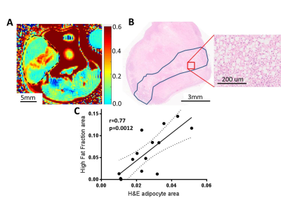 |
Quantitative imaging of adipocyte distribution in breast cancer xenograft models
Michal Tomaszewski1, William Dominguez Viqueira1, Bruna Victorasso Jardim Perassi1, Pravin Phadatare1, Robert J Gillies1, and Smitha Pillai1
1Cancer Physiology, H. Lee Moffitt Cancer Center, Tampa, FL, United States
Lipid deposition and metabolism is relevant for cancer prognosis and treatment response. Imaging characterisation of the adipocyte deposition patterns in breast cancer and its relationship with the microenvironment may therefore provide valuable insight into the tumour. A Chemical selected saturation based MR technique was optimised and validated in vivo and ex vivo to visualize adipocyte distribution in breast cancer xenografts. The measurements reveal clear differences in adipocyte accumulation between different breast models. In the future this technique will be applied to understand the role of adipocyte distribution in breast cancer.
|
|
4723.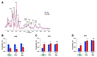 |
Early MR-detectable biomarkers of mutant IDH inhibition in patient-derived low-grade gliomas in vivo
Marina Radoul1, Chloé Najac1, Donghyun Hong1, Anne Marie Gillespie1, Pavithra Viswanath1, Russell O. Pieper2,3, Joseph F. Costello2, and Sabrina M. Ronen1,3
1Radiology and Biomedical Imaging, University of California San Francisco, San Francisco, CA, United States, 2Neurological Surgery, University of California San Francisco, San Francisco, CA, United States, 3Brain Tumor Research Center, University of California San Francisco, San Francisco, CA, United States
Low-grade glioma (LLG) patients have a relatively long-term survival of ~13 years but these tumors always recur. Since IDH mutations are present in >80% of LGGs, inhibition of mutant IDH activity is being tested as a new therapeutic approach. Here, we investigated response to mutant IDH inhibition by AG-881 in orthotopic LGG mouse models. Using in vivo 1H MRS we detected, in addition to a decrease in 2-HG, an early increase in both glutamate and glutamine/glutamate that were associated with subsequent tumor shrinkage. This identifies potential early metabolic biomarkers of LGG response to mutant IDH inhibition.
|
|
4724.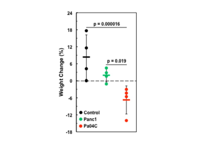 |
Brain and plasma choline compounds altered by cancer and cancer-induced cachexia
Santosh K Bharti1, Paul T Winnard1, Raj Kumar Sharma1, Marie-France Penet1,2, Balaji Krishnamachary1, and Zaver M. Bhujwalla1,2,3
1Div. of Cancer Imaging Research, The Russell H. Morgan Dept of Radiology and Radiological Science, The Johns Hopkins University School of Medicine,, Baltimore, MD, United States, 2Sidney Kimmel Comprehensive Cancer Center, The Johns Hopkins University School of Medicine, Baltimore, MD, United States, 33Radiation Oncology and Molecular Radiation Sciences, The Johns Hopkins University School of Medicine, Baltimore, MD, United States
Cancer induced cachexia is a multifactorial syndrome that results in unexplained weight loss in cancer patients. A major cause of morbidity and mortality, the syndrome is most prevalent in pancreatic cancer. Here we have identified changes in brain choline metabolites and plasma choline in human pancreatic cancer xenografts that induce cachexia (Pa04C) as well as noncachexia inducing pancreatic cancer xenografts (Panc1), compared to normal mice. These results highlight the systemic changes in choline metabolism that occur with cancer and with cancer induced cachexia that may lead to the development of early biomarkers as well to metabolic treatment strategies.
|
|
4725.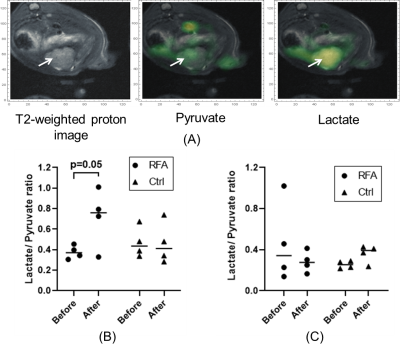 |
13C-MRI Detects Increased Lactate Flux in HCC Following Hepatic Thermal Ablation and Correlates with PFKFB3 Expression
Qianhui Dou1, Aaron K. Grant1, Cody Callahan1, David Mwin1, Muneeb Ahmed1, and Leo L. Tsai1
1Radiology, Beth Israel Deaconess Medical Center, Boston, MA, United States
Thermal ablation (TA) is commonly used to treat early hepatocellular carcinoma (HCC) and liver metastases. However, there is increasing clinical and experimental evidence that TA may sometimes induce more aggressive tumor biology within and outside the liver. We used hyperpolarized 13C MRI to detect increased HCC lactate flux following adjacent normal liver radiofrequency ablation in a rat model. This increased flux correlated with an increase in tumor expression of PFKFB3, a known modulator of glycolysis. PFKFB3 may be a potential therapeutic target and hyperpolarized 13C MRI may be used to track or predict treatment response.
|
|
4726.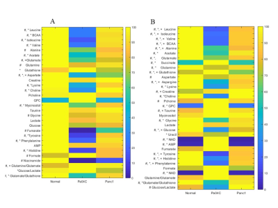 |
Lung and kidney metabolism altered by cancer-induced cachexia
Raj Kumar Sharma1, Santosh K. Bharti1, Paul T Winnard1, Marie-France Penet1,2, and Zaver M. Bhujwalla1,2,3
1Division of Cancer Imaging Research, The Russell H. Morgan Department of Radiology and Radiological Science, The Johns Hopkins University School of Medicine, Baltimore, MD, United States, 2Sidney Kimmel Comprehensive Cancer Center, The Johns Hopkins University School of Medicine, Baltimore, MD, United States, 3Radiation Oncology and Molecular Radiation Sciences, The Johns Hopkins University School of Medicine, Baltimore, MD, United States Poster Permission Withheld
To understand the impact of pancreatic cancers on body organs within the context of cancer induced cachexia we have, in ongoing studies, characterized the metabolism of organs in tumor bearing mice. Cancer-induced cachexia occurs in 80% of pancreatic ductal adenocarcinoma (PDAC) patients. Here we present data demonstrating the significant impact of cachexia-inducing Pa04C PDAC xenografts compared to non-cachexia inducing Panc1 xenografts and normal mice on kidney and lung metabolites as identified by 1H MRS of tissue extracts. These metabolic changes may contribute to increased morbidity and poor quality of life but equally may present novel targets for intervention.
|
|
4727.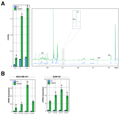 |
Ubiquitous Mitochondrial Creatine Kinase Drives Malignant Creatine Metabolite Profiles in Triple-Negative Breast Cancer Models
Vinay Ayyappan1, Caitlin Tressler1, Menglin Cheng1, Kanchan Sonkar1, Ruoqing Cai1, Michael T. McMahon1,2, and Kristine Glunde1,3
1he Russell H. Morgan Department of Radiology and Radiological Science, The Johns Hopkins University School of Medicine, Baltimore, MD, United States, 2F.M. Kirby Research Center for Functional Brain Imaging, Kennedy Krieger Institute, The Johns Hopkins University School of Medicine, Baltimore, MD, United States, 3The Sidney Kimmel Comprehensive Cancer Center, The Johns Hopkins University School of Medicine, Baltimore, MD, United States
Ubiquitous Mitochondrial Creatine Kinase (CKMT1) is a mitochondrial membrane protein that catalyzes the reversible conversion of creatine (Cr) to phosphocreatine (PCr). This study shows in a mouse model of triple-negative breast cancer that CKMT1-overexpression significantly increases tumor Cr and PCr levels while accelerating tumor growth. Since high CKMT1 predicts worsened survival in breast cancer patients, CKMT1 may hold promise as a potential diagnostic marker and/or treatment target whose expression can be monitored using magnetic resonance spectroscopy-detected Cr and PCr levels.
|
|
4728.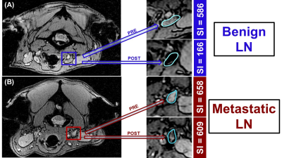 |
Lymphotropic Nanoparticle-Enhanced LNMRI for Accurate Prediction of Nodal Status in Canine Head and Neck Cancer Patients
Marina Stukova1, Lynn Griffin2, Bernard Seguin2, Chad Frank2, and Natalie Julie Serkova3
1Radiology, University of Colorado Anschutz Medical Campus, Aurora, CO, United States, 2Colorado State University, Fort Collins, CO, United States, 3Radiology, University of Colorado Anschutz, Denver, CO, United States
The objective of this study was to develop an optimal iron oxide enhanced lymph node MRI protocol and robust image analysis using pre-and-post ferumoxytol T2 and T2* scans in canine patients with HNC. Since those pet patients are naturally prone to HNC, they can serve as a great translational platform for development of novel non-invasive imaging modalities for LN staging and metastasis and to guide anti-lymphangiogenic therapies. The results indicate that Ferumoxytol can be used as T2 negative contrast agent for non-invasive LN staging 48 hrs post-injection. SI normalization should be performed to a phantom or to the brain.
|
|
4729.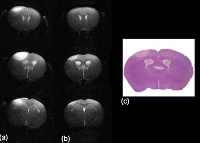 |
Imaging of Stroke in Rodents using a Clinical Scanner and Implanted Inductively Coupled Receiver Coils
Javier Istúriz1, María J. Nicolás2, Miguel Fernández3, Adriana Honrubia2, Pablo Domínguez3, Gorka Bastarrika3, Miguel Valencia2, and Maria A. Fernandez-Seara3
1NeosBiotec, Pamplona, Spain, 2Neuroscience, Fundación para la Investigación Médica Aplicada, Pamplona, Spain, 3Radiology, Clínica Universidad de Navarra, Pamplona, Spain
We and others have previously shown that clinical scanners can be employed to image small laboratory animals using wireless inductively coupled coils (ICC). In this work, we evaluated the use of implanted ICCs for the same purpose and demonstrated that they can provide high SNR images that allow longitudinal assessment of stroke lesions in rodents.
|
|
4730.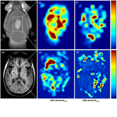 |
IAUC derived permeability index factor is a robust parameter for permeability visualization in breast cancer brain metastasis in mice & patients.
Daniele Procissi1, Supada Prakkamakul2,3, Sol Misener4, Nicola Bertolino4, Benjamin P Liu 2, and Irina Balyasnikova5
1Radiology & Biomedical Engineering, Northwestern University, Evanston, IL, United States, 2Radiology, Northwestern Memorial Hospital, Chicago, IL, United States, 3Diagnostic Radiology, Samitivej Hospital, Bangkok, Thailand, 4Radiology, Northwesten Unversity, Chicago, IL, United States, 5Neurological Surgery, Northwestern, Chicago, IL, United States
A iAUC derived tumor permeability index factor (TPIF) provides a reliable and robust reporter of breast cancer brain metastases (BCBM) tumor microenvironment permeability patterns without the confounding elements of model-based DCE-MRI analysis (i.e. low sensitivity, inability to fit, inter-subject data reliability). A comparison of results in both mice and patients suggests that this parameter can serve as an additional tool for improved, early clinical diagnosis and for therapeutic testing at the preclinical level.
|
|
4731.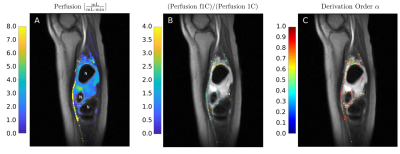 |
Fractional Calculus Tracer Kinetic Model for Tissue Blood Perfusion Quantification
Stefan Hindel1 and Lutz Lüdemann1
1University Hospital Essen, Essen, Germany
We present a one-compartment model with fractional elimination and time-dependent tracer input to estimate perfusion and its heterogeneity with intravascular contrast agent and DCE-MRI. Fractional order elimination leads to residue functions of a simple decreasing Mittag-Leffler type. The technique was validated with animal experiments and also tested clinically in a patient with lower limb sarcoma. Our results suggest that using intravascular contrast agent in combination with a fractional 1-compartment tracer kinetic model allows for the absolute perfusion quantification of low perfused organs like muscles by considering kinetic heterogeneity.
|
|
4732. |
The feasibility of ADC in optimizing chemotherapy for 5-FU-resistant gastric cancer with mouse model: when should replace 5-FU with paclitaxel?
Jia Sun1, Lai-yun Zhang1, Lei Yang1, Zhu-lin Li1, Qing-lei Shi2, and Tao Jiang1
1Beijing Chaoyang Hospital of Capital Medical Universit, Beijing, China, 2Siemens Healthcare,MR Scientific Marketing,Beijing China, Beijing, China
In this study, we verified the feasibility of apparent diffusion coefficient (ADC) values in predicting chemotherapy response and optimizing chemotherapy regimen for gastric cancer that is resistant 5-FU with mouse model. We found that based on the early changes of ADC to replace 5-FU with paclitaxel in advance can effectively reduce tumor burden which means it may be possible to use ADC values as a reliable bio-marker in predicting chemotherapy response and optimizing chemotherapy regimen for gastric cancer.
|
Session Topic: Cancer Imaging: Preclinical & Miscellaneous
Session Sub-Topic: Cancer Imaging: Quantitative
Digital Poster
Cancer
4733.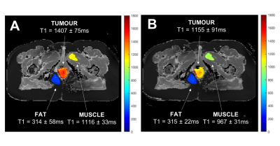 |
Oxygen-Enhanced MOLLI T1 Mapping during Chemoradiotherapy in Anal Squamous Cell Carcinoma
Emma Bluemke1, Daniel Bulte1, Ambre Bertrand1, Ben George2, Rosie Cooke2, Kwun-Ye Chu3, Lisa Durrant2, Vicky Goh4, Clare Jacobs2, Stasya Ng5, Victoria Strauss6, Maria Hawkins3, and Rebecca Muirhead2
1Institute of Biomedical Engineering, University of Oxford, Oxford, United Kingdom, 2Department of Oncology, Oxford University Hospitals Trust, Oxford, United Kingdom, 3Radiotherapy Department, Oxford University Hospitals NHS Foundation Trust, Oxford, United Kingdom, 4Department of Oncology, CRUK/MRC Oxford Institute for Radiation Oncology, Oxford, United Kingdom, 5Cancer Imaging, School of Biomedical Engineering & Imaging Sciences, King's College London, London, United Kingdom, 6Centre for Statistics in Medicine, NDORMS, University of Oxford, Oxford, United Kingdom
Early identification of patients in need of radiotherapy treatment intensification would allow tailored radiotherapy dose. We hypothesize that patients with poor vascularity or hypoxia will correlate with these patients, and T1 changes from oxygen-enhanced MRI could provide indications of tumour perfusion. We acquired T1-maps before and after 8-10 fractions of radiotherapy and examined whether the oxygen-enhanced MRI response relates to clinical outcome. There was a significant increase in tumour T1 across patients following chemoradiotherapy (p<0.001). Before chemoradiotherapy, OE-MRI showed no significant changes in tumour T1, however after receiving chemoradiotherapy, OE-MRI showed a significant decrease in tumour T1 (p<0.001).
|
|
4734.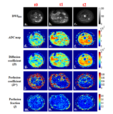 |
Response Evaluation in Osteosarcoma: RECIST1.1 criteria Versus Intravoxel Incoherent Motion
Esha Baidya Kayal1, Devasenathipathy Kandasamy2, Kedar Khare3, Sameer Bakhshi4, Raju Sharma2, and Amit Mehndiratta1,5
1Centre for Biomedical Engineering, Indian Institute of Technology, Delhi, New Delhi, India, 2Radio Diagnosis, All India Institute of Medical Sciences, New Delhi, New Delhi, India, 3Department of Physics, Indian Institute of Technology, Delhi, New Delhi, India, 4Department of Medical Oncology, Dr. B.R. Ambedkar Institute-Rotary Cancer Hospital (IRCH), All India Institute of Medical Sciences, New Delhi, New Delhi, India, 5Department of Biomedical engineering, All India Institute of Medical Sciences, New Delhi, New Delhi, India Poster Permission Withheld
Osteosarcoma (OS) is the most common primary malignant bone tumor in children and adolescents. Monitoring treatment response during chemotherapy might help in better and personalized therapeutic options improving overall therapeutic outcome. RECIST1.1 is the standard imaging based non-invasive treatment response evaluation criteria in solid tumors. Quantitative Intravoxel incoherent motion (IVIM) parameters and their histogram analysis were performed in Osteosarcoma in characterizing chemotherapy response with respect to RECIST1.1 criteria. IVIM parameters and its histogram analysis revealed clinically useful information in characterizing chemotherapy response in Osteosarcoma.
|
|
4735.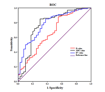 |
Quantitative Diagnosis of Microvascular Invasion of Hepatocellular Carcinoma in IVIM diffusion-weighted MR imaging
tianyuan zhang1, nan li1, xianyue quan1, yuan wang1, and xinquan chen1
1Zhujiang Hospital, Southern Medical University, Guangzhou, China
This study selected 48 HCC patients admitted to our hospital from January 2018 to June 2019, and divided them into 34 HCC patients in the MVI negative group and 14 HCC patients in the MVI positive group according to the postoperative pathological results. And then the round area of ROI was used to measure the lesion on the IVIM image. Finally, the diagnostic value of D, D*, f and ADC to MVI was analyzed by statistical software. The results showed that IVIM-DWI was helpful in evaluating the MVI of HCC, and D value had the best diagnostic efficacy.
|
|
4736.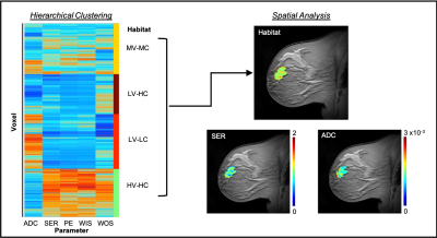 |
Quantifying heterogeneity in DW-MRI and DCE-MRI data of breast cancer for the prediction of treatment response: Preliminary results
Anum K Syed1, Chengyue Wu1, Angela M Jarrett1, Anna G Sorace2, John M Virostko1, and Thomas E Yankeelov1
1University of Texas at Austin, Austin, TX, United States, 2University of Alabama at Birmingham, Birmingham, AL, United States
We investigate multiparametric analysis of DW- and DCE-MRI data to identify tumor subregions indicative of response in breast cancer patients undergoing neoadjuvant therapy (NAT). The analysis showed significant increases in low vascular perfusion, low cellularity subregions early in the course of NAT for patients who achieved pathological complete response and significant decreases in high vascular perfusion, high cellularity subregions on the border with parenchyma. High-dimensional analysis of multimodal MRI data can be utilized to identify subregions of response within patients and these characterizations of intratumoral heterogeneity can potentially be used to guide therapy for improved patient outcome.
|
|
4737. |
Accuracy and precision of preclinical renal T2 mapping using monoexponential fitting and dictionary matching methods
Carlota M. Santos1, Andreia C. Freitas1, João S. Periquito2, Paula R. Delgado2, Thoralf Niendorf2, Andreas Pohlmann2, and Rita G. Nunes1
1ISR-Lisboa/LARSyS and Department of Bioengineering, Instituto Superior Técnico – Universidade de Lisboa, Lisbon, Portugal, 2Berlin Ultrahigh Field Facility (B.U.F.F.), Max Delbrück Center for Molecular Medicine in the Helmholtz Association, Berlin, Germany
Parametric mapping of T2* and T2 can be used to evaluate renal blood oxygenation. Efficient T2 mapping can be performed using a multi spin-echo sequence and fitting the pixel-wise signal with a monoexponential model although known to introduce T2 bias, or by matching it to pre-computed Echo Modulation Curves, accounting for all echo pathways. A Monte Carlo simulation was performed using a numerical kidney phantom to evaluate accuracy and precision of both approaches under the influence of noise. The results demonstrated an improved T2 accuracy using the EMC method compared to monoexponential fits and low dependence on tissue-type.
|
|
4738.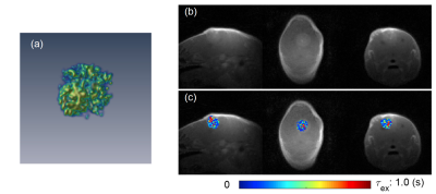 |
Comparison of tumor cellular-interstitial water exchange rates measured by DCE-MRI and diffusion time-dependent diffusion kurtosis imaging
Jin Zhang1, Karl Kiser1, Ayesha Bharadwaj Das1, Chongda Zhang1, and Sungheon Gene Kim1
1New York University School of Medicine, New York, NY, United States
Cellular-interstitial water exchange rate is an important property of tumor, potentially associated with tumor aggressiveness and treatment response. It is not trivial to measure the water exchange rate, not to mention the challenges in measuring it at a high spatial resolution. In this study, we use the 3D-UTE-GRASP method1 to generate water exchange rate maps and compare them with those measured using diffusion time-dependent diffusion kurtosis imaging2.
|
|
4739.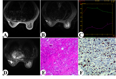 |
Evaluating Ki-67 status in breast cancer with DCE-MRI features
Jing Zhou1,2, Hongna Tan1,2, and Meiyun Wang1,2
1Department of Radiology, Henan Provincial People’s Hospital & Zhengzhou University People’s Hospital, Zhengzhou City, China, 2Imaging Diagnosis of Neurological Diseases and Research Laboratory of Henan Province, Zhengzhou City, China
Breast cancer is one most common cancer in female worldwide. Ki-67 index is an significant biomarker whose expression status is closely related with response to chemotherapy and the disease prognosis in patients with breast cancer. This study retrospectively investigated the relationship between DCE-MRI features and Ki-67 status in breast carcinoma. The results indicated the type of washout TIC and heterogeneous type of reinforcement is more common in high Ki-67 status in patients with breast cancer. The DCE-MRI imaging features can non-invasively assess Ki-67 status in breast carcinoma before surgery.
|
|
4740.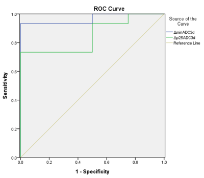 |
Whole volume histogram analysis of water diffusion predicts response to induction chemotherapy in patients with head and neck cancer
Xi Lin1, Kai-Lun Cheng2, Hsueh-Ju Lu3, Ying-Hsiang Chou1,4, Yeu-Sheng Tyan1,2, and Ping-Huei Tsai1,2
1Department of Medical Imaging and Radiological Sciences, Chung Shan Medical University, Taichung, Taiwan, 2Department of Medical Imaging, Chung Shan Medical University Hospital, Taichung, Taiwan, 3Division of Medical Oncology, Department of Internal Medicine, Chung Shan Medical University Hospital, Taichung, Taiwan, 4Department of Radiation Oncology, Chung Shan Medical University Hospital, Taichung, Taiwan
Head and neck squamous cell carcinoma (HNSCC) is one of the most common cancers worldwide. While quantified ADC value has been demonstrated to reveal information on the tumor microstructure, the capbility of predicting treatment response is still controversial. This study aim to assess the ability of whole-volume histogram analysis of water diffusion to predict response to induction chemotherapy in patients with HNSCC using multishot readout-segmented MRI. Our findings indicate that the ΔminADC and ΔADC25 values, could be potential biomarkers for predicting early response to induction chemotherapy in patients with HNSCC, which may facilitate the determination of further therapeutic strategy.
|
|
4741.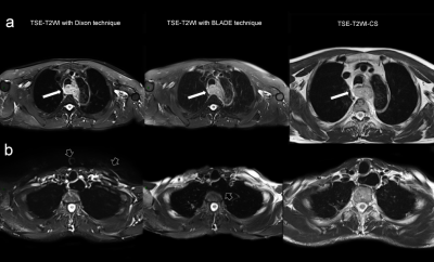 |
Optimizing TSE-T2WI MR imaging of esophageal cancer at 3T: evaluating the effects of chemical shift FS, Dixon and BLADE techniques to image quality
Yawen Wang1, Zhu Wang1, Ruobing Wang1, Dong Qu1, Qinglei Shi2, and Peihua Wu1
1Department of Imaging Diagnosis, National Cancer Center/Cancer Hospital, Chinese Academy of Medical Sciences, Beijing, China, 2Diagnostic Imaging, Siemens Healthcare Ltd, Beijing, China
This study evaluated the effects of chemical shift fat saturation (FS), Dixon and BLADE techniques to image quality in high-resolution imaging of esophageal cancer using a two-dimensional turbo spin echo (TSE) sequence. After quantitative and subjective quality comparing, TSE-T2WI-CS got higher image quality than T2WI Dixon and T2WI BLADE sequences, which demonstrated certain advantages in the diagnosis of esophageal cancer, while T2WI Dixon and T2WI BLADE sequences also has some advantages respectively.
|
|
4742.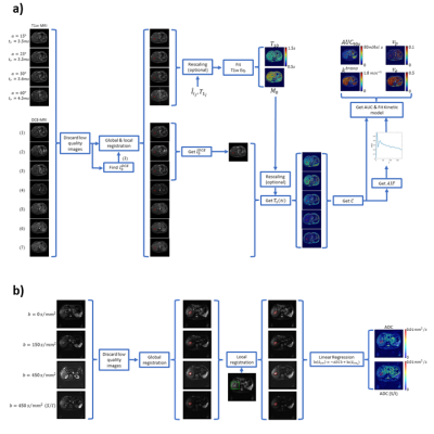 |
Quantitative MRI in a Phase 1 Clinical Study of the Vascular Disrupting Agent Crolibulin
Andres Arias Lorza1, Harshan Ravi1, Rohit Philip2, Jean-Philippe Galons2, Theodore Trouard2, Nestor Parra1, William Read3, Raoul Tibes4, Ronald Korn5, and Natarajan Raghunand1
1Moffitt Cancer Center, Tampa, FL, United States, 2University of Arizona, Tucson, AZ, United States, 3Emory University School of Medicine, Atlanta, GA, United States, 4Perlmutter Cancer Center, New York, NY, United States, 5Imaging Endpoints, Scottsdale, AZ, United States
Diffusion and DCE-MRI were performed at baseline and 2-3 days following Crolibulin (EPC2407) treatment in a phase 1 clinical study of this vascular disrupting agent (VDA). Several functional parameter maps were computed and co-registered across scan dates in 11 subjects with advanced solid tumors. We measured changes in these MRI parameters that indicate cell swelling and vascular reduction following treatment. We identified multivariate combinations of changes in these MRI parameters that are correlated with the dose, AUC and Cmax of Crolibulin, respectively, information that can guide Crolibulin dosing in clinical trials of this VDA in combination with cytotoxic drugs.
|
|
4743.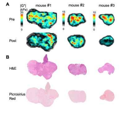 |
Biomechanical Response of Orthotopic Pancreatic Tumours to the Vascular Disrupting Agent ZD6126 Assessed by Magnetic Resonance Elastography
Jin Li1, Emma L. Reeves1, Jessica K.R. Boult1, Craig Cummings1, Jeffrey C. Bamber1, Ralph Sinkus2, Yann Jamin1, and Simon P. Robinson1
1Division of Radiotherapy & Imaging, The Institute of Cancer Research, London, United Kingdom, 2Division of Imaging Sciences and Biomedical Engineering, King's College London, King’s Health Partners, St. Thomas’ Hospital, London, United Kingdom
MRE-derived shear stiffness |G*| of orthotopic pancreatic tumours was sensitive to a heterogeneous treatment response induced by the vascular disrupting agent ZD6126.
|
|
4744.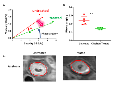 |
Magnetic Resonance Elastography Predicts Early Resistance to Chemotherapy in Cancer
Rami Mustapha1, Omar Darwish2, Peter Gordon1, Diana Cash3, Camilla Simmons3, Ralph Sinkus2,4, and Tony Ng1,5
1School of Cancer and Pharmaceutical Sciences, King's College London, London, United Kingdom, 2Division of Imaging Sciences and Biomedical Engineering, King's College London, London, United Kingdom, 3Department of Neuroimaging, Institute of Psychiatry, Psychology and Neuroscience, King's College London, London, United Kingdom, 4Laboratory of Vascular Translational Science, UMR1148,, INSERM-University Paris Diderot, Paris, France, 5UCL Cancer Institute, University College London, London, United Kingdom
There is an unmet need in cancer treatment for a non-invasive technique capable of identifying drug resistance very early in the therapeutic cycle. Our team has found that tumoral tissue changes its physical aspect as a response to therapeutic stress and that MRE can detect such changes by probing tissue biomechanics. Using a chemotherapy resistant pre-clinical model, we showed that tumors solidify as a resistance mechanism. Changes in the biomechanics were then correlated with changes Collagen and Hyaluronic acid depositions, both of which can affect the immune response.
|
|
4745.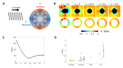 |
Extending an MR elastography based method for inferring total tumour pressure to multiple organs
Daniel Fovargue1, Jack Lee1, Marco Fiorito1, Adela Capilnasiu1, Siri Flogstad Svensson2, Kyrre Eeg Emblem2, Philippe Garteiser3, Aime Pacifique Manzi3, Gwenael Page3, Valerie Vilgrain4, Bernard Van-Beers3, Sweta Sethi5, Ayse Sila Dokumaci1,
Stefan Hoelzl1, Jurgen Henk Runge1,6, Jose de Arcos1, Keshthra Satchithananda7, David Nordsletten1,8, Arnie Purushotham7, and Ralph Sinkus1,3
1School of Biomedical Engineering and Imaging Sciences, King's College London, London, United Kingdom, 2Department for Diagnostic Physics, Oslo University Hospital, Oslo, Norway, 3INSERM, University Paris Diderot, Paris, France, 4Department of Radiology, Hopital Universitaire Beaujon, Clichy, France, 5Department of Research Oncology, Guy's and St Thomas' NHS Foundation Trust, London, United Kingdom, 6Radiology and Nuclear Medicine, Academic Medical Center, Amsterdam, Netherlands, 7Division of Cancer Studies, King's College London, London, United Kingdom, 8Department of Biomedical Engineering and Cardiac Surgery, University of Michigan, Ann Arbor, MI, United States
Both interstitial fluid pressure and solid pressure of tumours have been shown to correlate with decreased efficacy of treatment and potentially with poorer prognosis. This increased fluid and solid pressure causes tumours to push on surrounding tissue, leading to changes in tissue stiffness due to nonlinear effects. A previously presented method relates the magnitude of these changes, as measured by MR elastography, to pressure using a nonlinear biomechanical model. Here, this method is extended for use on both preliminary liver and brain data which show correlation between reconstructed pressure and invasion and tumour type, respectively.
|
|
4746.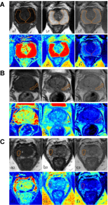 |
Quantitative mapping for prostate cancer detection in the peripheral zone using synthetic MRI
Weijing Zhang1, Huiming Liu2, Nian Lu1, Yanlin Zhu1, Long Qian3, Weiyin Liu3, Tie-bao Meng1, and Chuanmiao Xie2
1Department of Medical Imaging, Sun Yat-sen University Cancer Center, Guangzhou, China, 2Department of Radiology, Sun Yat-Sen University Cancer Center, Guangzhou, China, 3MR Research, GE Healthcare, Beijing, China
Our study aims to develop and evaluate an examination using Synthesis MRI based T1, T2, and proton density (PD) mapping for multiparametric characterization of prostate cancer. T1, T2 and PD mapping exhibited excellent differential diagnosis of normal prostate, prostatitis and Prostate cancer (PCa) and discrimination performance was better than ADC. T1, T2 and PD mapping of synthetic quantitative MRI seem to be a potential method in PCa detection. It may aid prostate biopsy by means of guiding and controlling localized therapy delivery. This preliminary study showed that synthetic MRI has a potential in the detection and characterization of PCa.
|
|
4747.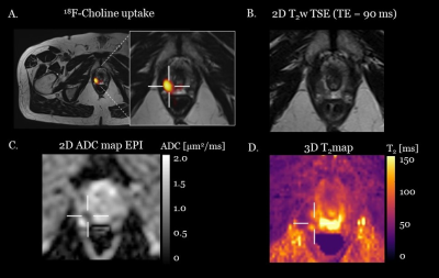 |
3D T2 mapping with dictionary-based matching in simultaneous PET/MR: a preliminary study in prostate cancer patients
Elisa Roccia1, Radhouene Neji1,2, Meghana Kulkarni3, Thomas Benkert4, James Stirling3, Sami Jeljeli3, Berthold Kiefer4, Gary Cook3, Vicky Goh3, and Isabel Dregely1
1Biomedical Engineering Department, School of Biomedical Engineering and Imaging Sciences, King's College London, London, United Kingdom, 2Siemens Healthcare Limited, Frimley, United Kingdom, 3Cancer Imaging Department, School of Biomedical Engineering and Imaging Sciences, King's College London, London, United Kingdom, 4Siemens Healthcare GmbH, Erlangen, Germany
In this work we aim to show the clinical feasibility of quantitative 3D T2 mapping for tissue characterization and with the potential for prostate cancer detection. We implemented a 3D T2-prepared multi-shot gradient echo sequence and a dictionary matching approach for quantitative T2 analysis. We tested it in three biopsy-proven prostate cancer patients who underwent a simultaneous 18F-Choline PET/MR scan. Quantitative T2 matched literature values and we observed reduced T2 in lesions compared to normal peripheral zone tissue even in only moderately differentiated cancers (Gleason 3+4). Lesion location matched uptake of simultaneously acquired 18F-Choline PET.
|
Session Topic: Cancer Imaging: Preclinical & Miscellaneous
Session Sub-Topic: Cancer Imaging: Miscellaneous 1
Digital Poster
Cancer
4748.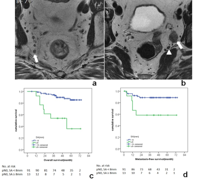 |
Baseline MRI detected lateral lymph node as a prognostic factor: a cohort study in pN0 low risk rectal cancer
Rui-Jia Sun1, Lin Wang1, Xiao-Ting Li1, Qiao-Yuan Lu1, Xiao-Yan Zhang1, and Ying-Shi Sun1
1Beijing Cancer Hospital, Beijing, China
Baseline MRI detected lateral lymph node was associated with poor prognosis in patients with pN0 low risk rectal cancer. For patients with MR detected lateral lymph node of a short axis at least 8mm, the present clinical strategies may be unlikely sufficient.
|
|
4749.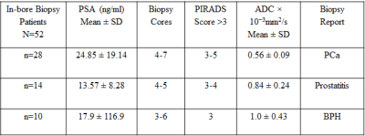 |
Prebiopsy mpMRI to Increase the Detection Rate and Reduce the Overdiagnosis of Prostate Cancer
Sujeet Kumar Mewar1, Sanjay Sharma2, Sanjay Thulka3, Ekta Dhamijia3, Pradeep Kumar1, Sridhar Panaiyadiyan4, Rama Jayasundar1, S. Senthil Kumaran1, S. Datta Gupta5, N.R. Jagannathan6, Rajeev Kumar4, and Virendra kumar1
1Department of NMR, All India Institute of Medical Sciences, New Delhi, India, Ansari nagar, India, 2Department of Radio-diagnosis, RPC, All India Institute of Medical Sciences, New Delhi, India, Ansari nagar, India, 3Department of Radio-diagnosis, IRCH, All India Institute of Medical Sciences, New Delhi, India, Ansari nagar, India, 4Department of Urology, All India Institute of Medical Sciences, New Delhi, India, Ansari nagar, India, 5Department of Pathology, All India Institute of Medical Sciences, New Delhi, India, Ansari nagar, India, 6Present address.Department of Radiology, Chettinad Academy of Research and Education, Kelambakkam, Tamil Nadu, India
The results of the study showed that mpMRI guided in-bore biopsy has the potential to increase the detection rate of PCa. Prebiopsy mpMRI of prostate was used for identification of suspicious areas of malignancy which were targeted for PCa detection and reducing unnecessary biopsies. The detection rate was compared with TRUS guided 12 core biopsy. The data also indicated that using prebiopsy mpMRI may also help to reduce unnecessary biopsies. The PCa detection rate of in-bore mpMRI targeted biopsy was 53.84 % compared to 37.27% for TRUS guided biopsy.
|
|
4750.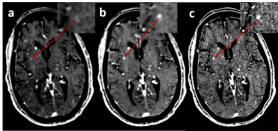 |
Improving tumor-tissue contrast by increasing spatial resolution
Jinghua Wang1,2, Kim M Cecil1, Mary Gaskill-shipley1,2, Lili He3,4, Bin Zhang5, Michael Lamba6, Lily Wang1, and Achala Vagal1
1Radiology, University of Cincinnati, Cincinnati, OH, United States, 2Brain Tumor Center, University of Cincinnati, Cincinnati, OH, United States, 3Pediatrics, University of Cincinnati, Cincinnati, OH, United States, 4The Perinatal Institute and Section of Neonatology, Perinatal and Pulmonary Biology, Cincinnati Children’s Hospital Medical Center, Cincinnati, OH, United States, 5Division of Biostatistics and Epidemiology, Cincinnati Children’s Hospital Medical Center, Cincinnati, OH, United States, 6Radiation Oncology, University of Cincinnati, Cincinnati, OH, United States
There is broad agreement that increased tumor-tissue contrast significantly improves tumor visibility. It is not yet clear whether increasing spatial resolution will improve tumor-tissue contrast. In this study, we found that the increasing spatial resolution can significantly increase tumor-tissue contrast when tumor size is comparable to the spatial resolution, but provides an invariant contrast when tumor size is much larger than the spatial resolution.
|
|
4751.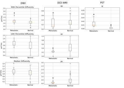 |
Assessment of Metastatic Lymph Node in Head and Neck Cancer Using Simultaneous 18F-FDG-PET, DCE-MRI, and DWI
Jenny Chen1, Mari Hagiwara1, Artem Mikheev1, Henry Rusinek1, Jean Logan1, Elcin Zan1, and Sungheon Gene Kim1
1Department of Radiology, NYU School of Medicine, New York, NY, United States
Detecting malignant lymph nodes in the neck remains a challenge such that most patients still have to consider aggressive treatment including nodal dissection. In this study, we used positron emission tomography with 18F-fluorodeoxyglucose (18F-FDG PET), dynamic contrast enhanced magnetic resonance imaging (DCE-MRI), and diffusion weighted imaging (DWI) to assess head and neck cancer cases and consider the feasibility of using all three imaging methods in synergy to assess the regional lymph nodes.
|
|
4752.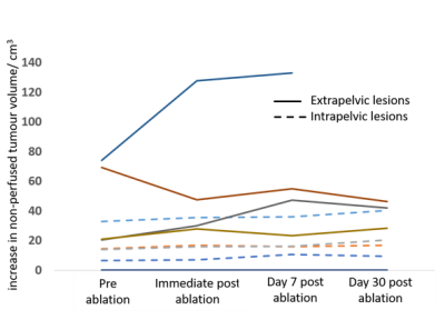 |
Imaging and pain response in extra vs intra-pelvic recurrent gynaecological tumours treated with MR guided HIFU: a pilot study
Georgios Imseeh1,2, Sharon L Giles3, Ian Rivens4, Alexandra Taylor2, Gail ter Haar4, Erica Scurr3, and Nandita M deSouza1,3
1Imaging, Institute of Cancer Research, Sutton, Surrey, United Kingdom, 2Clinical Oncology, The Royal Marsden NHS Foundation Trust, London, United Kingdom, 3MRI Unit, The Royal Marsden NHS Foundation Trust, Surrey, United Kingdom, 4Therapy Ultrasound, Institute of Cancer Research, Sutton, Surrey, United Kingdom
MR guided High Intensity Focused Ultrasound in 10 patients with symptomatic recurrent gynecological tumors treated for pain palliation resulted in higher temperatures in shallower extra-pelvic tumors compared to intra-pelvic ones (62.9±11.7oC vs. 49.4±2.8oC over median 16 sec sonication) despite lower energy delivery in them (39.1±23.6 kJ vs. 72.1±24.3 kJ). In 3 of 5 extra-pelvic tumors, an increase in the non-perfused tumor volume immediately after treatment was seen, indicating edema. At Day 30, 5 patients met the criteria of pain response, 3 with extra-pelvic tumors and 2 with intra-pelvic tumors.
|
|
4753.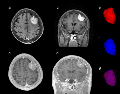 |
Identification of the dural tail sign of meningiomas with contrast-enhanced zero echo time (ceZTE) imaging
Lian Jian1, Weiyin Vivian Liu2, Xiaohuang Yang 1, and Xiaoping Yu1
1Radiology, Hunan Cancer Hospital, Hunan, China, 2MR Research, GE Healthcare, Beijin, China
The dural tail sign is often the pathological presence of meningiomas and recently reckoned as a response to vascular congestion and edema. Zero echo time (ZTE) imaging is capable of capturing signal of tissues with relatively short T2* such as pia mater right after onset of the radiofrequency pulse. The significant enhanced rim of meningioma on ecZTE showed the meningioma were supplied by both intracranial and extracranial arteries. Contrast-enhanced ZTE might be listed as a routine imaging sequence to improve accuracy of discriminating the tumor-cortex-dura interface and offer additional clinical information such as vascular growth and supply in meningiomas.
|
|
4754.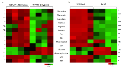 |
Hypoxia alters normal fibroblast metabolism towards a cancer associated fibroblast phenotype
Jesus Pacheco-Torres1, Tariq Shah1, William Nathaniel Brennen2, Flonne Wildes1, and Zaver M Bhujwalla1,2,3
1Division of Cancer Imaging Research, The Russell H. Morgan Department of Radiology and Radiological Science, The Johns Hopkins University School of Medicine, Baltimore, MD, United States, 2Sidney Kimmel Comprehensive Cancer Center, The Johns Hopkins University School of Medicine, Baltimore, MD, United States, 3Radiation Oncology and Molecular Radiation Sciences, The Johns Hopkins University School of Medicine, Baltimore, MD, United States
Fibroblasts play a pivotal role in cancer progression. In prostate cancer, fibroblasts have been shown to induce growth, confer castration-resistance, and increase metastatic potential. To further understand how fibroblasts respond to hypoxic tumor microenvironments that are frequently observed in prostate cancer, here we have characterized the effects of hypoxia on normal prostate fibroblast metabolites as detected by high resolution 1H magnetic resonance spectroscopy. We found that hypoxia induced metabolic changes in normal prostate fibroblasts that mimicked the metabolites detected in prostate cancer associated fibroblasts (CAFs), highlighting the potential role of hypoxia in the transition of normal fibroblasts to CAFs.
|
|
4755.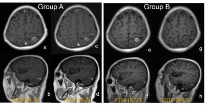 |
Performance of brain MR automatic slice positioning in Metastatic Brain Tumors
Jingjing Gao1, Min Du1, Shaodong Li1, Kai Xu1, and Zhongshuai Zhang2
1Department of Radiology, Affiliated Hospital of Xuzhou Medical University, Xuzhou, Xuzhou, China, 2SIEMENS Healthcare, Shanghai,China, Shanghai, China
This study evaluated the diagnostic sensitivities and confidence for monitoring the progression of brain metastases using brain auto-positioning and manually positioning methods. We found that automatic slice positioning do help in finding lesions by comparing initial imaging side-by-side, but the technique does not sacrifice the diagnostic sensitivity and the diagnostic confidence when comparing with the conventional manually positioning method for brain tumor.
|
|
4756.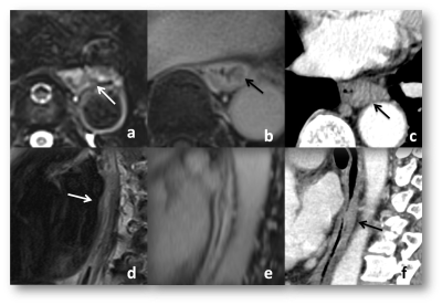 |
The feasibility of StarVibe in MR imaging and CT volume imaging in measuring the length of esophageal cancer after neo-adjuvant therapy on 3-T
Ruobing Wang1, Zhu Wang1, Yawen Wang1, Dong Qu1, Qinglei Shi2, and Peihua Wu1
1National Cancer Center/Cancer Hospital, Chinese Academy of Medical Sciences, Beijing, China, 2MR Scientific Marketing, Diagnostic Imaging, Siemens Healthcare Ltd, Beijing, China
This study evaluated the length of esophageal cancer lesions after neoadjuvant treatment with MR and thin-layer CT scanning using the length of gross specimens as the gold standard. MR sequence included T2WI and contrast-enhanced StarVibe (DCE StarVibe). In this study, two readers measured the length of esophageal cancer lesions on T2WI, DCE StarVibe and CT images. It was found that contrast-enhanced Starvibe and thin-slice CT can be used to measure the lenth of esophageal cancer after neoadjuvant treatment.
|
|
4757.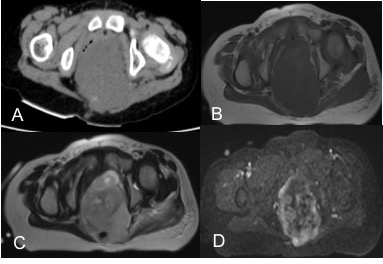 |
Radiopathological features of primary extragonadal endodermal sinus tumor
Fang fang Fu1, Cuiyun Chen1, Yan Bai1, Qiuyu Liu1, Kaiyu Wang2, and Meiyun Wang1
1Henan Provincial People’s Hospital, People’s Hospital of Zhengzhou University, Zhengzhou, China, 2Department of MR Research China, GE Healthcare, Beijing 100000, China., Beijing, China
The extragonadal endodermal sac tumor (EEST) is an extremely rare malignant germ cell tumor and has a poor prognosis. Therefore, familiarity of the radiological characteristics of EESTs could help preoperative diagnosis and improve surgical management of patients. In this study, we investigate CT and MRI characteristics of 25 patients with EESTs correlated with pathological results, for better understanding and cognition in the diagnosis of EEST. Finally, we found that imaging features coupled with clinical data and the serum AFP check are helpful for the diagnosis of EEST and could improve the accuracy of the preoperative diagnosis.
|
|
4758.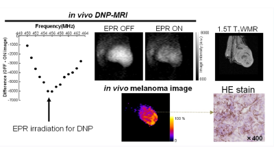 |
In vivo melanoma imaging based on melanin free radical using in vivo dynamic nuclear polarization magnetic resonance imaging
Fuminori Hyodo1, Shinichi Shoda1, Tomoko Nakaji2, Hinako Eto3, Tatsuya Naganuma2, Norikazu Koyasu1, Masaharu Murata3, and Masayuki Matsuo1
1Gifu University, Gifu, Japan, 2Japan Redox Inc., Fukuoka, Japan, 3Kyushu University, Fukuoka, Japan
Malignant melanoma is one of the most progressive tumors in humans with increasing incidence worldwide. Dynamic nuclear polarization (DNP)-MRI is a noninvasive imaging method to obtain the spatio-temporal information of free radicals. If endogenous free radicals in melanin pigment could be utilized as a bio-probe for DNP-MRI, this will be an advantage for the specific enhancement of melanoma tissues. We report that biological melanin pigment induced a in vivo DNP effect by interacting with water molecules. In addition, we demonstrated in vivo melanoma imaging based on the DNP effects of endogenous free radicals in the melanin pigment of living mice.
|
|
4759.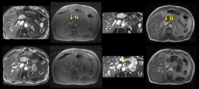 |
USPIO-Enhanced Imaging of Mediastinal Lymph Nodes in Esophageal Cancer with Sparse Radial MRI
Ivo Maatman1, Didi de Gouw2, John Hermans1, Kai Tobias Block3, Marnix Maas1, and Tom Scheenen1
1Department of Radiology and Nuclear Medicine, Radboud university medical center, Nijmegen, Netherlands, 2Department of Surgery, Radboud university medical center, Nijmegen, Netherlands, 3Department of Radiology, New York University, New York, NY, United States
Clinical imaging techniques are unable to outline metastatic spread of esophageal cancer to nearby lymph nodes. This deficiency is predominantly due to significant motion in the upper abdomen. We radially acquired MRI data with a contrast agent of USPIO nanoparticles to image the lymph nodes of an esophageal cancer patient. After retrospective gating the data into motion phases we created images with a compressed sensing reconstruction. We also acquired traditional multi-gradient echo sequences in breath-hold and apnea. The radially acquired, compressed sensing reconstructed and motion-gated images appear of similar diagnostic quality to those obtained with traditional modalities.
|
|
4760.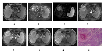 |
Value of Gd-EOB-DTPA-enhanced MRI in detecting residual hepatocellular carcinoma and evaluating regional hepatic function injury afterTACE
Hai-Feng Liu1, Yong-Sheng Xu2, Qing Wang1, Ji-Lei Zhang3, and Wei Xing1
1Third Affiliated Hospital of Soochow University, changzhou, China, 2First Hospital of Lanzhou University, lanzhou, China, 3Philips Healthcare, Shanghai, China, shanghai, China
In this study, we sought to investigate the value of Gd-EOB-DTPA-enhanced MRI in diagnosing residual hepatocellular carcinoma (HCC) and evaluating regional hepatic function injury after transarterial chemoembolization (TACE).Gd-EOB-DTPA-enhanced MRI is a sensitive imaging in diagnosing residual HCCs, especially for the imaging feature of arterial enhancement and DWI hyperintensity, and SNR value measured on HBP can be used as a valuable index to assess regional hepatic function injury after TACE.
|
|
4761.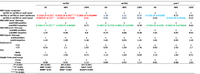 |
The value of novel magnetic resonance imaging tumor response score for locally advanced rectal cancer: a prospective and multicenter study
Zhen Guan1 and Ying shi Sun1
1Beijing cancer hospital, Beijing, China
Our study enrolled four possible predictive imaging parameters of the pTRG and establised the magnetic resonance imaging tumor response score for the theraputic evaluation and prognosis prediction of LARC. The objective and semi-quantitative mrTRS was established for the first time in this study, which is an independent factor of overall survival, distant metastasis and local recurrence.
|
|
4762.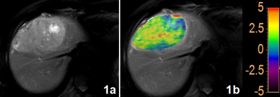 |
3D amide proton transfer MR imaging for evaluation of hepatocellular carcinoma
Nan Wang1, Qingwei Song1, Renwang Pu1, Mingli Gao1, Lihua Chen1, Jiazheng Wang2, Zhiwei Shen2, Liangjie Lin2, and Ailian Liu1
1The First Affiliated Hospital of Dalian Medical University, Dalian, China, 2Philips Healthcare, Beijing, China
Amide proton transfer weighted (APTw) imaging has been proved of great value in diagnoses of brain tumors, and has attracted increased interest for evaluation of abdomen diseases. Different regions of the hepatocellular carcinoma (HCC) are associated with different pathophysiological changes. The APTw-MRI was employed in this study to evaluate the protein and polypeptide molecular levels in different regions of HCC, and a significant difference was observed between APT values measured in the center of HCC and outside the tumor.
|
|
4763.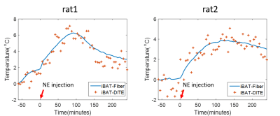 |
Validation of accuracy of fat-referenced PRFS thermometry on activated brown adipose tissue in rat in vivo
Chuanli Cheng1, Qian Wan1, Yangzi Qiao1, Min Pan2, Changjun Tie1, Hairong Zheng1, Xin Liu1, and Chao Zou1
1Shenzhen Institutes of Advanced Technology,Chinese Academy of Sciences, Shenzhen, China, 2Shenzhen Hospital of Guangzhou University of Chinese Medicine, Shenzhen, China
Brown adipose tissue (BAT) is a special adipose tissue which burns fat and dissipates energy in heat. As a result, heat production capacity of BAT is considered to represent its activity directly. In this study, we aimed to evaluate the accuracy of the proposed magnetic resonance fat-referenced PRFS method on BAT in vivo in rat after activated by Norepinephrine. The preliminary results show the PRFS based thermometry can describe the temperature change in activated BAT accurately, implying that magnetic resonance thermometry (MRT) is a useful tool to characterize the BAT activity.
|

 Back to Program-at-a-Glance
 View the Poster
 Watch the Video
 Back to Top