
Scientific Session
MRI of the Kidneys
Session Topic: MRI of the Kidneys
Session Sub-Topic: Kidney
Oral
Body
| Wednesday Parallel 3 Live Q&A | Wednesday, 12 August 2020, 15:15 - 16:00 UTC | Moderators: Pim Pullens |
Session Number: O-30
 |
0941.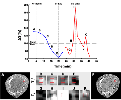 |
Single Nephron Glomerular Filtration and Macromolecular Dynamics in Perfused Kidneys using MRI
Edwin J. Baldelomar1, Scott C. Beeman2, Jennifer R. Charlton3, and Kevin M. Bennett4
1Radiology, Washington University in St. Louis, St. Louis, MO, United States, 2Biomedical Engineering, Arizona State University, Tempe, AZ, United States, 3Pediatrics, University of Virginia, Charlottesville, VA, United States, 4Radiology, Washington University in St. Louis, Saint Louis, MO, United States
In this work, we use contrast agents cationic ferritin and gadolinium-DTPA (Gd-DTPA) to visualize dynamics of macromolecules and freely filtering particles in individual nephrons throughout entire perfused rat kidneys. Further, we also look at dynamics in kidneys that received a vasoconstriction agent, angiotension II (AngII). Voxel time courses were fitted with a bi-exponential model for each experiment (Experiment I, CF infusion and Experiment II, Gd-DTPA bolus). From fitting we assess CF uptake rates and measure single nephron glomerular filtration rate (snGFR). CF uptake rates and values of snGFR were mapped spatially and observed to be heterogeneously distributed throughout the kidney.
|
0942.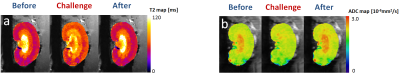 |
MRI Assessment of Renal Tubular Volume Fraction with an IVIM-NNLS Approach Under Increased Tubular Pressure
Joao Santos Periquito1, Kathleen Cantow2, Thomas Gladytz3, Bert Flemming2, Dirk Grosenick3, Erdmann Seeliger2, Thoralf Niendorf1, and Andreas Pohlmann1
1Max Delbrueck Center for Molecular Medicine, Berlin, Germany, 2Institute for Vegetative Physiology, Charité – Universitaetsmedizin Berlin, Berlin, Germany, 3Physikalisch-Technische Bundesanstalt (PTB), Berlin, Germany
The measurement of tubular volume fraction changes in the kidney may be valuable as a confounder of T2*-derived tissue oxygenation and as a potential biomarker. Diffusion weighted imaging provides information about in-vivo water mobility which can be linked to three sources: tissue water diffusion, blood perfusion within intrarenal microvasculature, and tubular fluid. In this work we explore the feasibility of assessing tubular volume fraction changes using the non-negative least squares (NNLS) approach under different physiological conditions.
|
|
0943.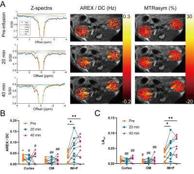 |
Delayed urea differential enhancement CEST (dudeCEST)-MRI with T1 correction for monitoring renal urea handling
Soo Hyun Shin1, Brandon Zhang1, K. L. Barry Fung1, Michael F. Wendland2, and Moriel H. Vandsburger1
1Department of Bioengineering, University of California, Berkeley, Berkeley, CA, United States, 2Berkeley Preclinical Imaging Core (BPIC),University of California, Berkeley, Berkeley, CA, United States
Urea recycling is a major component of renal tubular function and may provide an in vivo surrogate for tubular dysfunction in renal diseases. We demonstrate an approach of delayed urea differential enhancement CEST (dudeCEST)-MRI, which detects enhanced urea CEST contrast specific to the inner medulla and papilla of the mouse kidney at 20 minutes after urea injection. To enhance quantification while accounting for different T1 values within the kidney, apparent exchange-dependent relaxation (AREX) correction was applied. The combination of dudeCEST with AREX analysis will be a useful platform for assessment of renal urea recycling as a surrogate for tubular dysfunction.
|
|
0944.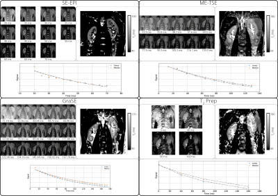 |
A Comparison of T2 Mapping Methods in the Kidneys
Alexander J Daniel1, Eleanor F Cox1, Charlotte E Buchanan1, and Susan T Francis1
1Sir Peter Mansfield Imaging Centre, University of Nottingham, Nottingham, United Kingdom
Renal T2 mapping is still in its infancy with little consensus on methodology between studies, this leads to a variation in T2 measurements between studies. Here four T2 mapping methods (Spin Echo-Echo Planar Imaging (SE-EPI), Multi-Echo Turbo Spin Echo (ME-TSE), Gradient Spin Echo (GraSE), and Carr-Purcell-Meiboom-Gill T2 preparation (T2 prep)) are compared on both a calibrated phantom and in-vivo kidneys. The GraSE technique was found to produce the most accurate maps relative to the phantom and form the clearest maps of the kidneys in-vivo, showing clear differences between cortical and medullary tissues.
|
|
0945.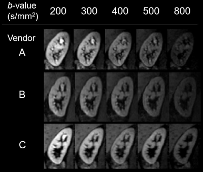 |
Travelling kidneys: Multicentre multivendor variability of renal diffusion-weighted imaging – preliminary results
Fabio Nery1, Charlotte Buchanan2, Andrew Priest3, João Sousa4, Michael Nation5, Iosif Mendichovszky3, Steven Sourbron6, Susan Francis2, and David Thomas7,8,9
1UCL Great Ormond Street Institute of Child Health, London, United Kingdom, 2Sir Peter Mansfield Imaging Centre, University of Nottingham, University Park, Nottingham, United Kingdom, 3Department of Radiology, Addenbrooke’s Hospital, Cambridge University Hospitals NHS Foundation Trust, Cambridge, United Kingdom, 4Imaging Biomarkers Group, Department of Biomedical Imaging Sciences, University of Leeds, Leeds, United Kingdom, 5Kidney Research UK, Peterborough, United Kingdom, 6Department of Infection, Immunity and Cardiovascular Disease, University of Sheffield, Sheffield, United Kingdom, 7Neuroradiological Academic Unit, UCL Queen Square Institute of Neurology, University College London, London, United Kingdom, 8Dementia Research Centre, UCL Queen Square Institute of Neurology, University College London, London, United Kingdom, 9Wellcome Centre for Human Neuroimaging, UCL Queen Square Institute of Neurology, University College London, London, United Kingdom
Multicentre validation studies are required to enable clinical translation of renal MRI biomarkers. Here, we report on the feasibility of standardising renal diffusion weighting imaging protocols and on the variability of renal apparent diffusion coefficient across a range of vendors. Results suggest feasibility of implementing near-identical renal diffusion weighted imaging acquisition protocols with product sequences and the potential of the apparent diffusion coefficient as a robust metric to characterise renal microstructure in multi-centre studies.
|
|
0946.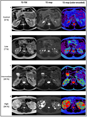 |
Novel magnetic resonance kidney biomarker Parenchyma-T2 for assessment of Autosomal dominant polycystic kidney disease
Florian Siedek1, Franziska Grundmann2, Kilian Weiss1, Daniel Pinto dos Santos1, Sita Arjune2, Stefan Haneder1, Thorsten Persigehl1, Roman-Ulrich Mueller2, and Bettina Baessler1
1Radiology, University of Cologne, Cologne, Germany, 2Department II of Internal Medicine, University of Cologne, Cologne, Germany
Novel biomarkers for a more sensitive and quick assessment of ADPKD patients especially in those cases where kidney function is still preserved and can be maintained is urgently needed. We analyzed in 139 patients and 10 healthy controls if magnetic resonance T2 mapping of the kidneys allows a sufficient differentiation of cyst fraction as a surrogate marker for disease severity. The new biomarker parenchyma-T2 showed the strongest correlation to renal cyst fraction and was faster to determine than the established biomarker htTKV. Consequently, parenchyma-T2 has the potential to serve as a novel predictive biomarker especially in early stages of disease.
|
|
|
0947. |
Diffusional Kurtosis Imaging of kidney in STZ-induced Diabetic Rats.
Youzhen Feng1, Zhongyuan Cheng1, Xiaoqiao Chen2, Xiaoqing Xiong1, Qiting Lin1, Dingkun SiTu1, Long Qian3, Huomei Chen1, and Xiangran Cai1
1Medical Imaging Center, First Affiliated Hospital, Jinan University, Guangzhou, Guangdong, China, Guangzhou, China, 2Medical Imaging Center, The Eighth Hospital of Sun Yat-sen University,shenzhen,China., Shenzhen, China, 3MR Research, GE Healthcare., Beijing, China
Diffusional kurtosis imaging (DKI) is an advanced diffusion model and could identify the heterogeneity of cellularity and microstructural complexity. To test whether the DKI could detect the functional changes of kidney in early Diabetic kidney disease (DKD), the STZ-induced diabetic rats were applied in current study. Further, the biochemical and pathological evidences would also be provided to compare with the DKI biomarker.
|
0948.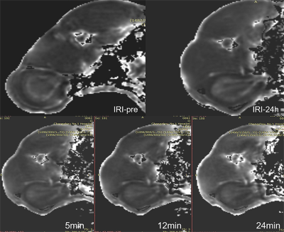 |
Evaluation of hypoxia with T2’ mapping in renal ischemia reperfusion injury
Jing gang Zhang1, Wei Xing1, Jie Chen1, and Weiqiang Dou2
1Radiology, Third Affiliated Hospital of Soochow University, Changzhou, China, 2MR Research China, GE Healthcare, Shanghai, China
The purpose was to explore if T2’mapping can assess renal oxygen in the ischemia-reperfusion injury (IRI). IRI models were established according to different ischemia time, followed by injection of furosemide 24 hours after IRI and consecutive MRI scans. Quantitative scores of oxygen were acquired with the hypoxic probe. We found that R2’ values of the inner and outer medulla were statistically significant. R2’ value of the outer medulla was highly correlated with oxygen scores. T2’mapping could serve as a quantitative biomarker to assess the renal oxygen and monitor the treatment in patients with IRI.
|
 Watch the Video