
Scientific Session
CEST, MT, Zero-TE and Relaxometry
Session Topic: CEST, MT, Zero-TE and Relaxometry
Session Sub-Topic: Relaxometry & Zero-TE
Oral
Contrast Mechanisms
| Tuesday Parallel 1 Live Q&A | Tuesday, 11 August 2020, 14:30 - 15:15 UTC | Moderators: Hai-Ling Cheng & Martijn Cloos |
Session Number: O-53
 |
0522.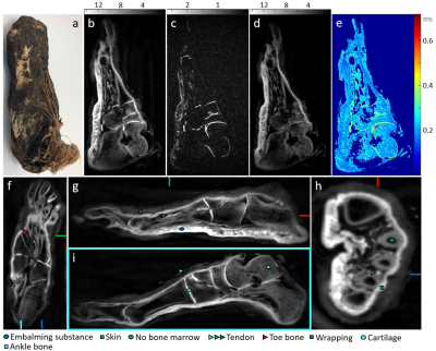 |
Short-T2 MRI of Ancient Egyptian Mummified Human Tissue
Emily Louise Baadsvik1, Markus Weiger1, Romain Froidevaux1, Manuela Barbara Rösler1, David Otto Brunner1, Lena Öhrström2, Patrick Eppenberger2, Frank J. Rühli2, and Klaas Paul Pruessmann1
1Institute for Biomedical Engineering, ETH Zurich and University of Zurich, Zurich, Switzerland, 2Institute of Evolutionary Medicine, University of Zurich, Zurich, Switzerland
Evolutionary medicine aims to study disease development over long timescales, and through the study of mummified human remains, tissue information dating back thousands of years becomes accessible. Due to their status as ancient relics, nonintrusive techniques are preferable, and to date CT imaging is the most common modality. However, CT images lack soft-tissue contrast, making complementary MRI data desirable. Due to the extensively dehydrated nature and short T2 times of mummified tissues, acquiring such data is challenging. This research explored the use of the zero echo-time sequences and a high-performance gradient in mummy MRI, yielding yet unparalleled image quality.
|
|
0523.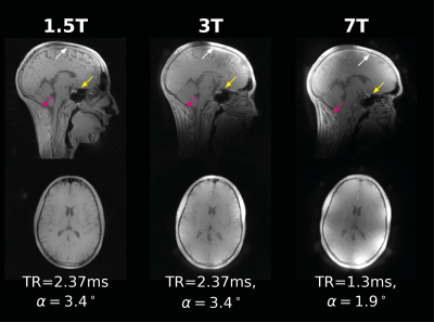 |
ZTE Imaging Across Field Strengths; Opportunities for Low-Field Imaging
Emil Ljungberg1, Brian Burns2, Tobias Wood1, Ana Beatriz Solana3, Peder E.Z. Larson4, Gareth J. Barker1, and Florian Wiesinger1,3
1Neuroimaging, King's College London, London, United Kingdom, 2ASL West, GE Healthcare, Menlo Park, CA, United States, 3ASL Europe, GE Healthcare, Munich, Germany, 4Univeristy of California, San Francisco, San Francisco, CA, United States
Zero Echo Time (ZTE) imaging enables ultra-fast, near silent data acquisition. In this work we demonstrate how contrast-to-noise, between white and gray matter in the brain, with a ZTE acquisition changes with field strength. At low field strength, maximum contrast is achievable with low RF power, which is promising for implementation on low field systems. We demonstrate through in vivo experiment that ZTE imaging can be performed at 1.5T/3T/7T, and how variable flip angle data at, for instance, 1.5T can be used for synthesising high quality T1-weighted MR images.
|
0524.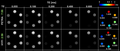 |
Efficient mapping of ultra-fast T2* decay
Romain Froidevaux1, Markus Weiger1, Manuela Barbara Rösler1, David Otto Brunner1, Benjamin Emanuel Dietrich1, and Klaas Paul Pruessmann1
1Institute for Biomedical Engineering, ETH Zurich and University of Zurich, Zurich, Switzerland With recent developments in gradient hardware even tissues with T2s down to tens of microseconds have become accessible for MRI. Hence, mapping signal decay or imaging short-T2 tissues selectively is of particular interest. This can be performed using ultra-short echo time imaging with multiple TEs. However, for typical resolutions this approach is limited to T2s down to hundreds of microseconds. In this work, the PETRA and HYFI techniques are utilized to map the signal decay of samples with T2s down to 54 μs. Considerably larger scan efficiency is obtained for the HYFI approach. |
|
0525.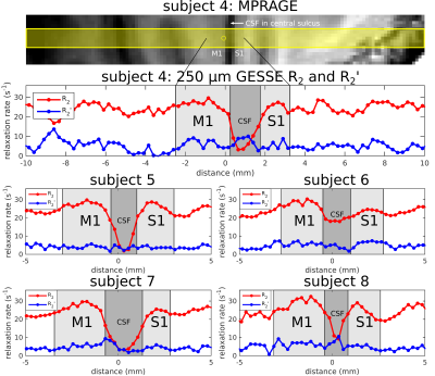 |
In vivo irreversible and reversible transverse relaxation rates in human cerebral cortex via line scans at 7T with 250 micron radial resolution
Mukund Balasubramanian1,2, Robert V. Mulkern1,2, and Jonathan R. Polimeni1,3,4
1Harvard Medical School, Boston, MA, United States, 2Department of Radiology, Boston Children's Hospital, Boston, MA, United States, 3Athinoula A. Martinos Center for Biomedical Imaging, Department of Radiology, Massachusetts General Hospital, Charlestown, MA, United States, 4Harvard-MIT Division of Health Sciences and Technology, Massachusetts Institute of Technology, Cambridge, MA, United States
A novel “line-scan GESSE” pulse sequence was used to measure irreversible and reversible transverse relaxation rates—R2 and R2´, respectively—in the cerebral cortex of eight healthy human subjects, scanned at 7T with extremely high resolution (250 μm) in the radial direction, i.e., perpendicular to the cortical surface. Within primary visual (V1), motor (M1) and somatosensory (S1) cortex, we observed patterns of R2 versus cortical depth that were quite consistent across subjects. These patterns are also consistent with the intracortical non-heme iron content in these areas, known from prior histology studies.
|
|
|
0526.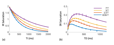 |
Field-Dependence of White Matter T1 Through Macromolecular Relaxation and Magnetization Transfer
Yicun Wang1, Peter van Gelderen1, Jacco A. de Zwart1, and Jeff H. Duyn1
1AMRI, LFMI, NINDS, National Institutes of Health, Bethesda, MD, United States
Brain tissue T1 predominately reflects local macromolecular content and is magnetic field strength dependent. In this study, we quantified the field dependence of macromolecular proton T1 (or rate Rm) in white matter by evaluating its effect exerted on the water signal through magnetization transfer. Inversion recovery and saturation recovery experiments were performed on a group of eight volunteers at 0.55, 1.5, 3 and 7 T, and were jointly analyzed using a two-pool exchange model. Rm was found to be close to inversely proportional to B0, consistent with previous in vitro findings at very low fields.
|
|
0527.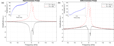 |
Adiabatic Inversion and T1 Relaxation of Bovine White Matter
Luke A. Reynolds1, Alex L. MacKay1,2,3, and Carl A. Michal1
1Physics & Astronomy, University of British Columbia, Vancouver, BC, Canada, 2Radiology, University of British Columbia, Vancouver, BC, Canada, 3MRI Research Centre, University of British Columbia, Vancouver, BC, Canada
Adiabatic pulses are commonly used in clinical MRI due to their insensitivity to B1 inhomogeneity and uniform flip angle over a selected bandwidth. When applied to white matter, they are generally assumed to saturate the magnetization of the non-aqueous protons in myelin. We performed adiabatic inversion recovery experiments on bovine brain in vitro using a solid state NMR spectrometer to directly observe the effects of adiabatic inversions on the non-aqueous signal. Substantial non-aqueous magnetization remains after typical adiabatic pulses. The state of the non-aqueous magnetization seriously impacts measurement of T1, yielding values dependent on the form of inversion pulse used.
|
0528.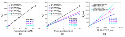 |
Short T1 Measurement Using an Inversion Recovery Prepared Three-Dimensional Ultrashort Echo Time Cones (3D IR-UTE-Cones) Method
Zhao Wei1,2,3, Yajun Ma1, Hyungseok Jang1, Wenhui Yang3, and Jiang Du1
1Department of Radiology, UC San Diego, San Diego, CA, United States, 2University of Chinese Academy of Sciences, Beijing, China, 3Institute of Electrical Engineering, Chinese Academy of Sciences, Beijing, China
In magnetic resonance imaging (MRI), T1 is an important biomarker for many diseases and plays a key role in affecting image contrast. We propose a novel T1 measurement method combining adiabatic inversion recovery with 3D ultrashort echo time cones pulse sequences (3D IR-UTE-Cones). This study aimed to verify the feasibility of using 3D IR-UTE-Cones to accurately calculate T1s of short T2* tissues. The results indicated that this method could precisely measure a broad range of T1s and that it performed better than commonly used clinical protocols in ultrashort T1 measurement.
|
|
 |
0529.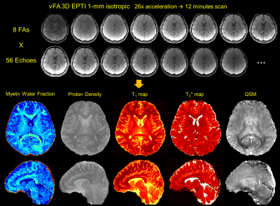 |
Variable Flip Angle 3D Echo Planar Time-Resolved Imaging (vFA 3D-EPTI) for Fast Multi-Compartment Quantitative Mapping
Zijing Dong1,2, Fuyixue Wang1,3, Kwok-Shing Chan4, Timothy G. Reese1, Berkin Bilgic1, José P. Marques4, and Kawin Setsompop1,3
1Athinoula A. Martinos Center for Biomedical Imaging, Massachusetts General Hospital, Charlestown, MA, United States, 2Department of Electrical Engineering and Computer Science, MIT, Cambridge, MA, United States, 3Harvard-MIT Health Sciences and Technology, MIT, Cambridge, MA, United States, 4Donders Institute for Brain, Cognition and Behaviour, Radboud University, Nijmegen, Netherlands
Multi-compartment models have been developed to detect the microstructure properties of brain tissue using multi-modal MRI, but are limited by the long scan time of multi-contrast multi-parametric acquisition. In this work, a novel variable flip angle EPTI (vFA 3D-EPTI) technique is developed to quickly acquire rich multi-contrast information for multi-compartment analysis. The optimized ‘temporal variant CAIPI’ sampling was used, and an augmented subspace reconstruction with multi-compartment modelling is also developed to accurately reconstruct complex signal evolution. Through this approach, myelin water fraction, proton density, multi-compartment T1, T2* maps can be acquired simultaneously in 12 minutes at 1-mm isotropic resolution.
|
|
0530.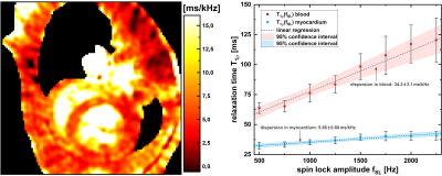 |
Synthetic T1rho dispersion imaging for improved myocardial tissue characterization using dispersion reconstruction
Maximilian Gram1,2, Daniel Gensler1,3, Patrick Winter1,2, Michael Seethaler2,3, Peter Jakob2, and Peter Nordbeck1,3
1Department of Internal Medicine I, University Hospital Würzburg, Würzburg, Germany, 2Experimental Physics 5, University of Würzburg, Würzburg, Germany, 3Comprehensive Heart Failure Center (CHFC), University Hospital Würzburg, Würzburg, Germany
T1ρ dispersion imaging is a very time-consuming process because full T1ρ-mapping at different spin-lock amplitudes is required. Due to this issue, investigation of T1ρ dispersion is hardly feasible in the limited measurement time of a small animal experiment. In this work, we present a novel approach for the rapid measurement of cardiac T1ρ dispersion called dispersion reconstruction. With our new concept a T1ρ dispersion image is generated by only acquiring a fraction of the required mapping data. Phantom and in vivo experiments confirm the applicability of our new method as part of a conventional protocol for small animal studies.
|
|
0531.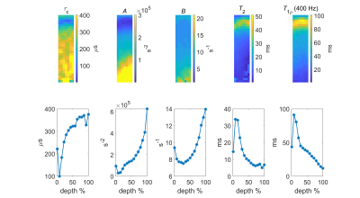 |
Correlation Time as a New MRI Contrast
Hassaan Elsayed1,2, Jouni Karjalainen1,2, Nina Hänninen1,3, Isabel Stavenuiter3, Stefan Zbyn1,4, Mikko Nissi1,3, Miika T. Nieminen1,2,5, and Matti Hanni1,2,5
1Research Unit of Medical Imaging, Physics and Technology, University of Oulu, Oulu, Finland, 2Medical Research Center, University of Oulu and Oulu University Hospital, Oulu, Finland, 3Department of Applied Physics, University of Eastern Finland, Kuopio, Finland, 4Center for Magnetic Resonance Research, Department of Radiology, University of Minnesota, Minneapolis, MN, United States, 5Department of Diagnostic Radiology, Oulu University Hospital, Oulu, Finland
In this study, we determined correlation time ($$$\tau_c$$$ ), a parameter that we propose as MRI contrast indicative of the structure of articular cartilage. $$$T_{1\rho}$$$ dispersion data were acquired from intact patellar bovine cartilage samples and $$$\tau_c$$$ maps were obtained by fitting a Lorentzian model function to the $$$T_{1\rho}$$$ dispersion. The association between $$$\tau_c$$$ and the tissue properties was assessed by correlation analysis between $$$\tau_c$$$ and histology. The results suggest that the proposed parameter $$$\tau_c$$$ as well as other fitting parameters can reveal cartilage structure. More investigation is needed to establish correlation between $$$\tau_c$$$ and histology.
|
 Watch the Video