Scientific Session
RF Technologies
| Wednesday Parallel 5 Live Q&A | Wednesday, 12 August 2020, 13:45 - 14:30 UTC | Moderators: Lucia Navarro de Lara & Manuela Rösler |
0760.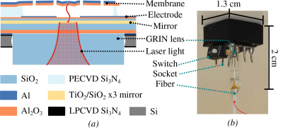 |
Magnetic resonance imaging with direct optical detection
Anders Simonsen1, Juan D. Sánchez-Heredia2, Sampo Saarinen1, Jan H. Ardenkjær-Larsen2, Albert Schliesser1, and Eugene S. Polzik1
1Niels Bohr Institute, University of Copenhagen, Copenhagen, Denmark, 2Department of Health Technology, Technical University of Denmark, Kgs. Lyngby, Denmark
A new approach to MRI detection is reported, where an optomechanical transducer directly converts and amplifies the MR signal to amplitude modulation of laser light. The transducer, which is simultaneously a capacitor and an optical cavity, is connected directly in parallel to the receive coil. The mechanical Q-factor of the transducer is 1500, providing a 3 dB bandwidth of 1 kHz. We show here for the first time that this technology can be used to directly acquire a 13C MRI at 3T (32.13 MHz), and reconstruct it using the standard pipeline of a commercial MR scanner.
|
|
|
|
||
0761.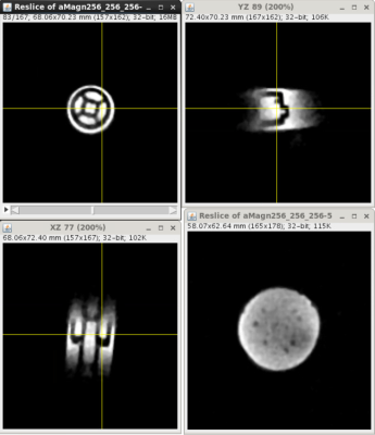 |
A Non-Magnetic Staggered Commutation Based Circulator to Achieve Time-Efficient Simultaneous Transmit and Receive (STAR) MRI
Hazal Yuksel1, Lance DelaBarre2, Djaudat Idiyatullin3, Julie Kabil4, Gregor Adriany3, Sung-Min Sohn5, Harish Krishnaswamy 1, John Thomas Vaughan 4, and Michael Garwood3
1Electrical Engineering, Columbia University, New York, NY, United States, 2Center for Magnetic Resonance Research, University of Minnesota, Minneapolis, MN, United States, 3University of Minnesota, Minneapolis, MN, United States, 4Columbia University, New York, NY, United States, 5Arizona State University, Tempe, AZ, United States
Traditional MRI relies on the temporal separation of the receiver (RX) and transmitter (TX) to solve the problem of self-interference. Often, the TX signal is billions of times larger than the RX signal, and T/R switches are used so the TX does not saturate or destroy the RX. This leads to an inefficient method of acquiring imaging data for especially fast decaying signals. We propose a magnetic-free, PCB based circulator to remove the T/R switch and achieve simultaneous transmit and receive MRI. We present images of a phantom acquired with a continuous SWIFT sequence to validate the concept.
|
|
 |
0762.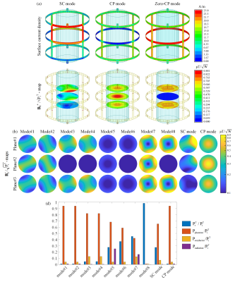 |
Design of transmit array coils for MRI by minimizing the modal reflection coefficients
Ehsan Kazemivalipour1,2, Alireza Sadeghi-Tarakameh1,2, and Ergin Atalar1,2
1Electrical and Electronics Engineering Department, Bilkent University, Ankara, Turkey, 2National Magnetic Resonance Research Center (UMRAM), Bilkent University, Ankara, Turkey
We propose a general analysis based on minimization of modal reflection coefficients, providing a simple tool for quantifying the performance of transmit array (TxArray) coils in terms of power efficiency. We investigate the performance of various dual-row birdcage TxArrays, with an additional degree of freedom to correct B1+-field inhomogeneities by adding RF shimming ability in the longitudinal-direction, together with simulations and experiments. The chosen structure of the TxArray allows the coil to act like degenerate birdcage coils. We demonstrate when TxArrays are properly designed, in some critical excitation modes such as circularly-polarized (CP) mode, the total reflection becomes negligibly small.
|
 |
0763.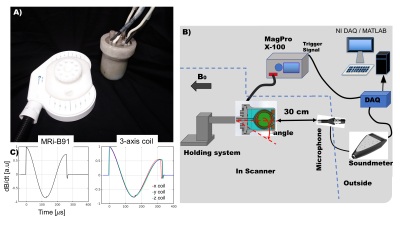 |
Assessment of MR compatibility for multichannel stimulation using three-axis TMS coil elements
Lucia Navarro de Lara1,2, Mohammad Daneshzand1,2, Anthony Mascarenas3, Douglas Paulson3, Sergey Makarov1,4, Jason Stockmann1,2, Larry Wald1,2,5, and Aapo Nummenmaa1,2
1Radiology, A.A Martinos Center for Biomedical Imaging/MGH, Charlestown, MA, United States, 2Harvard Medical School, Boston, MA, United States, 3Tristan Technologies, San Diego, CA, United States, 4Department of Electrical and Computer Engineering, A.A Martinos Center for Biomedical Imaging/MGH, Worcester, MA, United States, 5HST/MIT, Cambridge, MA, United States We investigate the feasibility of using small-diameter three-axis TMS coil as a basis for constructing a simultaneous stimulation and imaging array. We present an MR-compatible 3-axis TMS coil prototype comprising of three orthogonal coil X/Y/Z units. We assess the influence of the TMS coil element on the MRI images and measure the sound pressure levels with systematically varying the current amplitudes and coil orientation with respect to the magnetic field. Supported by simulations, we conclude that construction of a large-scale multichannel; system using such a 3-axis TMS elements as a basis appears feasible but the acoustic properties should be improved |
0764.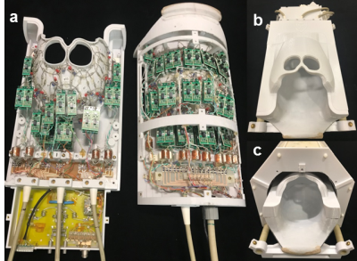 |
A 64-Channel 7T array coil for accelerated brain MRI
Azma Mareyam1, John E. Kirsch1,2, Yulin Chang3, Gunjan Madan3, and Lawrence L. Wald1,2
1Athinoula A. Martinos Center for Biomedical Imaging, Massachusetts General Hospital, Charlestown, MA, United States, 2Harvard Medical School, Boston, MA, United States, 3Siemens Medical Solutions USA, Boston, MA, United States
We construct and test a prototype 64-channel brain array coil for 7T and compare it to a 32-channel coil of similar design. Coil characteristics like signal to noise ratio, noise correlation matrix, and noise amplification (G-factor) for parallel imaging are described as well as and a comparison of the B1+ maps to assess birdcage coil efficiency and homogeneity. The coil was designed on a split-half former with a sliding top half to facilitate patient entry and utilizes a sliding birdcage coil for transmit
|
|
0765.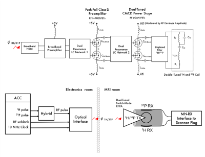 |
Dual-Tuned Optically Controlled On-Coil Switch-Mode Amplifier
Natalia Gudino1, Stephen J Dodd1, Steve Li2, and Jeff H Duyn1
1LFMI, NINDS, National Institutes of Health, Bethesda, MD, United States, 2MIB, NIMH, National Institutes of Health, Bethesda, MD, United States
Optically controlled on-coil amplifiers have been presented for the practical implementation of pTx system at ultra-high field MRI. We present a new prototype that can transmit power to excite 1H and X-nuclei. We show bench and MRI data with a dual-tuned on-coil amplifier implementation for 1H and 31P excitation at 7T. We expect this technology can allow a simpler and more versatile implementation of a multinuclear multichannel hardware compared to the traditional multinuclear setup based on 50 Ω broadband voltage mode amplification.
|
|
 |
0766.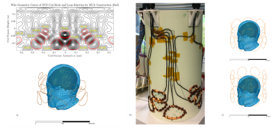 |
First prototype of a Stream-Function-based Multi-Coil Array dedicated to human brain shimming at Ultra-High-Field
Bruno Pinho Meneses1,2, Jason Stockmann3,4, and Alexis Amadon1
1Neurospin/CEA-Saclay, Gif-sur-Yvette, France, 2Université Paris-Saclay, Saclay, France, 3Athinoula A. Martinos Center for Biomedical Imaging, Charlestown, MA, United States, 4Harvard Medical School, Boston, MA, United States
Using Singular Value Decomposition of optimal Stream Functions computed from a database of 100 B0 fieldmaps, a 13-channel cylindrical optimized Multi-Coil Array for shimming of the human brain is built and tested in an experimental setup for field measurement at 7T. Such measurements are compared to expected performances, serving as proof of concept for this novel design methodology. Performance is compared to what would be achieved by matrix multi-coil array designs patterned on a cylinder.
|
 |
0767.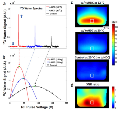 |
Permittivity-Tunable Ceramic Technology for Largely Improving B1 fields and SNR for Broad MR Imaging Applications
Byeong-Yeul Lee1, Xiao-Hong Zhu1, Hannes M. Wiesner1, Maryam Sarkarat2, Sebastian Rupprecht3, Michael T Lanagan2, Qing X Yang3, and Wei Chen1
Video Permission Withheld
1CMRR, Radiology, University of Minnesota, Minneapolis, MN, United States, 2Department of Engineering Science and Mechanics, Pennsylvania State University, University Park, PA, United States, 3Center for NMR Research, Neurosurgery, Pennsylvania State College of Medicine, Hershey, PA, United States
We present an innovative technique of tunable ultrahigh dielectric constant (tuHDC) ceramics incorporating RF coil(s) for MR imaging applications. The ceramic has a very high permittivity tunability of 2000-15000 by varying the ceramic temperature between few to 40 °C to achieve optimal performance at the nuclide Larmor frequency of interest, resulting in larger B1 field and SNR improvements for 1H MRI at 1.5T clinic scanner and 17O MRSI at 10.5T human scanner. We found a large denoising effect in 17O MRSI, which further boosts the SNR gain. The technology should benefit for biomedical research and clinical diagnosis.
|
 |
0768.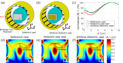 |
Design and demonstration of an artificial dielectric for 7 T MRI
Vsevolod Vorobyev1, Alena Shchelokova1, Irena Zivkovic2, Alexey Slobozhanyuk1, Juan Domingo Baena3, Juan Pablo del Risco4, Redha Abdeddaim5, Andrew Webb2,6, and Stanislav Glybovski1
1Department of Nanophotonics and Metamaterials, ITMO University, Saint Petersburg, Russian Federation, 2Department of Radiology, C.J.Gorter Center for High Field MRI, Leiden University Medical Center, Leiden, Netherlands, 3Department of Physics, Universidad Nacional de Colombia, Bogota, Colombia, 4School of Exact Sciences and Engineering, Universidad Sergio Arboleda, Bogota, Colombia, 5Aix Marseille University, CNRS, Centrale Marseille, Institut Fresnel, Marseille, France, 6Stevens Research Center, Carle Foundation Hospital, Urbana, IL, United States
We present a new approach replacing high-permittivity water-based dielectric pads with a non-resonant low-cost artificial dielectric to improve MR image quality by modifying the interferences present in the radiofrequency field at 7T MRI. The artificial dielectric comprises a stack of metal patches printed on dielectric substrates. Numerical studies and imaging of a head-phantom with the proposed structure showed the same increase in the transmit radiofrequency field distribution at the area of interest as for conventional dielectric pads. The advantages of the new structure include ease of manufacture and long-term stability.
|

 Back to Program-at-a-Glance
 Watch the Video
 Back to Top