Oral - Power Pitch Session
Spinal Cord, Head and Neck
| Thursday Parallel 2 Live Q&A | Thursday, 13 August 2020, 15:05 - 15:50 UTC | Moderators: Kristin O'Grady |
1171.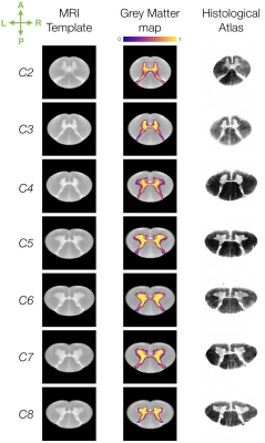 |
Ex vivo MRI template of the human cervical cord at 80μm isotropic resolution
Charley Gros1, Abdullah Asiri2,3, Benjamin De Leener4, Charles Watson5, Gary Cowin6, Marc Ruitenberg7, Nyoman Kurniawan2, and Julien Cohen-Adad1,8
1NeuroPoly Lab, Institute of Biomedical Engineering, Polytechnique Montreal, Montreal, QC, Canada, 2Centre for Advanced Imaging, The University of Queensland, Brisbane, Australia, 3Radiology department, College of applied medical sciences, Najran University, Najran, Saudi Arabia, 4Department of Computer and Software Engineering, Polytechnique Montreal, Montreal, QC, Canada, 5Faculty of Health Sciences, Curtin University of Technology, Perth, Australia, 6National Imaging Facility, Centre for Advanced Imaging, The University of Queensland, Brisbane, Australia, 7School of Biomedical Sciences, The University of Queensland, Brisbane, Australia, 8Functional Neuroimaging Unit, CRIUGM, Université de Montréal, Montreal, QC, Canada
Spinal cord MRI templates allow reproducible and large scale atlas-based studies. However, current templates may have suboptimal resolutions (~0.5mm isotropic) to analyse high resolution data acquired at ultra-high field (e.g. 7T scanners). We generated a 3D human cervical cord template at 80µm isotropic resolution, from 13 ex vivo specimens, with a reference based on spinal levels. Further, the template was registered to the existing in vivo PAM50 template. Results showed consistency with histological studies in terms of grey matter morphology, and template generation achieved high accuracy (mean distance error: 0.10±0.01mm). The template and related scripts will be made publicly available.
|
|
1172.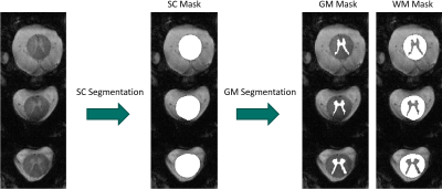 |
Spinal Cord Segmentation and T2*-relaxation times of GM and WM within the Spinal Cord at 9.4T
Ole Geldschläger1, Dario Bosch1, Nikolai Avdievitch1, Klaus Scheffler1,2, and Anke Henning1,3
1High-field Magnetic Resonance, Max-Planck-Institut for biolog. Cybernetics, Tübingen, Germany, 2Institute for Biomedical Magnetic Resonance, University Hospital Tübingen, Tübingen, Germany, 3Advanced Imaging Research Center, University of Texas Southwestern Medical Center, Dallas, TX, United States
This study presents the first investigations with algorithmic spinal cord-segmentation, as well as gray matter/white matter-segmentation within the spinal cord, at the ultrahigh field strength of 9.4T. On multi-echo gradient-echo acquisitions from three subjects, the tested algorithms perform the segmentations correctly. Based on these multi-echo data, pixel-wise T2*-relaxation time maps were calculated. By means of the segmentations, averaged T2*-times of 24.88ms +- 6.68ms for gray matter and 19.37ms +- 8.66ms for white matter, were calculated.
|
|
1173.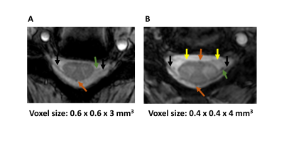 |
A multi-element transceive array for cervical spinal cord imaging at 7T
Ece Ercan1, Thomas Ruytenberg1, Kristin P. O’Grady2,3, Seth A. Smith2,3,4, Andrew Webb1, and Irena Zivkovic1
1C.J. Gorter Center for High Field MRI, Department of Radiology, Leiden University Medical Center, Leiden, Netherlands, 2Vanderbilt University Institute of Imaging Science, Nashville, TN, United States, 3Department of Radiology and Radiological Sciences, Vanderbilt University Medical Center, Nashville, TN, United States, 4Department of Biomedical Engineering, Vanderbilt University, Nashville, TN, United States
Spinal cord imaging at 7T MRI is challenging and limited by the need for dedicated RF coils. In this study, we present a flexible coil design for cervical spinal cord imaging at 7T. B1+ inhomogeneities were addressed by using multichannel array and phased-based RF shimming. Dorsal and ventral nerve roots, denticulate ligaments, and blood vessels were visible on axial T2*-weighted images. Cross-sectional area measurements from C3-C4 cervical levels were consistent with literature values.
|
|
1174.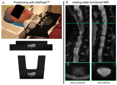 |
Effect of non-protonated perfluorocarbon liquid-filled SatPads on spinal cord MR imaging
Benjamin De Leener1,2, Linda Soltrand Dahlberg2, Ali Khatibi2,3, Julien Cohen-Adad4,5, and Julien Doyon2
1Department of computer engineering and software engineering, Polytechnique Montreal, Montreal, QC, Canada, 2Montreal Neurological Institute, McGill University, Montreal, QC, Canada, 3Center of Precision Rehabilitation for Spinal Pain (CPR Spine), University of Birmingham, Birmingham, United Kingdom, 4NeuroPoly Lab, Institute of Biomedical Engineering, Polytechnique Montreal, Montreal, QC, Canada, 5Functional Neuroimaging Unit, CRIUGM, Université de Montréal, Montreal, QC, Canada
Acquiring high-quality functional MRI data of the spinal cord is challenging due to large susceptibility artifacts and high physiological noise, causing signal dropout and distorsions, particularly in the cervical region. This study demonstrated the beneficial effect of using non-protonated perfluorocarbon liquid-filled SatPadsTM during fMRI acquisition. Indeed, results show an increase of 31.51% for the global signal and 36.59% for the temporal signal-to-noise ratio for resting-state fMRI data acquired in the cervical spinal cord.
|
|
1175.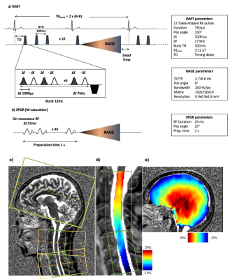 |
Towards minimal T1 and B1 contributions in cervical spinal cord inhomogeneous magnetization transfer imaging
Arash Forodighasemabadi1,2,3,4, Thomas Troalen5, Lucas Soustelle1,2, Guillaume Duhamel1,2, Olivier Girard1,2, and Virginie Callot1,2,4
1Aix-Marseille Univ, CNRS, CRMBM, Marseille, France, 2APHM, Hopital Universitaire Timone, CEMEREM, Marseille, France, 3Aix-Marseille Univ, IFSTTAR, LBA, Marseille, France, 4iLab-Spine International Research Laboratory, Marseille-Montréal, France, 5Siemens Healthcare SAS, Saint-Denis, France
Inhomogeneous Magnetization Transfer (ihMT) is a promising MRI technique, sensitive to myelinated tissue that can be used to study demyelinating pathologies such as MS. But the conventional MT and ihMT ratio metrics could be sensitive to T1 and B1 variations, especially in the context of spinal cord imaging. In order to minimize these effects, this study focuses on 3D ihMT-RAGE sequence with high FA reference acquisition and ihMTR inverse metric computation. The quantifications within GM and WM along the cervical spinal cord demonstrate that this technique is promising for investigating SC pathologies.
|
|
1176.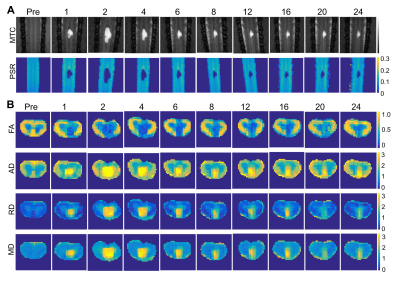 |
Regional and longitudinal changes of multiple MRI parameters correlate with behavioral impairment and recovery after spinal cord injury
Feng Wang1,2, Tung-Lin Wu1, Pai-Feng Yang1,2, Nellie E. Byun1, Li Min Chen1,2, and John C. Gore1,2,3
1Vanderbilt University Institute of Imaging Science, Vanderbilt University Medical Center, Nashville, TN, United States, 2Radiology and Radiological Sciences, Vanderbilt University Medical Center, Nashville, TN, United States, 3Biomedical Engineering, Vanderbilt University, Nashville, TN, United States
Quantitative magnetization transfer (qMT) and diffusion tensor imaging (DTI) may detect and track compositional and structural changes in spinal cords before and after injury and during repair. This study aims to systematically evaluate the abilities of the qMT-derived pool size ratio (PSR) and DTI-derived diffusion parameters to assess injury-associated regional changes in spinal cords of monkeys, and to correlate them to specific sensorimotor behaviors. An overall goal is to evaluate the relationships between longitudinal changes in different regional MRI measures and sensorimotor behavioral impairment and recovery following spinal cord injury over a long period of time (months).
|
|
1177. |
Preoperative evaluation of multimodal spinal MRI in patients with acute traumatic spinal cord injury
Yuan Liu1, Fengzhao Zhu2, Xiangchuang Kong1, Jiazheng Wang3, Xiaodong Guo2, and Yang Lian1
1Radiology, Union Hospital, Wuhan, China, 2orthopedics, Union Hospital, Wuhan, China, 3Philips Healthcare, Beijing, China
Baseline MRI was recommended in acute spinal cord injury for clinical decision making and outcome prediction. The study presented a new quantitative method for evaluating the spinal cord severity to grade the retained fiber tracks by zoom DTI in pre-operation. For patients with ASIA A, no ASIA grade got promoted in FTClass A1 with completely fibers interruption and 3 out of 6 patients converted to C within 6-month follow-up in FTclass A2 with partially retention. The retained spinal cord fibers were critical for postoperative functional recovery. Multimodal MRI, especially accurate DTI provide potential quantitative predictive indicators for prognosis.
|
|
1178.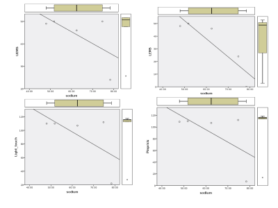 |
Sodium concentration alterations in spinal cord injury and associations to motor and sensory function
Bhavana Shantilal Solanky1, Ferran Prados1, Carmen Tur1, Selma Al-Ahmad2, Xixi Yang1, Baris Kanber3, David Choi2, Jalesh N Panicker4, and Claudia A M Gandini Wheeler-Kingshott1
1NMR Research Unit, Queen Square MS Centre, Department of Neuroinflammation, NMR Research Unit, Queen Square MS Centre, Department of Neuroinflammation, UCL Queen Square Institute of Neurology, Faculty of Brain Sciences, UCL, London, United Kingdom, 2National Hospital For Neurology and Neurosurgery, Queen Square, London, United Kingdom, 3Translational Imaging Group, Centre for Medical Image Computing, Department of Medical Physics and Biomedical Engineering, University College London, London, United Kingdom, 4Department of Uro-neurology, National Hospital for Neurology and Neurosurgery, London, United Kingdom Sodium retention as a consequence of spinal cord injury is thought to impair the regenerative ability of neurons but also reduce damage. Studies have shown that sodium-blockers can lead to improved outcomes in some SCI patients. Here alterations in spinal cord total sodium concentrations in spinal cord injury patients and healthy controls were investigated using sodium MRS. The association of sodium concentration to cross sectional area and ASIA score was also explored.
|
|
1179.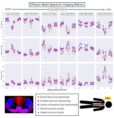 |
Inhomogeneous Magnetization Transfer and DBSI detect downstream white matter damage in post-mortem human cervical spinal cord injury
Sarah Rosemary Morris1,2,3, Andrew Yung1,3,4, Valentin Prevost1,3,4, Shana I George5, Piotr Kozlowski1,2,3,4, Andrew Bauman1,3,4, Farah Samadi1,6, Caron Fournier1,6, Lisa Parker7, Kevin Dong1, Femke Streijger1, G.R. Wayne Moore1,6,7,8, Brian Kwon1,9,10,
and Cornelia Laule1,2,3,6
1International Collaboration on Repair Discoveries, Vancouver, BC, Canada, 2Physics & Astronomy, University of British Columbia, Vancouver, BC, Canada, 3Radiology, University of British Columbia, Vancouver, BC, Canada, 4UBC MRI Research Centre, Vancouver, BC, Canada, 5Carson Graham Secondary School, Vancouver, BC, Canada, 6Pathology & Laboratory Medicine, University of British Columbia, Vancouver, BC, Canada, 7Vancouver General Hospital, Vancouver, BC, Canada, 8Medicine, University of British Columbia, Vancouver, BC, Canada, 9Vancouver Spine Surgery Institute, Vancouver, BC, Canada, 10Orthopaedics, University of British Columbia, Vancouver, BC, Canada
Spinal cord injuries are heterogeneous, with complex microstructure which changes over time. We used 7T Diffusion Tensor Imaging (DTI), Diffusion Basis Spectrum Imaging (DBSI) and inhomogeneous Magnetization Transfer (ihMT) to investigate microstructural damage in post-mortem human spinal cord injury tissue. We measured sharp decreases in DTI fractional anisotropy and DBSI fiber fraction at the injury epicentre of the three cords with the most severe injuries. We found evidence for downstream demyelination (ihMT) and axonal loss (DTI FA, DBSI fiber fraction) in the two cords with the longest injury-death interval suggesting a time-frame for the detection of Wallerian degeneration by MRI.
|
|
1180.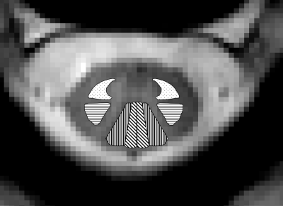 |
Is recovery from whiplash influenced by macromolecular changes in spinal cord white matter?
Mark Andrew Hoggarth1,2, James Elliott2,3, Mary Kwasny4, Marie Wasielewski2, Kenneth Weber5, and Todd Parrish1,6
1Biomedical Engineering, Northwestern University, Chicago, IL, United States, 2Physical Therapy and Human Movement Sciences, Northwestern University, Chicago, IL, United States, 3Northern Sydney Local Health District & Faculty of Health Sciences, The University of Sydney, Sydney, Australia, 4Preventive Medicine, Northwestern University, Chicago, IL, United States, 5Anesthesiology, Perioperative and Pain Medicine, Stanford University School of Medicine, Palo Alto, CA, United States, 6Radiology, Northwestern University, Chicago, IL, United States
Whiplash injuries are the most common outcome from non-fatal motor vehicle collisions, affecting nearly four million people in the United States each year. The purpose of this cross-sectional study was to investigate the macromolecular environment of cervical spinal cord white matter in participants with persistent whiplash. This investigation of 76 individuals demonstrated changes in cervical white matter integrity following whiplash injuries using magnetization transfer imaging. Significant differences in the magnetization transfer ratio homogeneity of large cervical white matter tracts were observed in females with poor clinical outcome, indicating a spinal cord insult may contribute to chronic pain after whiplash injury.
|
|
1181.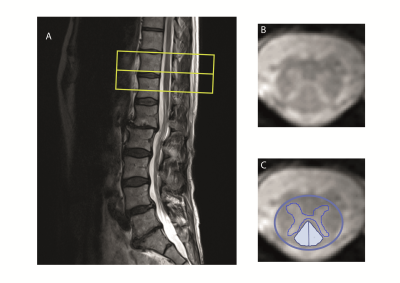 |
Structural MRI investigation caudal to degenerative cervical myelopathy: a clinical MRI application
Kevin Vallotton1, Maryam Seif1, Markus Hupp1, Armin Curt1, and Patrick Freund1,2,3
1Spinal Cord Injury Center, University Hospital Balgrist, University of Zurich, Zurich, Switzerland, 2Department of Neurophysics, Max Planck Institute for Human Cognitive and Brain Sciences, Leipzig, Germany, 3Wellcome Trust Centre for Neuroimaging, UCL Institute of Neurology, London, United Kingdom
Degenerative cervical myelopathy (DCM) is the most common form of non-traumatic spinal cord injury (SCI) and induces neurodegeneration in the cervical cord at and above the primary stenosis level. However, whether similar neurodegeneration occurs at the lumbar level remains unclear. We therefore applied high resolution T2*-weighted MRI in the lumbar cord in both DCM patients and healthy controls to investigate potential injury-induced structural changes. Significant atrophy was found in mild DCM patients and its magnitude was associated with sensory impairment.
|
|
1182.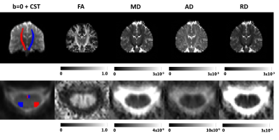 |
Spinal cord and brain DTI alterations in cervical spondylotic myelopathy (CSM)
Rebecca Sara Samson1, Jonathan Stutters1, Muhammad Ali Akbar2, Armin Curt3, Julien Cohen-Adad4,5, Michael Fehlings2,6, Patrick Freund3,7,8, Blair Innerarity1, Maryam Seif3, Carmen Tur1, and Claudia A. M. Gandini Wheeler-Kingshott1,9,10
1NMR Research Unit, Queen Square MS Centre, Department of Neuroinflammation, University College London, London, United Kingdom, 2Institute of Medical Science, University of Toronto, Toronto, ON, Canada, 3Spinal Cord Injury Center Balgrist, University of Zurich, Zurich, Switzerland, 4NeuroPoly Lab, Institute of Biomedical Engineering, Polytechnique Montreal, Montreal, QC, Canada, 5Functional Neuroimaging Unit, CRIUGM, Université de Montréal, Montreal, QC, Canada, 6Krembil Research Institute, University Health Network, Toronto, ON, Canada, 7Department of Neurophysics, Wellcome Trust Centre for Neuroimaging, University College London, London, United Kingdom, 8Max Planck Institute for Human Cognitive and Brain Sciences, Leipzig, Germany, 9Department of Brain and Behavioural Sciences, University of Pavia, Pavia, Italy, 10Brain MRI 3T Center, IRCCS Mondino Foundation, Pavia, Italy
We explored diffusion tensor imaging (DTI) metrics along the corticospinal tract (CST) from the cervical cord to the motor cortex, measured using separate brain and cervical cord DTI protocols in healthy subjects and cervical spondylotic myelopathy (CSM) patients at two sites. Instead of looking at either brain or cord separately, here, we combine brain and cord measurements and examine how the CST is affected in CSM, in addition to exploring correlations with clinical measures. Statistically significant changes were observed between CSM and HC when comparing cord and brain CST data, demonstrating the sensitivity of CST metrics to cord pathology.
|
|
1183.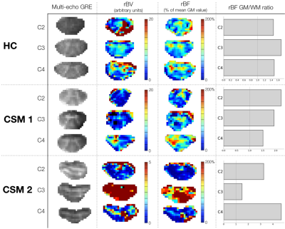 |
Dynamic Susceptibility Contrast imaging at 7T for spinal cord perfusion mapping in Cervical Spondylotic Myelopathy patients
Simon Lévy1,2,3,4, Pierre-Hugues Roche4,5, and Virginie Callot1,2,4
1Aix-Marseille Univ, CNRS, CRMBM, Marseille, France, 2APHM, Hopital Universitaire Timone, CEMEREM, Marseille, France, 3Aix-Marseille Univ, IFSTTAR, LBA, Marseille, France, 4iLab-Spine International Research Laboratory, Marseille-Montreal, QC, France, 5Neurosurgery Department, APHM, Hopital Nord, Marseille, France
The performance of Dynamic Susceptibility Contrast imaging at 7T for spinal cord perfusion mapping within clinical constraints was investigated. A cardiac-gated spin-echo EPI sequence with 0.7x0.7mm2 in-plane resolution was used in one healthy volunteer and two Cervical Spondylotic Myelopathy patients. Relative blood volume and flow maps successfully revealed the higher perfusion of gray matter versus white matter for the volunteer and one patient. Results were limited for the patient with greater functional impairment and disadvantageous acquisition conditions. Although human spinal cord perfusion has never been mapped as precisely, several issues remain to address (image distortions, Specific-Absorption-Rate limitations, Arterial Input Function).
|
|
1184.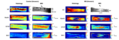 |
New potential MRI markers of glial scarring and tissue damage in multiple sclerosis spinal cord pathology using diffusion MRI
Marco Palombo1, Francesco Grussu1,2, Torben Schneider1,2,3, Gabriele C. DeLuca4, Daniel C. Alexander1, Claudia A. M. Gandini Wheeler-Kingshott2,5,6, and Hui Zhang1
1Centre for Medical Image Computing, Department of Computer Science, University College London, London, United Kingdom, 2NMR Research Unit, Queen Square MS Centre, Queen Square Institute of Neurology, Faculty of Brain Sciences, University College London, London, United Kingdom, 3Philips UK, Guildford, Surrey, United Kingdom, 4Nuffield Department of Clinical Neurosciences, University of Oxford, Oxford, United Kingdom, 5Department of Brain and Behavioural Sciences, University of Pavia, Pavia, Italy, 6Brain MRI 3T Center, IRCCS Mondino Foundation, Pavia, Italy
Multiple sclerosis (MS) is characterized by demyelination, extra-cellular matrix disruption, inflammation and astrocytic scarring of WM lesions. This study investigates the use of a recently introduced MRI technique called SANDI (Soma And Neurite Density Imaging) to provide histologically meaningful estimates of cell body (namely soma) density in MS spinal cord pathology. Our results on ex-vivo human spinal cord specimens show significant positive correlation between SANDI metrics (fneurite and fsoma) and histological markers of myelination (plp) and astrocytes reactivity (gfap), respectively. The study suggests SANDI metrics as complementary imaging markers of demyelination (fneurite), astrocytic scarring (fsoma) and extra-cellular matrix disruption (fextra).
|
|
1185.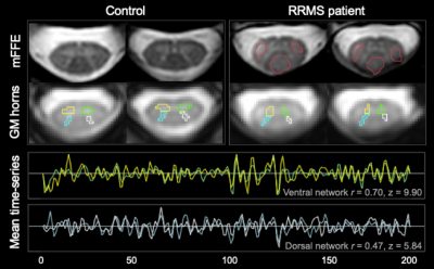 |
Cervical cord resting-state fMRI shows preserved functional connectivity in low disability relapsing-remitting multiple sclerosis
Anna Combes1,2, Baxter P. Rogers1,2, Mereze Visagie2, Kristin P. O'Grady1,2, Richard D. Lawless2,3, Sanjana Satish2, Atlee Witt2, Shekinah Malone4, Colin D. McKnight2, Francesca R. Bagnato5, John C. Gore1,2,3, and Seth A. Smith1,2,3
1Radiology & Radiological Sciences, Vanderbilt University Medical Center, Nashville, TN, United States, 2Vanderbilt University Institute of Imaging Science, Nashville, TN, United States, 3Department of Biomedical Engineering, Vanderbilt University, Nashville, TN, United States, 4School of Medicine, Meharry Medical College, Nashville, TN, United States, 5Clinical Neurology, Vanderbilt University Medical Center, Nashville, TN, United States
Functional connectivity (FC) in the cervical spinal cord can be assessed with 3T resting-state fMRI. FC strength in the ventral and dorsal networks was measured in a group of relapsing-remitting multiple sclerosis (MS) patients with low disability, high cervical lesion load, and mildly impaired sensorimotor function and was found similar to matched healthy controls. There was no impact of the presence of cord lesions, suggesting FC is preserved even in the presence of structural damage. Future work will explore the longitudinal trajectories of cord FC in support of intact or impaired sensorimotor function in MS.
|

 Back to Program-at-a-Glance
 Watch the Video
 Back to Top