
Oral - Power Pitch Session
Novel Clinical Applications of CMR
Session Topic: Novel clinical applications of CMR
Session Sub-Topic: Cardiovascular Power Pitch: Applications
Oral - Power Pitch
Cardiovascular
| Thursday Parallel 3 Live Q&A | Thursday, 13 August 2020, 15:05 - 15:50 UTC | Moderators: Daniel Herzka |
Session Number: PP-08
 |
1204.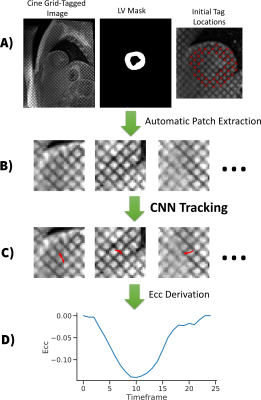 |
Measuring Cardiac Strain in Duchenne Muscular Dystrophy with a Convolutional Neural Net Tag Tracking Method
Michael Loecher1,2, Luigi E Perotti3, Patrick Magrath4, and Daniel B Ennis1,2,5,6
1Radiology, Stanford, Palo Alto, CA, United States, 2Radiology, Veterans Administration Health Care System, Palo Alto, CA, United States, 3Mechanical Engineering, University of Central Florida, Orlando, FL, United States, 4Radiology, University of California Los Angeles, Los Angeles, CA, United States, 5Cardiovascular Institute, Stanford, Palo Alto, CA, United States, 6Center for Artificial Intelligence in Medicine & Imaging, Stanford, Palo Alto, CA, United States
The objective of this work was to demonstrate the feasibility of using a convolutional neural net (CNN) based tag tracking algorithm for deriving strain measurements in grid tagged cardiac MR images. The method was tested in 23 subjects. When compared to commercial software the CNN-based method produces similar measurements for peak Ecc and shows lower strain in boys with DMD compared to healthy subjects [CNN = -0.15±0.03 vs -0.21±0.03] and [Conventional = -0.16±0.03 vs -0.21 ± 0.02] (p < .001). Peak Ecc was not significantly different within cohorts when compared between methods [DMD cohort: p=0.32, Healthy cohort: p=0.99]
|
 |
1205.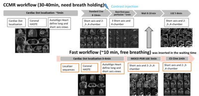 |
Clinical Value of an Almost Automated Fast Free-breathing Cardiac Magnetic Resonance Workflow
Keyan Wang1, Michaela Schmidt2, Jing An3, and Xiaoming Bi4
11st affiliated hospital of zhengzhou university, Zhengzhou, China, 2Siemens Healthcare GmbH, Erlangen, Germany, 3Siemens Shenzhen Magnetic Resonance Ltd, Shenzhen, China, 4Siemens Healthineers, Los Angeles, CA, United States
The application of cardiac magnetic resonance (CMR) is limited in patients with arrhythmia or poor breath holding ability. In this study, clinical value of a rapid free-breathing workflow was assessed by employing real-time compressed-sensing for cardiac function and motion-corrected LGE embedded in a workflow engine for cardiac assessment. Results indicated that the proposed free-breathing fast workflow allowed in a short acquisition time to assess the morphological features of the heart and left ventricular function, especially in patients with severe heart failure.
|
|
1206.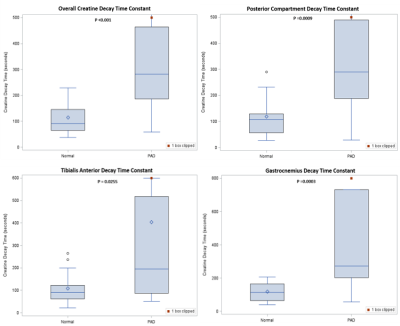 |
CrCEST energetics of calf muscle groups at 3T distinguish patients with peripheral arterial disease from age-matched normals
Helen Sporkin1, Christopher Schumann2, Roshin Mathew2, Christopher Kramer2,3, and Craig Meyer1,3
1Biomedical Engineering, University of Virginia, Charlottesville, VA, United States, 2Cardiology, University of Virginia, Charlottesville, VA, United States, 3Radiology and Medical Imaging, University of Virginia, Charlottesville, VA, United States
In Peripheral Arterial Disease, arterial occlusions in the lower limbs lead to tissue ischemia, which can lead to claudication pain or the need for amputation. Creatine CEST (CrCEST) imaging can indirectly image creatine as it exchanges protons with free water during metabolism after exercise. Ischemia in the skeletal reduces available oxygen, slowing metabolism. CrCEST data of three major calf muscle groups were fit to a monoexponential function in order to compare energetics between PAD patients and age-matched normal subjects. We have so far imaged 22 aged-matched subjects and 19 PAD patients, and found a significant increase in the decay constant.
|
1207.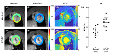 |
ECG-free, free-breathing myocardial T1/ECV mapping at high heart rates using MR Multitasking: A feasibility study in a HFpEF rat model
Pei Han1,2, Rui Zhang3,4, Anthony Christodoulou2, Shawn Wagner2, Yibin Xie2, Eugenio Cingolani3, Eduardo Marban3, and Debiao Li1,2,5
1Department of Bioengineering, UCLA, Los Angeles, CA, United States, 2Biomedical Imaging Research Institute, Cedars-Sinai Medical Center, Los Angeles, CA, United States, 3Smidt Heart Institute, Cedars-Sinai Medical Center, Los Angeles, CA, United States, 4Department of Cardiology, Xin-Hua Hospital Affiliated to Shanghai Jiao Tong University School of Medicine, Shanghai, China, 5Department of Medicine, UCLA, Los Angeles, CA, United States
CMR T1 and ECV quantification can be used to characterize focal or diffuse myocardial fibrosis. However, it is technically challenging to acquire high-quality maps in small animals for preclinical research because of high heart rates and high respiration rates. In this study, we developed an ECG-free, free-breathing MR Multitasking T1 mapping method on a 9.4T small animal MRI system. The feasibility of characterizing diffuse myocardial fibrosis was tested in a HFpEF rat model. Elevated ECV found in the HFpEF group is consistent with previous human studies and shows strong correlation with the histological data.
|
|
1208.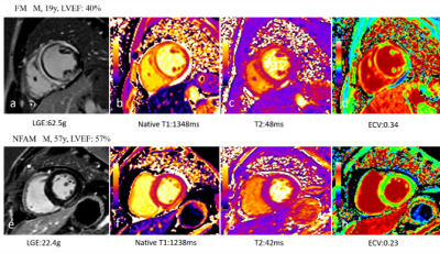 |
Tissue characterization by mapping and strain cardiac magnetic resonance imaging to evaluate myocardial inflammation in fulminant myocarditis
Hui Zhu1, Haojie Li1, Zhaoxia Yang1, and Liming Xia1
1Radiology, Tongji Hospital, Tongji Medical College, Huazhong University of Science and Technology, Wuhan, China
Fulminant myocarditis show significant different LGE patterns, increased edema and decreased strain measurements compared with non-fulminant acute myocarditis and global peak circumferential and radial strain were closely correlated with quantitative parameters of myocardial edema.
|
|
1209.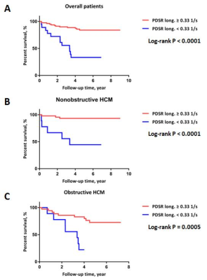 |
Global longitudinal diastolic strain rate predict adverse outcomes in hypertrophic cardiomyopathyas assessed with CMR tissue tracking
Chunchao Xia1, Xiaoyue Zhou2, and Zhenlin Li1
1West China Hospital, Chengdu, China, 2MR Collaboration, Siemens Healthineers Ltd., Shanghai, China
During hypertrophic cardiomyopathy (HCM), left ventricular (LV) diastolic dysfunction is regarded as one of the primary mechanisms responsible for the main adverse cardiovascular events (MACEs). Early evaluation of LV diastolic function is of great importance to risk stratification and management optimization in HCM patient populations. Our study indicated that the cardiac magnetic resonance tissue tracking (CMR-TT)–derived longitudinal global diastolic strain rate (PDSR) is a novel and easy-to-perform index for evaluating LV diastolic dysfunction and predicting adverse outcomes in HCM patient populations, which would also be beneficial for risk stratification.
|
|
1210.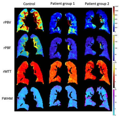 |
Assessing lung perfusion in pulmonary hypertension
Paul J.C. Hughes1, Andrew J. Swift1,2, Frederick J. Wilson3, Marcella Cogliano1, Fasial AA Alandejani1, Anthony Cahn3, Lindsay Kendall3, David G. Kiely2,4,5, and Jim M. Wild1,2
1POLARIS, Department of Infection, Immunity and Cardiovascular Disease, The University of Sheffield, Sheffield, United Kingdom, 2Insigneo Institute for in silico Medicine, The University of Sheffield, Sheffield, United Kingdom, 3GlaxoSmithKline R&D Ltd, Stevenage, United Kingdom, 4Sheffield Pulmonary Vascular Disease, Sheffield Teaching Hospitals NHS Trust, Sheffield, United Kingdom, 5Department of Infection, Immunity and Cardiovascular Disease, The University of Sheffield, Sheffield, United Kingdom
Pulmonary arterial hypertension (PAH) is a condition that impacts on lug perfusion and right ventricular function. This work aimed to assess i) the diagnostic utility of relative pulmonary perfusion parameters to distinguish patients with PAH from healthy controls and ii) changes in lung perfusion in 2 patient groups with PAH: newly diagnosed patients initiating and patients escalating treatment and clinically stable patients who had no escalation of treatment.
|
|
1211.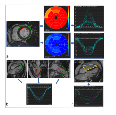 |
The relationship between CMR–derived myocardial strain and late gadolinium enhancement in asymptomatic heart transplant patients
Xuehua Shen1, Yating Yuan1, Ming Yang1, Xiaoyue Zhou2, Jing Wang1, Wei Sun3, Mingxing Xie3, Li Zhang3, and Bo Liang1
1Department of Radiology, Union Hospital, Tongji Medical College, Huazhong University of Science and Technology, Wuhan, Hubei, China, 2MR Collaboration, Siemens Healthineers Ltd., Shanghai, China, Shanghai, China, 3Department of Ultrasound, Union Hospital, Tongji Medical College, Huazhong University of Science and Technology, Wuhan, Hubei, China
No study has explored the relationship between CMR-derived myocardial strain and the extent of LGE in asymptomatic HT patients. The purpose of this study was to evaluate this relationship using DRA and FT strain analysis. In this study, there were strong correlation and good reproducibility between DRA and FT strain modalities. HT patients with LGE had reduced LVGLS and preserved LVGCS. CMR-derived LVGLS was significantly and independently correlated with LGE.
|
|
1212.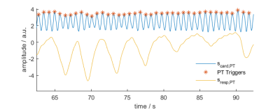 |
Retrospective Analysis of Pilot Tone Derived Cardiac and Respiratory Motion Information in a Patient Cohort
Mario Bacher1,2, Lorenzo Di Sopra1, Peter Speier2, Davide Piccini3, Anna-Giulia Pavon1, Christopher Roy1, Juerg Schwitter1, Jérôme Yerly1, and Matthias Stuber1
1Department of Radiology, Lausanne University Hospital and University of Lausanne, Lausanne, Switzerland, 2Siemens Healthineers, Erlangen, Germany, 3Siemens Healthineers, Lausanne, Switzerland
The Pilot Tone is a novel motion sensing method capable of simultaneous, sequence independent measurement of respiratory and cardiac motion. Here, we show that Pilot Tone motion data can be used as an alternative to self-gating in a free-breathing, cardiac- and respiration resolved CMRI sequence. We demonstrate in a patient cohort that Pilot Tone motion information correlates well with ECG ground-truth and self-gating respiratory signals.
|
|
|
1213.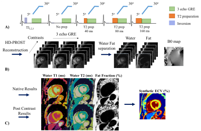 |
Reproducibility, Repeatability and Preliminary Clinical Results of Dixon Cardiac MRF: T1, T2, ECV and fat fraction tissue characterization
Olivier Jaubert1, Gastao Cruz1, Aurelien Bustin1, Torben Schneider2, Georgios Georgiopoulos1, Mariya Doneva3, Pier-Giorgio Masci1, Rene Michael Botnar1, and Claudia Prieto1
1Biomedical Engineering Department, School of Biomedical Engineering and Imaging Sciences, King's College London, London, United Kingdom, 2Philips Healthcare, London, United Kingdom, 3Philips Research Hamburg, Hamburg, Germany
Dixon cardiac Magnetic Resonance Fingerprinting (DcMRF) has been recently proposed to enable simultaneous water T1, water T2 and fat fraction (FF) quantification in a single breath-hold scan. Here we investigate the reproducibility, repeatability and clinical feasibility of DcMRF in comparison to reference MOLLI, T2GRASE and 6 echo proton density FF measurements. Reproducibility and repeatability were investigated in healthy subjects, whereas native T1, T2 and FF, and post contrast T1 and synthetic ECV measurements were performed in patients with suspected cardiovascular disease.
|
1214.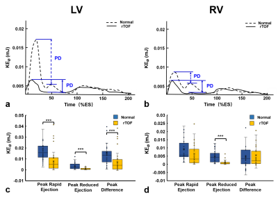 |
Evaluate Myocardial Circumferential Kinetic Energy for Patients with Repaired Tetralogy of Fallot
Shi-Ying Ke1, Meng-Chu Chang1, Ming-Ting Wu2, Ken-Pen Weng3,4, and Hsu-Hsia Peng1
1Department of Biomedical Engineering and Environmental Sciences, National TsingHua University, Hsinchu, Taiwan, 2Department of Radiology, Kaohsiung Veterans General Hospital, Kaohsiung, Taiwan, 3Department of Pediatrics, Kaohsiung Veterans General Hospital, Kaohsiung, Taiwan, 4Department of Pediatrics, National Yang-Ming University, Taipei, Taiwan
This study aims to evaluate biventricular myocardial circumferential kinetic energy (KEØ) for assessment of regional myocardial function in repaired tetralogy of Fallot (rTOF) patients with preserved global cardiac function. The tissue phase mapping (TPM) was acquired in the basal, mid, and apical slices in the left and right ventricles. We found the altered KE values and abnormal distribution of three-directional %KE in rTOF patients. In conclusion, the altered myocardial KEØ may provide useful information for assessment of regional myocardial function in rTOF patients with preserved global cardiac function.
|
|
1215.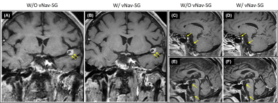 |
Motion-Compensated 3D TSE for More Robust Intracranial MR Vessel Wall Imaging
Zhehao Hu1,2, Fei Han3, Andre J.W. Van der Kouwe4,5, Xiaoming Bi3, Bin Sun6, Jiayu Xiao1, Junzhou Chen1,2, Shlee S. Song7, Marcel M. Maya8, Debiao Li1,2,9, and Zhaoyang Fan1,2,9
1Biomedical Imaging Research Institute, Cedars-Sinai Medical Center, Los Angeles, CA, United States, 2Bioengineering Department, University of California, Los Angeles, Los Angeles, CA, United States, 3Siemens Healthineers, Los Angeles, CA, United States, 4A. A. Martinos Center for Biomedical Imaging, Massachusetts General Hospital, Charlestown, MA, United States, 5Department of Radiology, Harvard Medical School, Boston, MA, United States, 6Department of Radiology, Fujian Medical University Union Hospital, Fuzhou, China, 7Department of Neurology, Cedars-Sinai Medical Center, Los Angeles, CA, United States, 8Department of Imaging, Cedars-Sinai Medical Center, Los Angeles, CA, United States, 9Department of Medicine, University of California, Los Angeles, Los Angeles, CA, United States
While underexplored to date, motion susceptibility may critically undermine clinical translation of 3D intracranial MR vessel wall imaging (VWI). Motion artifacts observed in intracranial VWI are either caused by head bulk motion or internally localized movement. By combing volumetric navigators (vNav) and self-gating (SG) strategies, we propose a novel motion compensation approach that can simultaneously address these two motion issues. Our preliminary studies demonstrated the potential of using this technique to improve robustness of 3D intracranial MR VWI.
|
 Watch the Video