Oral - Power Pitch Session
High Resolution fMRI
Session Topic: High resolution fMRI
Session Sub-Topic: fMRI Applications
Oral - Power Pitch
fMRI
| Thursday Parallel 4 Live Q&A | Thursday, 13 August 2020, 15:05 - 15:50 UTC | Moderators: Giovanna Diletta Ielacqua & Scott Peltier |
Session Number: PP-29
 |
1235.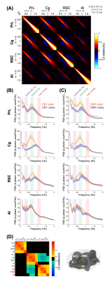 |
Simultaneous fMRI and multi-channel, spectrally resolved fiber-photometry reveals the neural basis of default mode network modulation in rats
Tzu-Hao Harry Chao1, Li-Ming Hsu1, Martin MacKinnon1, and Yen-Yu Shih1
1University of North Carolina at Chapel Hill, Chapel Hill, NC, United States
This study establishes a novel MR-compatible multi-channel fiber-photometry platform and demonstrates 1) photometry-CBV measured in PrL and Cg co-fluctuate with global DMN signal derived from CBV-fMRI, 2) significantly enhanced 0.6-0.8 Hz GCaMP power in PrL, Cg, and RSC, but not AI, between two DMN states, 3) significantly enhanced 0.25-0.45 Hz GCaMP power in PrL, Cg and RSC precedes DMN activation peak by 3-5 s, but not AI, and 4) significantly enhanced 0.6-0.8 Hz GCaMP power in AI precedes DMN deactivation valley by 11 s.
|
 |
1236.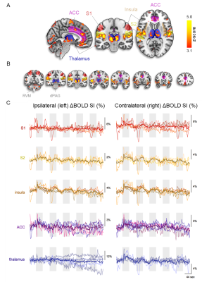 |
Deciphering the Pain-Matrix with Ultrahigh functional MRI: En Route to Objective Biomarkers for Pain
Gijs Jurjen Heij1, Thoralf Niendorf1,2, and Henning Matthias Reimann1
1Berlin Ultrahigh Field Facility (B.U.F.F.), Max-Delbrück-Center for Molecular Medicine, Berlin, Germany, 2Experimental and Clinical Research Center, a joint cooperation between the Charité Medical Faculty and the Max Delbrück Center for Molecular Medicine in the Helmholtz Association, Berlin, Germany
Despite millions of people suffering from chronic pain, clinicians still rely on self-reports for the assessment of pain. In attempts to identify objective biomarkers, fMRI studies have revealed an assembly of regions that consistently activates in response to noxious stimuli, but also to non-noxious equally salient stimuli. Since most studies have been performed at 3T or lower, higher field strengths might be able to differentiate pain from saliency. As a first step, we aimed to identify the pain-matrix at ultrahigh fields using an ON/OFF heat stimulation paradigm and show here that the regions associated with the pain-matrix could be identified.
|
 |
1237.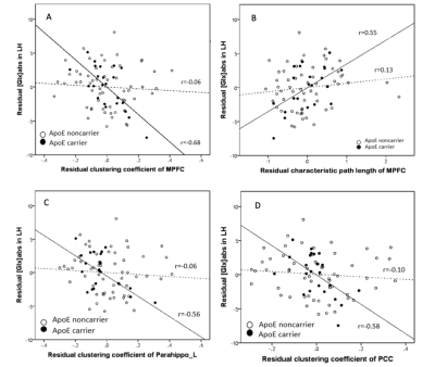 |
Disrupted Small-World Networks and Differences in Metabolite Concentration in Healthy Adults with Low and High Genetic Risk
Hui Zhang1,2, Pui Wai Chiu1,3, Isaac Ip4, Tianyin Liu5, Gloria Hoi Yan Wong5, You-Qiang Song6, Savio Wai Ho Wong4, Queenie Chan7, Karl Herrup8, and Henry Ka Fung Mak1,2,3
1Department of Diagnostic Radiology, The University of Hong Kong, Hong Kong, Hong Kong, 2Alzheimer's Disease Research Network, Hong Kong, Hong Kong, 3State Key Laboratory of Brain and Cognitive Sciences, Hong Kong, Hong Kong, 4Department of Educational Psychology, the Chinese University of Hong Kong, Hong Kong, Hong Kong, 5Department of Social Work and Administration, The University of Hong Kong, Hong Kong, Hong Kong, 6Department of Biochemistry, The University of Hong Kong, Hong Kong, Hong Kong, 7Philips Healthcare, Hong Kong, Hong Kong, 8Alzheimer Disease Research Centre, University of Pittsburgh, Pittsburgh, PA, United States
To identify the relationship between the topological properties and glutamate in genetic-related subgroups (ApoE4 carriers and non-ApoE4 carriers), combined resting state fMRI (rs-fMRI) and MRS were applied in this study. Graph theory metrics of subgroups were calculated and compared. In the results, ApoE4 carriers had worse network segregation and integration. However, there was significant correlation between [Glx]abs in left hippocampus and topological metrics in high-risk group. We postulated that glutamatergic synaptic transmission modulates rs-fMRI activities in ApoE4 carriers.
|
 |
1238.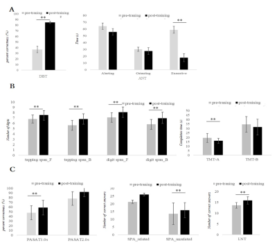 |
Combined Working Memory and Attention Training Improves Cognition via Task-Specific and Transfer Effects
Daisuke Sawamura1,2, Ryusuke Suzuki3, Keita Ogawa4, Shinya Sakai2, Xinnan Li1, Hiroyuki Hamaguchi1, and Khin Khin Tha5,6
1Department of Biomarker Imaging Science, Graduate School of Biomedical Science and Engineering, Hokkaido University, Sapporo, Japan, 2Department of Functioning and Disability, Faculty of Health Sciences, Hokkaido University, Sapporo, Japan, 3Departments of Medical Physics, Hokkaido University Hospital, Sapporo, Japan, 4Department of Rehabilitation, Hokkaido University Hospital, Sapporo, Japan, 5Department of Diagnostic and Interventional Radiology, Hokkaido University, Sapporo, Japan, 6Global Station for Quantum Medical Science and Engineering, Hokkaido University, Sapporo, Japan
Little is known about how neurocognition is modulated upon combined computerized cognitive training (CCT). We developed a combined CCT program designed to improve several cognitive functions simultaneously, and evaluated its effect on neurocognitive performance and functional connectivity (FC) of the brain. The results suggest that the CCT improves not only the targeted functions but also the other aspects of neurocognition via augmentation of transfer effect. The LPFC and fronto-parieto-occipital networks are thought to play role.
|
 |
1239.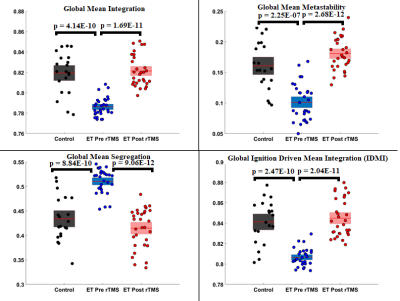 |
Repetitive TMS increases whole brain metastability and dynamic integrity in Essential Tremor
SUJAS BHARDWAJ1, RAJANIKANT PANDA2, SHWETA PRASAD1, SUNIL KUMAR KHOKHAR3, SNEHA RAY3, ROSE DAWN BHARATH3, and PRAMOD KUMAR PAL1
1NEUROLOGY, NIMHANS, Bengaluru, India, 2Coma Science Group, Universitè de Liège, Liège, Belgium, 3NI & IR, NIMHANS, Bengaluru, India
We assessed the effect of repetitive transcranial magnetic stimulation (rTMS) on whole brain dynamics using fMRI. MRI was acquired before and after a single session of rTMS in 30 patients of Essential Tremor and 20 age matched healthy controls. Whole brain dynamic synchronization - “metastability”, and propagation of integration - “intrinsic ignition”, were studied in order to assess brain network topological fluctuations following rTMS. Single subject and group wise whole brain metastability, integration and ignition driven integration was found improve with a single session of rTMS.
|
 |
1240.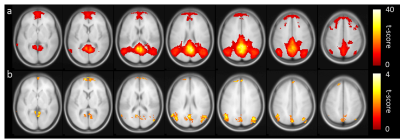 |
Diminished default mode network connectivity in older individuals is associated with aberrant brain metabolism
Xirui Hou1, Zixuan Lin1, Peiying Liu1, Corinne Pettigrew2, Anja Soldan2, Marilyn Albert2, and Hanzhang Lu1
1The Russell H. Morgan Department of Radiology, Johns Hopkins University School of Medicine, Baltimore, MD, United States, 2Department of Neurology, Johns Hopkins University School of Medicine, Baltimore, MD, United States
Cerebral metabolic rate of oxygen (CMRO2), the rate at which O2 is consumed in the brain, is thought to be a direct index of energy hemostasis and brain health. Recent studies have suggested that CMRO2 is elevated but functional connectivity is declined with age. In this work, we demonstrated the diminished default network was associated with aberrant CMRO2 in older healthy subjects. Network analysis indicated that the increasing amount of CMRO2 was used to compensate for the inefficiency of degraded networks.
|
 |
1241.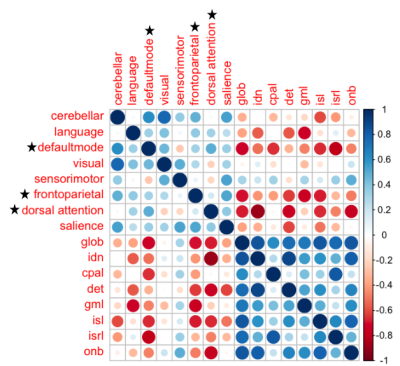 |
Functional connectivity is associated with radiotherapy-induced vascular injury and cognitive impairment in young brain tumor survivors
Melanie Morrison1, Angela Jakary1, Erin Felton2, Schuyler Stoller2, Sabine Mueller2, and Janine Lupo1
1Radiology & Biomedical Imaging, University of California San Francisco, San Francisco, CA, United States, 2Department of Pediatrics, University of California San Francisco, San Francisco, CA, United States
While radiation therapy plays an essential role in the management of brain tumor patients, exposure to radiation has been known to lead to declines in neurocognitive performance and vascular injury. As there remains a need for a reliable marker and predictor of patient outcome, this study explores the usefulness of functional connectivity measurements derived from 7T rsfMRI. We found that temporal properties, specifically low-frequency signals of some large-scale brain networks,are associated with more severe cognitive impairment and vascular injury, highlighting the potential benefit of using rsfMRI for treatment planningand prediction of patient outcome after RT.
|
1242.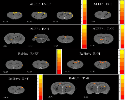 |
Effects of EGR3 transfection on behavior and resting-state fMRI in rats and evaluation of risperidone treatment in schizophrenia model
Xiaowei Han1,2, Guolin Ma1, and Lizhi Xie3
1Department of Radiology, China-Japan Friendship Hospital, Beijing, China, 2Graduate School of Peking Union Medical College, Beijing, China, 3GE Healthcare, MR Research China, Beijing, China
Schizophrenia is a neurodevelopmental psychiatric disorder with unclear etiology and no effective treatment. In this study, we established a new schizophrenia model in rats using early growth response (EGR3) gene transfection which was injected into the hippocampus and dentate gyrus of rats. The model was examined by evaluating the behavioral impact and cerebral alterations of schizophrenia model rats using behavioral phenotyping and resting-state functional magnetic resonance imaging (rs-fMRI). In addition, the efficacy of risperidone therapy was also evaluated in treated group rats. Briefly, we found several regional alterations in the cerebrum, which were consequently partially reversed by risperidone.
|
|
 |
WITHDRAWN | |
1244.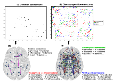 |
Estimation of stable whole-brain effective-connectivity characterization of mental disorders
Lipeng Ning1,2 and Yogesh Rathi1,2
1Brigham and Women's Hospital, Boston, MA, United States, 2Harvard Medical School, Boston, MA, United States
We propose an algorithm to estimate whole-brain effective connectivity measures by integrating structural connectivity matrix between brain regions and resting-state functional MRI data. Our algorithm first uses the Lyapunov inequality from control theory to ensure that the estimated whole-brain dynamic system is stable and physically meaningful. Then, the effective connectivity measure is characterized by a novel conditional causality measure. We applied the proposed algorithm to a public dataset which consisted of healthy controls (n=94), patients with schizophrenia (n=45), bipolar (n=44) and ADHD (n=37). Our results show that the proposed approach provides reliable estimation brain-network features of these brain disorders.
|
|
1245.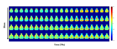 |
Altered patterns of neural activity and functional connectivity revealed by dynamic rsfMRI in the Q175 mouse model of Huntington's disease
Tamara Vasilkovska1, Bram Callewaert1, Somaie Salajeghe1, Dorian Pustina2, Longbin Liu2, Mette Skinbjerg2, Celia Dominguez2, Ignacio Munoz-Sanjuan2, Annemie Van der Linden1, and Marleen Verhoye1
1Department of Biomedical Sciences, University of Antwerp, Antwerp, Belgium, 2CHDI Foundation, Princeton, NJ, United States
Static FC changes in neurodegeneration can indicate underlining neural mechanism pathology present in pre-manifest disease stage. In addition to the spatial FC component, quasi-periodic patterns (QPPs) implement spatiotemporal information of neural activity, allowing integrated assessment of possible initial changes in large-scale brain dynamics. We measured Low Frequency (LF) BOLD changes using rsfMRI in Q175 HD mouse model at 3 and 12 months of age. Results indicate decreased FC between specific regions in heterozygous compared to wild-type mice at 12 months. Both at 3 and 12 months, additional QPPs are present in the heterozygous group, deviating from the wild type group.
|
|
 |
1246.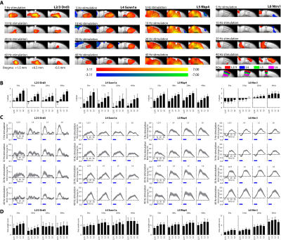 |
Laminar fMRI using layer-specific optogenetic stimulations
Russell W Chan1, Mazen Asaad2, Bradley J Edelman1, Hyun Joo Lee1, Hillel Adesnik3, David Feinberg3, and Jin Hyung Lee1,4,5,6
1Neurology and Neurological Sciences, Stanford University, Stanford, CA, United States, 2Molecular and Cellular Physiology, Stanford University, Stanford, CA, United States, 3Helen Wills Neuroscience Institute, University of California, Berkeley, CA, United States, 4Bioengineering, Stanford University, Stanford, CA, United States, 5Neurosurgery, Stanford University, Stanford, CA, United States, 6Electrical Engineering, Stanford University, Stanford, CA, United States
We attempted to establish the mesoscale layer-specific fMRI representation of neuronal activity using layer-specific Cre-driver mouse lines, optogenetic stimulations, fMRI and electrophysiological recordings. Although laminar fMRI responses were distinct during L2/3, L4, L5 and L6 stimulations, all fMRI responses increased along the cortical depth. This phenomenon was, however, not observed in LFP and spike recordings. This discrepancy between fMRI, LFP and spiking may be due to the draining veins transporting deoxyhemoglobin from the deeper layers to the superficial layers. Future studies may take into account of neurovasculature to elucidate the exact mechanisms of mesoscale layer-specific neurovascular coupling.
|
1247.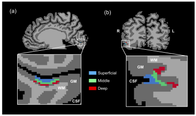 |
Layer-dependent repetition suppression in the human visual cortex
Uk-Su Choi1,2, Seiji Ogawa3, and Ikuhiro Kida1,2
1Center for Information and Neural Networks, NICT, Suita, Japan, 2Graduate School of Frontier Biosciences, Osaka University, Suita, Japan, 3Kansei Fukushi Research Institute, Tohoku Fukushi University, Sendai, Japan
To examine the layer-dependent fMRI responses of face processing in human visual cortex, we acquired submillimeter functional and anatomical data at 7T. Here, we measured fMRI responses to left single and paired faces with various interstimulus intervals; furthermore, we calculated the repetition suppression (RS) ratio from three cortical layers in the right primary visual cortex (V1) and occipital face area (OFA). The fMRI response and RS ratio of superficial and middle layers of the right V1 were similar to those of all right OFA layers. Hence, the layers in different hierarchical visual areas were modulated in the same manner.
|
|
1248.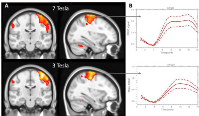 |
Comparing stimulus and resting state fMRI at 3T and 7T reveal a superiority of 7T in detecting changes in subcortical networks
Silke Kreitz1,2, Angelika Mennecke1, Laura Cristina Konerth 2, Armin Nagel3, Frederic Laun3, Michael Uder3, Arnd Doerfler1, and Andreas Hess2
1Department of Neuroradiology, University Hospital Erlangen, Friedrich-Alexander-University Erlangen-Nürnberg, Erlangen, Germany, 2Institute of Experimental and Clinical Pharmacology and Toxicology, Friedrich-Alexander University Erlangen-Nurnberg, Erlangen, Germany, 3Department of Radiology, University Hospital of the Friedrich-Alexander University Erlangen-Nürnberg, Erlangen, Germany
7T MRI is hoped to improve diagnostics, therapy and research of neurological diseases. Here, we characterize the influence of field strength on fMRI approaches including task based and RS-fMRI. Quality metrics revealed a basic separation of 3T and 7T fMRI data, mainly by tSNR, MDI, CNR and EFC. 7T fMRI showed higher BOLD response amplitudes, more functional connections and higher connectivity strength especially in inferior brain regions. Though higher variability between subjects at 7T likely requires enhanced statistical power in group comparisons, intra individual fMRI measurements might detect subtle connectivity changes at 7T useful for diagnosis and therapy.
|
|
1249.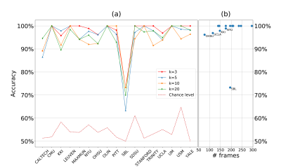 |
Classifying Autism Spectrum Disorder Patients from Normal Subjects using a Connectivity-based Graph Convolutional Network
Lebo Wang1, Kaiming Li2, and Xiaoping Hu1,2
1Department of Electrical and Computer Engineering, University of California, Riverside, Riverside, CA, United States, 2Department of Bioengineering, University of California, Riverside, Riverside, CA, United States
Traditional deep learning architectures have met with limited performance improvement on fMRI data analysis. Our connectivity-based graph convolutional network modeled fMRI data as graphs and performed convolutions within connectivity-based neighborhood. We demonstrate that our approach is substantially more robust in classifying Autism Spectrum Disorder (ASD) patients from normal subjects compared with those in published work. Extracting spatial features and averaging across frames are beneficial in reducing variance and improving classification accuracy.
|

 Back to Program-at-a-Glance
 Watch the Video
 Back to Top