Oral - Power Pitch Session
Novel Hardware
| Thursday Parallel 5 Live Q&A | Thursday, 13 August 2020, 15:05 - 15:50 UTC | Moderators: Stephan Orzada |
1270.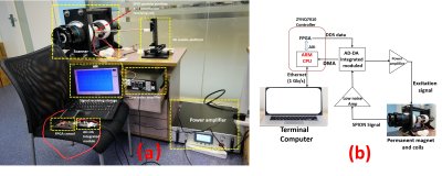 |
Implementation of Low-cost FPGA-based Magnetic Particle Imaging System
Congcong Liu1,2, Caiyun Shi1, Jianguo Cui2, Xin Liu1, Hairong Zheng1, Dong Liang1, and Haifeng Wang1
1Shenzhen Institutes of Advanced Technology,Chinese Academy of Sciences, Shenzhen, China, 2Chongqing University of Technology, Chongqing, China
Conventional Magnetic Particle Imaging (MPI) systems are expensive and bulky, and most of them use CPU and MPU as controllers. Field Programmable Gate Arrays (FPGAs) have recently become widely utilized as controllers in many systems for flexibility and speed. In this paper, we proposed an implementation scheme of a low-cost, small-volume MPI system based on FPGA architecture. The experimental results showed that the complete operation of the MPI system signal link could be realized by the low-cost FPGA-based control system (≤$200), and the distribution of the superparamagnetic iron oxide nanoparticles (SPION) could be imaged by the signal of the particle.
|
|
1271.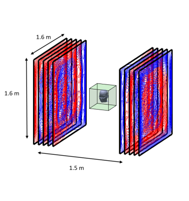 |
A wearable 50-channel magnetoencephalography (MEG) system
Ryan Hill1, Elena Boto1, Niall Holmes1, Gillian Roberts1, Jim Leggett1, Zelekha Seedat1, Molly Rea 1, Tim Tierney2, Stephanie Mellor 2, Vishal Shah 3, James Osborne3, Gareth Barnes4, Richard Bowtell1, and Matthew Brookes1
1Sir Peter Mansfield Imaging Centre, University of Nottingham, Nottingham, United Kingdom, 2Wellcome Centre for Human Neuroimaging, UCL, London, United Kingdom, 3QuSpin Inc., Louisville, CO, United States, 4Wellcome Trust Centre for Neuroimaging, UCL, London, United Kingdom
We have a developed a wearable magnetoencephalography (MEG) system comprising 50, miniaturised optical pumped magnetometers (OPMs) fixed on an EEG-type cap. Large, bi-planar field and field-gradient coils sited inside a magnetically shielded room reduce the field around the head to < 1nT. This allows the subject to move their head during experiments without confounding the OPM signals. We report results from a simple finger abduction paradigm and a motor learning experiment in which a subject learns to play the ukulele. The results demonstrate the potential of OPM-MEG to overcome some limitations of neuroimaging investigations using fixed, cumbersome scanners.
|
|
1272.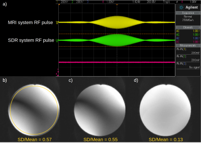 |
Triggered software defined radio for parallel transmission MRI research
Fred Tam1, Benson Yang1, and Simon J Graham1,2
1Physical Sciences, Sunnybrook Research Institute, Toronto, ON, Canada, 2Department of Medical Biophysics, University of Toronto, Toronto, ON, Canada
Building a flexible setup for parallel transmission (PTx) MRI research is still challenging. A commercial software defined radio device was tested for suitability in a revised setup. Software was developed to make the device generate radiofrequency (RF) bursts in response to triggers. Preliminary bench tests showed quick and reliable triggering, consistent amplitude and phase across channels, and successful runtime adjustment. An initial PTx MRI demonstration showed capability for RF shimming in echo planar imaging of a phantom. Further troubleshooting is planned to reduce observed phase jitter, but the setup is already capable of a range of PTx research applications.
|
|
1273.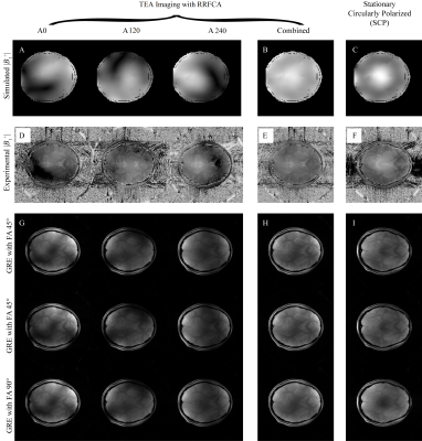 |
Time-multiplexed Excitation and Acquisition (TEA) with Rotating RF Coil Array (RRFCA)
Jin Jin1,2,3, Zhentao Zuo4,5, Mingyan Li2, Ewald Weber2, Aurelien Destruel2, Feng Liu2, Rong Xue4,5, and Stuart Crozier2
1Siemens Healthcare Pty Ltd, Brisbane, Australia, 2The University of Queensland, St Lucia, Australia, 3University of Southern California, Los Angeles, CA, United States, 4Institute of Biophysics, Chinese Academy of Sciences, Beijing, China, 5Sino-Danish College, University of Chinese Academy of Sciences, Beijing, China
RF shimming, by means of adjusting the relative amplitudes and phases of the independent transmit channels of a parallel transmit system, is widely used in ultra-high-field imaging to improve transmit homogeneity but has limited capabilities especially for large fields of view. This work designed and tested an 8-channel rotating transceiver array to provide more degrees-of-freedom for both transmission and reception. During transmission, the array achieved a uniform effective transmit magnetic field in a time-multiplexed fashion; during reception, the array provided more unique sensitivity profiles, facilitating higher image SNR. Parallel-imaging-like reconstruction was developed, assisted by a robust self-calibrated sensitivity estimation technique.
|
|
1274.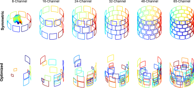 |
An optimized multi-coil shim setup matching inhomogeneity distribution in the human brain; positive and negative aspects
Ali Aghaeifar1, Jiazheng Zhou1, Feng Jia2, Maxim Zaitsev2, and Klaus Scheffler1
1Max Planck Institute for Biological Cybernetics, Tuebingen, Germany, 2Dept. of Radiology, Medical Physics, Medical Center University of Freiburg, Faculty of Medicine, University of Freiburg, Freiburg, Germany
Multi-coil shim setup is a popular choice for B0 shimming. In contrast to conventional regular arrangement of the shim coils, one can effectively position the shim coil to match inhomogeneity distribution in the human brain. In this work, a comparison between regular and optimized arrangement of the local coils in a multi-coil shim setup is performed and the pros and cons of each design are evaluated.
|
|
1275.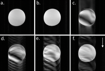 |
Imaging artefacts during simultaneous in-beam MR imaging and proton pencil beam irradiation
Sebastian Gantz1,2, Volker Hietschold3, Sergej Schneider1,2,4, and Aswin Louis Hoffmann1,2,5
Video Permission Withheld
1Institute of Radiooncology-OncoRay, Helmholtz-Zentrum Dresden - Rossendorf, Dresden, Germany, 2OncoRay – National Center for Radiation Research in Oncology, Faculty of Medicine and University Hospital Carl Gustav Carus, Technische Universität Dresden, Helmholtz-Zentrum Dresden-Rossendorf, Dresden, Germany, 3Department of Radiology, Faculty of Medicine and University Hospital Carl Gustav Carus, Technische Universität Dresden, Dresden, Germany, 4Technische Universität Dresden, Carl Gustav Carus Faculty of Medicine, Dresden, Germany, 5Department of Radiotherapy and Radiation Oncology, Faculty of Medicine and University Hospital Carl Gustav Carus, Technische Universität Dresden, Dresden, Germany
The targeting precision of proton therapy is expected to benefit from real-time MRI guidance. We developed a setup of a first prototype in-beam MRI scanner with a proton pencil beam scanning nozzle. Dipole magnets in the nozzle used for beam steering produce time-dependent magnetic fringe fields that may interfere with the MR image acquisition. In this study, we show that vertical beam steering shows no degradation of the MR image quality, whereas horizontal beam steering introduces severe ghosting artefacts in phase encoding direction. The origin of these artefacts is unraveled and strategies to eliminate or correct these artefacts are proposed.
|
|
1276.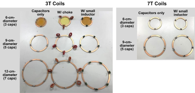 |
Low-profile AC/DC coils without RF Chokes
Jixin Xia1,2, Charlotte R. Sappo1,3, William A. Grissom1,3,4, and Xinqiang Yan3,4
1Department of Biomedical Engineering, Vanderbilt University, Nashville, TN, United States, 2Department of Electrical Engineering, Vanderbilt University, Nashville, TN, United States, 3Vanderbilt University Institute of Imaging Science, Nashville, TN, United States, 4Department of Radiology, Vanderbilt University Medical Center, Nashville, TN, United States
Large RF chokes increase the inductance/resistance of the DC loop and thus lead to unwanted power dissipation and bulk. We theoretically analyzed the added coil loss induced by bridge inductors in AC/DC coils and found that large bridge chokes can be replaced by low-profile inductors with much smaller values, if the capacitance is adjusted to compensate the resonance frequency shift. This will reduce the footprint of AC/DC coils as well as the inductance/resistance and thus power dissipation, with a cost of slightly higher coil loss.
|
|
1277.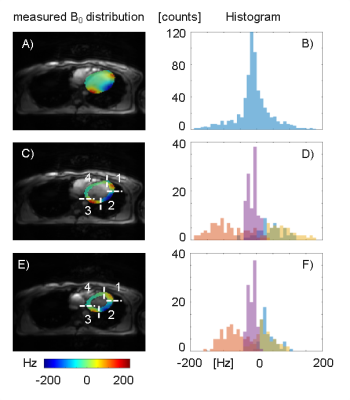 |
Considerations for Cardiac Phase-Specific B0 Shimming at 7 T
Michael Hock1, Maxim Terekhov1, Markus Johannes Ankenbrand1, David Lohr1, Theresa Reiter1,2, Christoph Juchem3, and Laura Maria Schreiber1
1Chair of Cellular and Molecular Imaging, Comprehensive Heart Failure Center (CHFC), University Hospital Wuerzburg, Wuerzburg, Germany, 2Department of Internal Medicine I, University Hospital Wuerzburg, Wuerzburg, Germany, 3Departments of Biomedical Engineering and Radiology, Columbia University, New York City, NY, United States
Spatio-temporal inhomogeneities of the static magnetic (B0) field are a major limiting factor in cardiac magnetic resonance applications at 7T. A previously developed shim strategy was demonstrated to correct spatial myocardial B0-field inhomogeneities in a preliminary in vivo implementation. To correct localized spots of B0-inhomogeneities, third-order terms were found to be beneficial. Cardiac phase-specific shimming was evaluated in simulations based on the in vivo field map data, and optimal shim settings were shown to differ between cardiac phases. Future work will address the application of a shim averaged over all cardiac phases to each individual phase.
|
|
1278.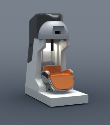 |
A compact vertical 1.5T human head scanner with shoulders outside the bore and window for studying motor coordination
Michael Garwood1, Michael Mullen1, Naoharu Kobayashi1, Lance delaBarre1, Steven Suddarth1, Djaudat Idiyatullin1, John Strupp1, Gregor Adriany1, Jarvis Haupt1, Alex Gutierrez1, Taylor Froelich1, Russell Lagore1, Benjamin Parkinson2,
Konstantinos Bouloukakis2, Mark Hunter2, Mathieu Szmigiel2, Mailin Lemke2, Edgar Rodriguez-Ramirez2, Robin de Graaf3, Chathura Kumaragamage3, Scott McIntyre3, Terry Nixon3, Christoph Juchem4, Sebastian Theilenberg4, Yun Shang4, Jalal Ghazouani4,
Alberto Tannús5, Mateus José Martins5, Edson Vidoto5, Fernando Paiva5, Daniel Pizetta5, Maurício Falvo5, Diego Turibio5, Christian Bones5, Eduardo Falvo5, John Thomas Vaughan4, Julie Kabil4, Hazal Yüksel4, Harish Krishnaswamy4,
Sung-Min Sohn6, and Ramon Gilberto Gonzalez7
1Center for Magnetic Resonance Research, University of Minnesota, Minneapolis, MN, United States, 2Victoria University of Wellington, Wellington, New Zealand, 3Yale University, New Haven, CT, United States, 4Columbia University, New York, NY, United States, 5Centro de Imagens e Espectroscopia por Ressonância Magnética, Universidade de São Paulo in São Carlos, São Carlos, Brazil, 6Arizona State University, Tempe, AZ, United States, 7MGH/Harvard, Boston, MA, United States
A multi-disciplinary team of researchers in a multi-institutional consortium have designed and are building an easily relocatable head-only 1.5T MRI scanner weighing only ~500 kg. The goal is to develop a radically new type of MRI scanner that will enhance brain research, and ultimately, enable the diagnosis of neurological diseases in underserved populations throughout the world where MRI scanners are currently unavailable. To image with this system, pulse sequences have been developed and implemented to generate images using a highly inhomogeneous B0.
|
|
1279. |
Improvement of SNR in MRgFUS with Strategic Design of Bath Medium and Transducer Ground Plane
Christopher M. Collins1,2, Ryan Brown1, and Daniel K. Sodickson1
1New York University School of Medicine, New York, NY, United States, 2Center of Advanced Imaging, Innovation, and Research (CAI2R)), New York, NY, United States
By adjusting the electrical permittivity of the material in the bath and adding slots to the conductive ground for the ultrasound array in MR-guided focused ultrasound it is possible to go from a situation where the ultrasound array and associated fluid bath detrimentally affect the RF fields for MRI and prohibit effective imaging in the Region of Interest, to where they actually enhance it, thereby improving image quality.
|
|
1280.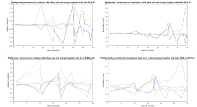 |
Motion detection using reflected signals from an eight channel parallel transmit head coil at 7T
Hans Hoogduin1, Mark Gosselink1, Giel Mens1, Wim Prins2, Tijl van der Velden1, and Dennis Klomp1
1UMCU, Utrecht, Netherlands, 2Philips, Best, Netherlands
Directional couplers are used to measure reflected waves from an eight channel PTx coil to detect head motion at 7T. The method doesn't require any changes to pulse sequences and has no time penalty. A general linear model is used to predict head motion from the signals measured at the couplers.
|
|
1281. |
Multi-Channel RF Power and Phase Supervision Systems Technology for Thermal Magnetic Resonance: Development, Evaluation and Application
Haopeng Han1, Thomas Wilhelm Eigentler1, Eckhard Grass2,3, and Thoralf Niendorf1,4,5
1Berlin Ultrahigh Field Facility (B.U.F.F.), Max Delbrück Center for Molecular Medicine in the Helmholtz Association, Berlin, Germany, 2IHP – Leibniz-Institut für innovative Mikroelektronik, Frankfurt (Oder), Germany, 3Institute of Computer Science, Humboldt-Universität zu Berlin, Berlin, Germany, 4Experimental and Clinical Research Center (ECRC), a joint cooperation between the Charité Medical Faculty and the Max Delbrück Center for Molecular Medicine, Berlin, Germany, 5MRI.TOOLS GmbH, Berlin, Germany Thermal Magnetic Resonance makes use of the physics of radio frequency waves applied at ultrahigh field-MRI. To achieve precise energy focal point formation, accurate thermal dose control and safety management, the transmitted RF signal amplitude and phase need to be supervised and regulated in real-time. In this work, a multi-channel power and phase supervision module was developed, evaluated and applied as an integral part of the Thermal MR hardware system. Preliminary experiments were conducted to demonstrate that the proposed module is suitable and essential for RF heating using a hybrid Thermal MR approach. |
|
1282.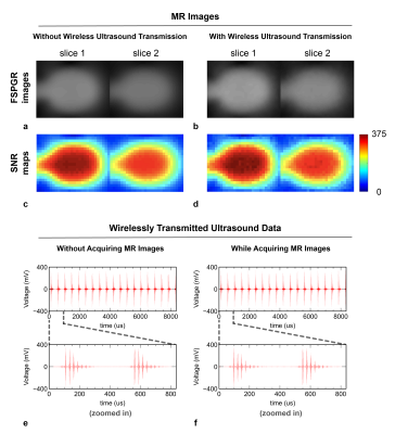 |
Integrated RF/Wireless Coil and Ultrasound-Based Sensors to Enable Wireless Physiological Motion Monitoring in MRI
Devin Willey1,2, Julia Bresticker1,2, Trong-Kha Truong1,2, Allen Song1,2, Bruno Madore3, and Dean Darnell1,2
1Brain Imaging and Analysis Center, Duke University, Durham, NC, United States, 2Medical Physics Graduate Program, Duke University, Durham, NC, United States, 3Department of Radiology, Brigham and Women's Hospital, Harvard Medical School, Boston, MA, United States
An integrated RF/wireless coil, used to wirelessly transmit data and acquire MR images, and small ultrasound-based sensors, used to monitor physiological motion and correct MRI artifacts, were combined to enable wireless transmission of 1-MHz ultrasound data acquired from the sensors. Experiments were performed to validate that the coil could wirelessly transmit ultrasound data 1) while taking MR images and 2) from inside and outside the bore to a computer. This integrated system improves the mobility of the OCM sensors, thereby enabling them to accompany patients throughout the hospital, and demonstrates the coil’s ability to transmit high-fidelity analog data while imaging.
|
|
1283.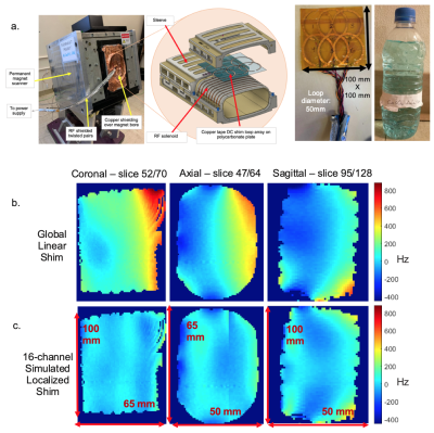 |
B0 Shim Array Integrated into a Solenoid TRX Coil for Localized B0 in Permanent Magnet Scanners
Rafael Limgenco Calleja1, Celine Jeyda Veys1, and Michael Lustig1
1Electrical Engineering and Computer Sciences, University of California, Berkeley, Berkeley, CA, United States
Low-to-mid field systems have nearly been abandoned for high-field, high-performance systems. However, recent improvements in MR hardware, algorithms and computation are stimulating a resurgence in interest of upgrading lower-end magnets to achieve high-performance at significantly lower cost, improved accessibility, portability and siting. Here, we look at upgrading a wrist/animal 1T permanent-magnet system. We integrate a simple B0 array into a solenoid TRX coil for reducing B0 through localized targeted shimming. We demonstrate potential for improved field homogeneity with simulations, and demonstrate homogeneity improvements using a 6-channel prototype array with negligible effects on the transmit field and received SNR.
|

 Back to Program-at-a-Glance
 Watch the Video
 Back to Top