Elisabetta Pagani1, Laura Cacciaguerra1,2, Gianna C. Riccitelli1, Marta Radaelli2, Massimo Filippi1,2,3, and Maria A. Rocca1,2
1Neuroimaging Research Unit, Institute of Experimental Neurology, Division of Neuroscience, IRCCS San Raffaele Scientific Institute, Milan, Italy, 2Neurology Unit, IRCCS San Raffaele Scientific Institute, Milan, Italy, 3Vita-Salute San Raffaele University, Milan, Italy
1Neuroimaging Research Unit, Institute of Experimental Neurology, Division of Neuroscience, IRCCS San Raffaele Scientific Institute, Milan, Italy, 2Neurology Unit, IRCCS San Raffaele Scientific Institute, Milan, Italy, 3Vita-Salute San Raffaele University, Milan, Italy
Hippocampal
dentate gyrus hypertrophy was found in multiple sclerosis but not in neuromyelitis optica spectrum disorders
patients, and correlated with measures of hippocampal disconnection, suggesting
the role of processes other than inflammation.

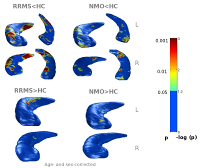
Figure
2. Surface distribution of hippocampal atrophy and hypertrophy
found in relapsing-remitting multiple sclerosis (RRMS) and neuromyelitis optica spectrum (NMO) patients compared
to healthy controls (HC). Logarithmic p values for each
comparison are color coded and displayed on the average hippocampal shape.

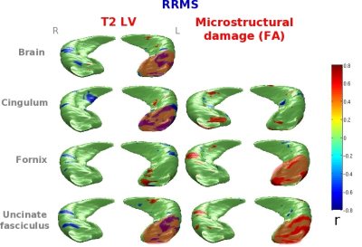
Figure
3. Correlations between regional hippocampal atrophy and MRI measures of inflammation and hippocampal disconnection in
relapsing-remitting multiple sclerosis (RRMS) patients. T2 lesion volume (LV) and
average fractional anisotropy (FA) within fiber bundles are reported. For
significant correlations, r values
are color coded and displayed on the average hippocampal shape.
