Septian Hartono1,2, Isabel Hui Min Chew3, Amanda Jieying Lee3, Joey Xin Yi Oh3, Leon Qi Rong Ooi4, Yao-Chia Shih3, Jongho Lee5, Zheyu Xu1,2, Eng King Tan1,2, and Ling Ling Chan2,3
1Department of Neurology, National Neuroscience Institute, Singapore, Singapore, 2Duke-NUS Medical School, Singapore, Singapore, 3Department of Diagnostic Radiology, Singapore General Hospital, Singapore, Singapore, 4Department of Electrical & Computer Engineering, National University of Singapore, Singapore, Singapore, 5Department of Electrical & Computer Engineering, Seoul National University, Seoul, Korea, Republic of
1Department of Neurology, National Neuroscience Institute, Singapore, Singapore, 2Duke-NUS Medical School, Singapore, Singapore, 3Department of Diagnostic Radiology, Singapore General Hospital, Singapore, Singapore, 4Department of Electrical & Computer Engineering, National University of Singapore, Singapore, Singapore, 5Department of Electrical & Computer Engineering, Seoul National University, Seoul, Korea, Republic of
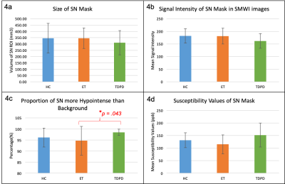
SMWI differentiated between ET and TDPD groups by quantifying the proportion of voxels in the SN more hyperintense than the background. This could be useful to aid differentiation between ET and TDPD patients when clinical doubt exists.

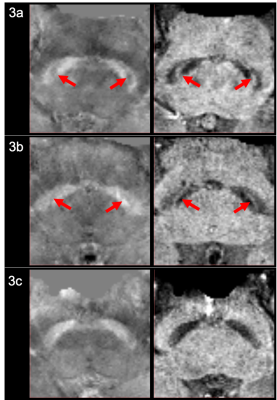
Representative QSM & SMWI images of HC, ET and TDPD Subjects. The left and right columns show the oblique axial QSM and SMWI images respectively for a representative HC (3a), ET (3b) and PD (3c) patient. The presence of the Nigrosome-1, which appears as the thin curvilinear area of hypointensity or hyperintensity within the Substantia Nigra on QSM and SMWI images respectively for HC (3a) and ET (3b) subjects, are indicated by the red arrows. There is loss of the Nigrosome-1 in the Substantia Nigra of the PD patient (3c).

Comparison of Mean Substantia Nigra Metrics on SMWI and QSM images amongst HC, ET and TDPD Subjects. On SMWI (4a-c), the SN mask was smallest (4a) and most markedly hypointense (4b) in TDPD patients. The SN mask in TDPD patients contained the most voxels more hypointense than the background (4c), signifying highest Nigrosome-1 loss. This was significantly higher than that in ET patients. Corroborating evidence of the highest iron content (4d) in the SN of TDPD patients was noted on QSM. [SN=substantia nigra, SI=signal intensity]