Jae-Hyuk Shim1 and Hyeon-Man Baek1
1Gachon University, Incheon, Republic of Korea
1Gachon University, Incheon, Republic of Korea
Basal ganglia nuclei of Parkinson's disease patients and controls were segmented using 7T MRI T1w images. Tractography between each nuclei were generated and compared between Parkinson's and controls
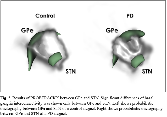
Fig.
2. Results
of PROBTRACKX between GPe and STN. Significant differences of basal ganglia
interconnectivity was shown only between GPe and STN. Left shows probabilistic tractography
between GPe and STN of a control subject. Right shows probabilistic
tractography between GPe and STN of a PD subject.
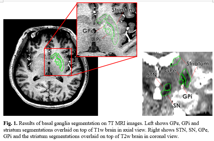
Fig.
1. Results
of basal ganglia segmentation on 7T MRI images. Left shows GPe, GPi and
striatum segmentations overlaid on top of T1w brain in axial view. Right shows STN,
SN, GPe, GPi and the striatum segmentations overlaid on top of T2w brain in
coronal view.