Jitender Saini1, Sarbesh Tiwari2, Sanjib Sinha3, Ravindranadh Chowdary M3, and Raghavendra K3
1Nueroimaging and Interventional Radiology, National Institute of Mental Health and, Bangalore, India, 2Diagnostic and Interventional Radiology, All India Institute of Medical Sciences,, Jodhpur, India, 3Neurology, National Institute of Mental Health and, Bangalore, India
1Nueroimaging and Interventional Radiology, National Institute of Mental Health and, Bangalore, India, 2Diagnostic and Interventional Radiology, All India Institute of Medical Sciences,, Jodhpur, India, 3Neurology, National Institute of Mental Health and, Bangalore, India
In this study, we have reported that ASL
perfusion could be incorporated into the presurgical evaluation of all patients
with localisation related epilepsy as it may reveal seizure induced alteration
in brain perfusion and may help identifying the location and extent of the
epileptogenic zone.

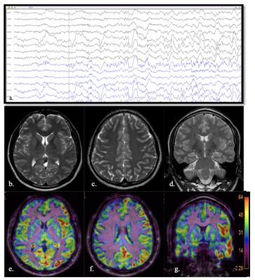
Figure 1: The first row demonstrate the video-EEG
(Image a.) showed ictal onset from the left frontocentral region.
Second row- The sMRI images [(b, c)-
axial & (d)-
coronal] was interpreted as normal on
consensus.
ASL-T1MPRAGE coregistered images [(e, f) axial and (g) coronal] revealed hyperperfusion along left cerebral
hemisphere with increased perfusion predominantly
at left frontal and temporal lobes.
Thus, though structural MRI was normal, the seizure could be lateralized both on
ASL and video-EEG
