Wen-Tung Wang1, Dzung Pham1, and John A Butman1,2
1Center for Neuroscience and Regenerative Medicine, NIH/USU, Bethesda, MD, United States, 2Radiology and Imaging Sciences, NIH, Bethesda, MD, United States
1Center for Neuroscience and Regenerative Medicine, NIH/USU, Bethesda, MD, United States, 2Radiology and Imaging Sciences, NIH, Bethesda, MD, United States
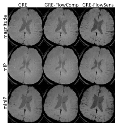
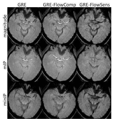
By including flow sensitization gradients in SWI
sequence, signals from moving protons are suppressed, and dark
vessels are delineated at correct spatial locations.

