Shuai Yuan1, Larry Davis1, Petrice Cogswell2, Spencer Waddle3, Lori Jordan1, and Manus Donahue4
1Vanderbilt University Medical Center, Nashville, TN, United States, 2Mayo clinic, Rochester, MN, United States, 3Vanderbilt University, Nashville, TN, United States, 4Vanderbilt University Medical Center Department of Radiology, Nashville, TN, United States
1Vanderbilt University Medical Center, Nashville, TN, United States, 2Mayo clinic, Rochester, MN, United States, 3Vanderbilt University, Nashville, TN, United States, 4Vanderbilt University Medical Center Department of Radiology, Nashville, TN, United States
Vessel wall imaging of basilar
artery shows concentric wall thickening in sickle cell disease with an inverse
relationship to hematocrit. Vessel wall thickness measured by vessel wall imaging MRI may provide an additional
marker of cerebrovascular impairment in SCD patients.
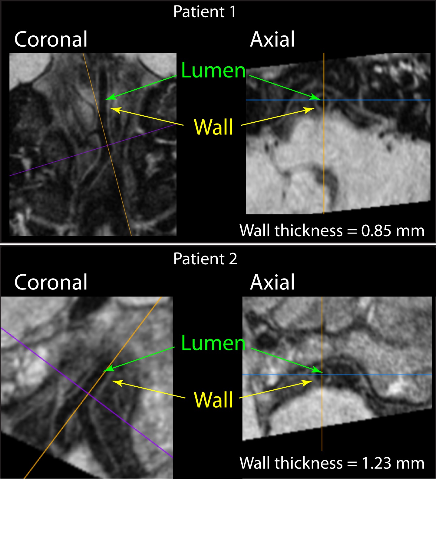
Figure 2. Representative basilar vessel wall measurements from SCD patients with low
(above) and high (below) vessel wall thickness. For each participant, 3D data
were resliced along the course of the vessel of interest by a board-certified
radiologist and measurements were made in the transverse plane.
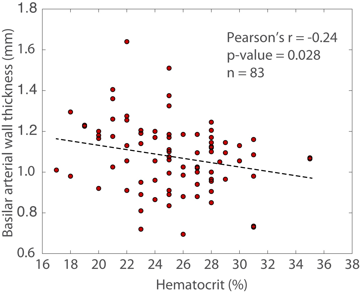
Figure 4. Inverse relationship between wall thickness and hematocrit
is observed in sickle cell patients, consistent with the extent of anemia and reduced oxygen carrying
capacity leading to concentric wall thickening. Damage to
the vessel wall likely arises from increased flow velocities and potentially
more turbulent flow, despite largely preserved lumen diameters.