Francesca Inglese1, Myriam G. Jaarsma-Coes1, Gerda M. Steup-Beekman2, Tom Huizinga2, Mark A. van Buchem1, Itamar Ronen1, and Jeroen de Bresser1
1Department of Radiology, Leiden University Medical Center, Leiden, Netherlands, 2Department of Rheumatology, Leiden University Medical Center, Leiden, Netherlands
1Department of Radiology, Leiden University Medical Center, Leiden, Netherlands, 2Department of Rheumatology, Leiden University Medical Center, Leiden, Netherlands
NP-SLE patients more often had deep white
matter hyperintensities compared to non-NP-SLE patients. This finding may
contribute to the diagnosis of NP-SLE.
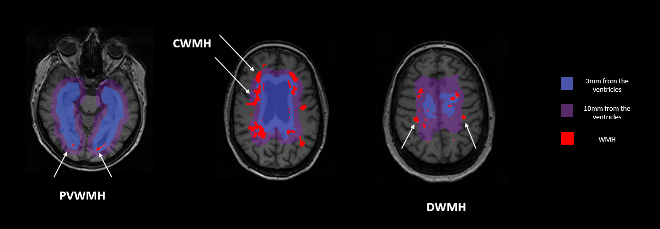
Figure 2. White matter hyperintensity types. In
blue 3 mm space distance from the lateral ventricles. In purple 10 mm space
distance from the lateral ventricles. In red white matter hyperintensities
(WMH).
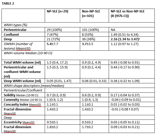
TABLE 2. Comparison between NP-SLE and non-NP-SLE patients.
Variables are shown in terms of: mean±SD in case of normally distributed
variables; or median (10-90 CI) in case of not normally distributed variables.
Non normally distributed variables were multiplied by 1000 and then natural log
transformed before the logistic or linear regression analysis. DWMH are more
often present in NP-SLE patients compared to non-NP-SLE patients. NA = not applicable.