Sirui Li1, Wenbo Sun1, Yuan Zheng2, Qing Wei3, Samo Lasic4, Shihong Han3, Shuheng Zhang3, Danielle van Westen5, Karin Bryskhe4, Daniel Topgaard4,5, and Haibo Xu1
1Zhongnan Hospital of Wuhan University, Wuhan, China, 2UIH America, Inc., Houston, TX, United States, 3United Imaging Healthcare, Shanghai, China, 4Random Walk Imaging, Lund, Sweden, 5Lund University, Lund, Sweden
1Zhongnan Hospital of Wuhan University, Wuhan, China, 2UIH America, Inc., Houston, TX, United States, 3United Imaging Healthcare, Shanghai, China, 4Random Walk Imaging, Lund, Sweden, 5Lund University, Lund, Sweden
Diffusion kurtosis decomposition (DKD) was used to assess cell shapes
and density heterogeneity in gliomas via specific metrics that are inextricably
entangled in conventional DTI and DKI. DKD accurately describes microstructural
changes related to tumor grade.

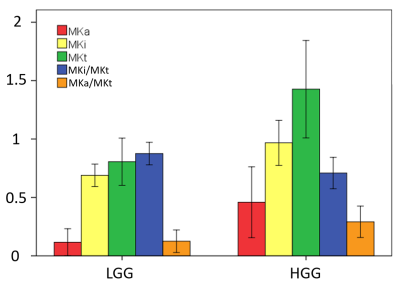
Figure 3: Bar graphs showing MKi, MKa, and MKt, as well as the ratios MKa/MKt and
MKi/MKt in low and high grade gliomas (LGG and HGG) averaged over all studied subjects.
Standard deviation across subjects were shown as error bars. Significant
differences were observed between all groups.

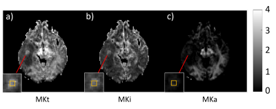
Figure 2: DKD parameters maps of a grade IV glioma case. The total, isotropic, and
anistropic mean kurtosis (MKt, MKi and MKa) are shown in a), b), c)
respectively. The insets show the zooms of the tumor. The metrics in a 3×3 ROI indicated
by the yellow box are: MKt = 2.09 ± 0.19, MKi = 1.70 ± 0.14, MKa = 0.38 ± 0.11.
The comparatively high value of MKi reveals cell density heterogeneity, while
the low but finite values of MKa shows that the cells in the tumor are more
elongated than in its immediately surroundings.