Fatiha Andoh1, Marion Tardieu2, Claire Barakat Pellot3, and Xavier Maître1
1Univ. Paris Saclay, IR4M Laboratory, Orsay, France, 2Institut du Cancer de Montpellier, ICM, Montpellier, France, 3INSERM, IMIV Laboratory, Orsay, France
1Univ. Paris Saclay, IR4M Laboratory, Orsay, France, 2Institut du Cancer de Montpellier, ICM, Montpellier, France, 3INSERM, IMIV Laboratory, Orsay, France
Mechanical parameters may be underestimated by MRE if data
quality is not properly monitored to discarded inaccurate and unprecised biased
data. This study proposes to evaluate and compare MRE parameters (wave data sampling λ/a and quality factor Q) for 2 excitation freuquencies.
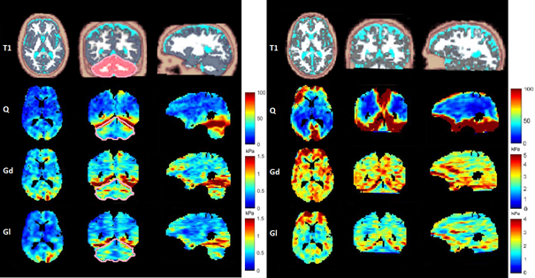
Figure 1: Brain MRE at
45 Hz (left) and at 113 Hz (right); From top to bottom: Segmented-T1
(white matter white, grey matter grey, CSF cyan and cerebellum in red segmented
from T1 map by SPM12 for data at 45Hz [6]), Q maps, Gd maps and Gl maps
of the full brain in axial (1st column), coronal (2nd
column), and sagittal (3rd column) view.
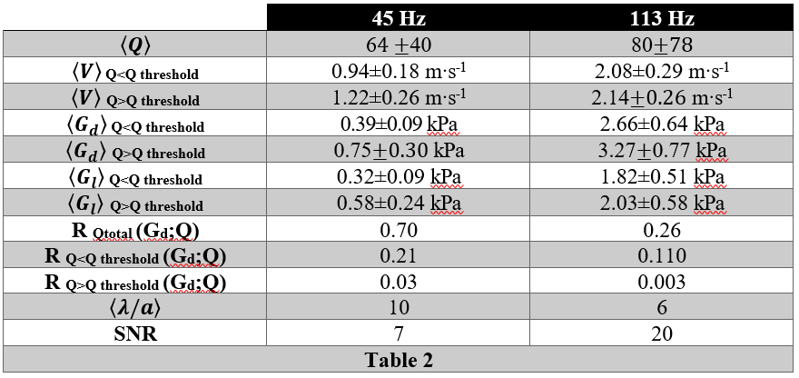
Table
2: Mean global