Li Chen1, Duygu Baylam Geleri 1, Jie Sun 1, Hiroko Watase 1, Jiarui Cai1, Yin Guo1, Niranjan Balu 1, Dongxiang Xu 1, Thomas Hatsukami 1, Yongjun Wang 2, Jenq-Neng Hwang 1, and Chun Yuan 1
1University of Washington, Seattle, WA, United States, 2Beijing Tiantan Hospital, Capital Medical University, Beijing, China
1University of Washington, Seattle, WA, United States, 2Beijing Tiantan Hospital, Capital Medical University, Beijing, China
A visualization technique including image registration,
artery tracing and reformation was developed for improving multi-contrast/timepoint
vessel wall review, providing better sensitivity and higher accuracy for plaque
identification and quantification than traditional review.
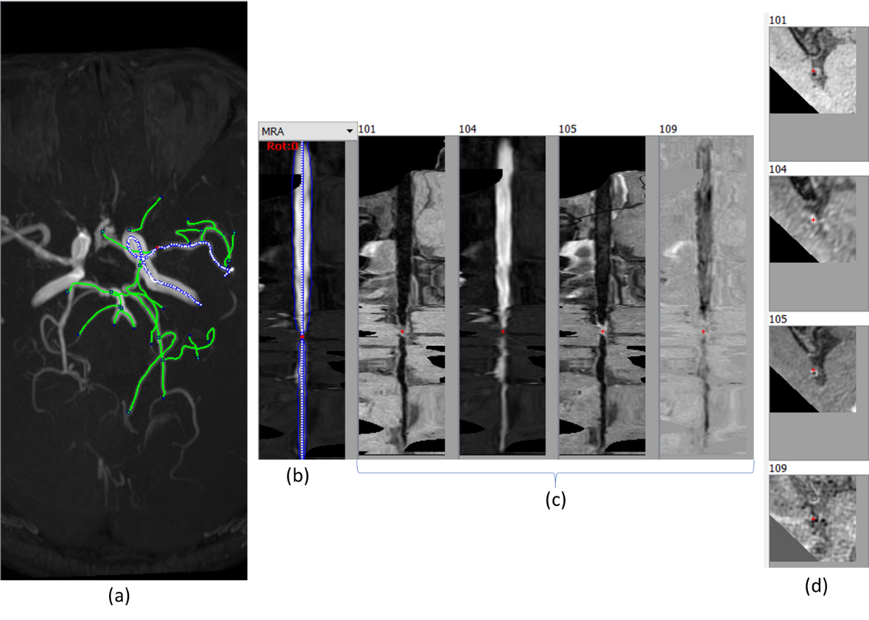
Figure 2 MOCHA for intracranial artery. (a) MIP
of intracranial MRA. Centerlines (blue) generated from artery tracing in a
region of interest, and the selected one for analysis is shown with the dotted
blue line. (b) Centerline (blue line) shown in MPR with the red cross
indicating the plaque location. (c) MPR for the registered sequences. 101: T1
VISTA, 104: ToF MRA, 105: contrast enhanced T1 VISTA, 109: SNAP (d)
Cross-sectional images of the plaque highlighted in the MPR views (red cross).
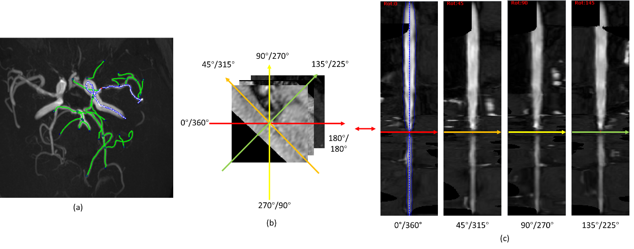
Figure 1 Illustration of MPR generation (a) Maximum
intensity projection (MIP) of intracranial MRA. The artery of interest is shown
with the dotted blue line. (b) Series of cross-sectional planes along the
selected artery centerline with a gap of 1 pixel. Profiles of colors indicate
different angles. (c) MPR images generated at different angles of view.
Multiple profiles at a certain angle from consecutive cross-sectional images
stack together vertically to form the MPR images.