Nikhil Deveshwar1 and Peder E. Z. Larson1
1Department of Radiology and Biomedical Imaging, University of California, San Francisco, San Francisco, CA, United States
1Department of Radiology and Biomedical Imaging, University of California, San Francisco, San Francisco, CA, United States
Ultrashort-T2* measurements can provide new characterizations of white matter anatomy in comparison to classic DTI measurements. Signal magnitude and frequency shift are significantly different between white matter with thick versus thin myelin sheaths.
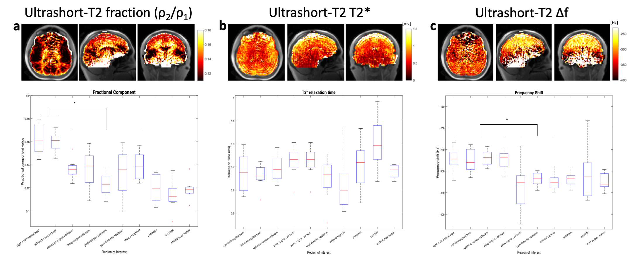
Whole-brain representative parameters maps and boxplots of the ultrashort-T2* signal components including a) fractional component, b) T2* relaxation time and c) frequency shift across 8 healthy volunteers. Ultrashort-T2* fractional component is significantly higher in both corticospinal tracts compared to other white matter structures. Frequency shift is also significantly higher in both corticospinal tracts and the body and splenium of the corpus callosum.
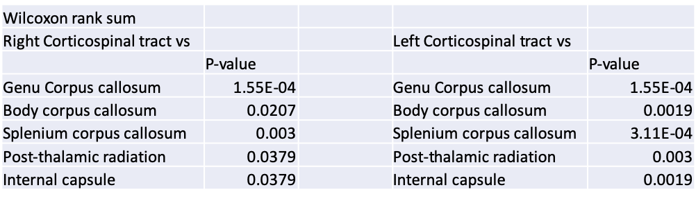
Wilcoxon rank sum test performed comparing measured ultrashort-T2* component fraction in right and left corticospinal tracts to other white matter structures. Fraction is significantly higher for both corticospinal tracts when compared to other white matter anatomy.