Anna Crawford1, Stephen Jones1, and Mark Lowe1
1Imaging Institute, Cleveland Clinic Foundation, Cleveland, OH, United States
1Imaging Institute, Cleveland Clinic Foundation, Cleveland, OH, United States
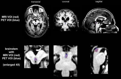
Some brain structures including
brainstem nuclei are difficult to identify with MR imaging. We have come up
with a “recipe” for defining the VTA and DRN using 3 anatomic atlases and
confirmed with PET imaging. Our method allows for robust, reproducible ROIs
using only a high resolution T1w image.

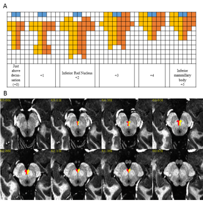
Figure 2: Definition of the ventral tegmental
area (VTA): A) Axial 1mm3 grid used to define VTA where yellow and
orange show the left and right VTA and blue is the interpeduncular fossa, B)
Example VTA viewed on MP2RAGE axial image that is re-oriented to defined axis
(Middle of mammillary body – superior colliculus) at 0.75mm3
spacing.

