Evan Hunter Stanton1, Niklas Daniel Åke Persson1, Björn Sigurdsson1, Tuomas Lilius1, Humberto Mestre2, Maiken Nedergaard1, and Yuki Mori1,3
1Division of Glial Disease and Therapeutics, University of Copenhagen, Center for Translational Neuromedicine, Copenhagen, Denmark, 2Division of Glial Disease and Therapeutics, University of Rochester Medical Center, Center for Translational Neuromedicine, Rochester, NY, United States, 3Faculty of Health and Medical Sciences, University of Copenhagen, Copenhagen, Denmark, Panum NMR Core Facility, Copenhagen, Denmark
1Division of Glial Disease and Therapeutics, University of Copenhagen, Center for Translational Neuromedicine, Copenhagen, Denmark, 2Division of Glial Disease and Therapeutics, University of Rochester Medical Center, Center for Translational Neuromedicine, Rochester, NY, United States, 3Faculty of Health and Medical Sciences, University of Copenhagen, Copenhagen, Denmark, Panum NMR Core Facility, Copenhagen, Denmark
The
present study describes the development of 3D-DCE glymphatic MRI with high
temporal resolution. Utilizing FISP, detailed tracer distributions were shown
to vary across anesthetics, with more influx in K/X and deep cervical node shunting
of CSF in isoflurane anesthesia.
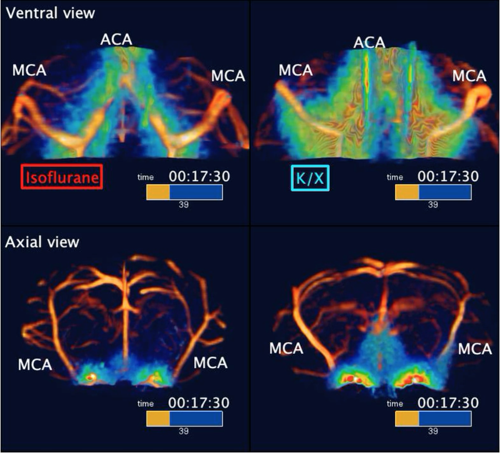
Contrast agent distribution with FISP-based
DCE-MRI. Anesthesia alters the spatial and temporal distribution of contrast
agent in the circle or willis and middle cerebral artery (MCA).
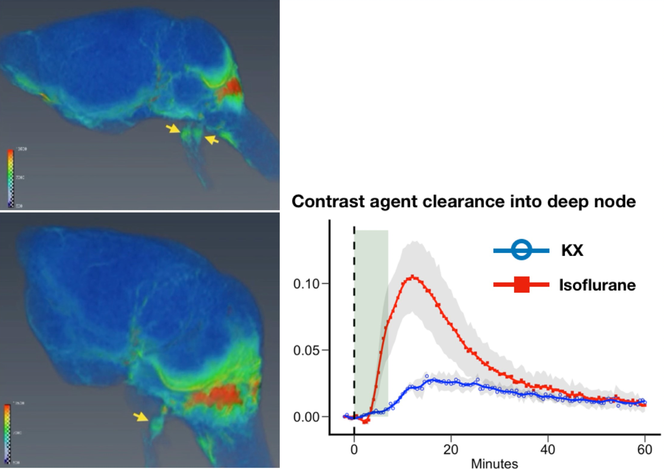
4D visualization of contrast agent
distribution and CSF clearance into deep node. CSF clearance via
deep node can be seen under isoflurane anesthesia, while cannot be seen under
K/X.