Grzegorz Kwiatkowski1, Georgios Louloudis1, Jan Klohs1, and Sebastian Kozerke2
1Institute for Biomedical Engineering, ETH Zurich, Zurich, Switzerland, 2ETH Zurich, Zurich, Switzerland
1Institute for Biomedical Engineering, ETH Zurich, Zurich, Switzerland, 2ETH Zurich, Zurich, Switzerland
A comparison between changes
in quantitative Magnetization Transfer MR and standard MR contrast are reported for the mouse brain after transient
cerebral ischemia.
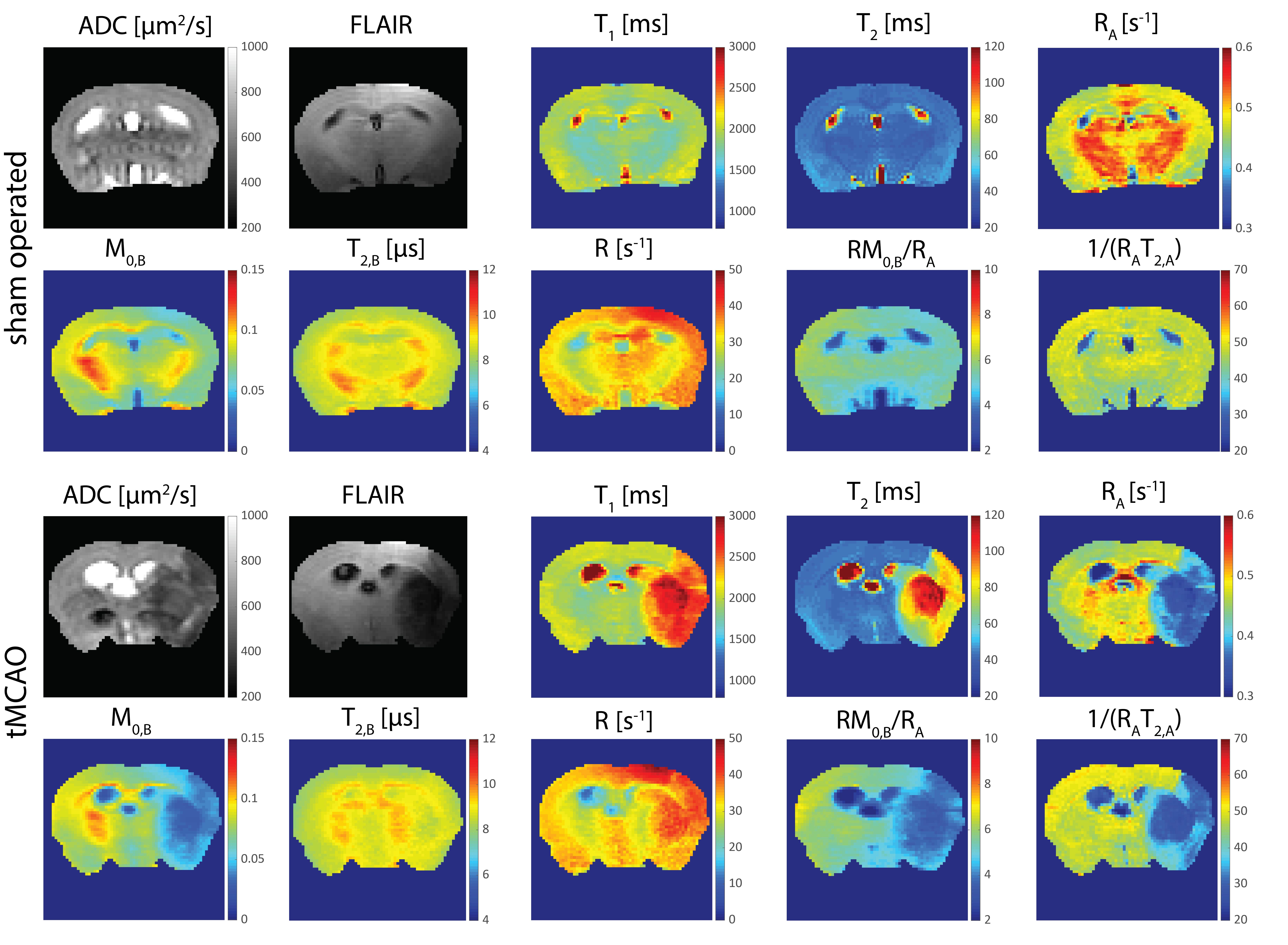
Figure 2: Examples
of MR metric maps obtained for the sham-operated and tMCAO animals, 24h post-reperfusion.
Each MR contrast was plotted with the same scale between different animal
groups.
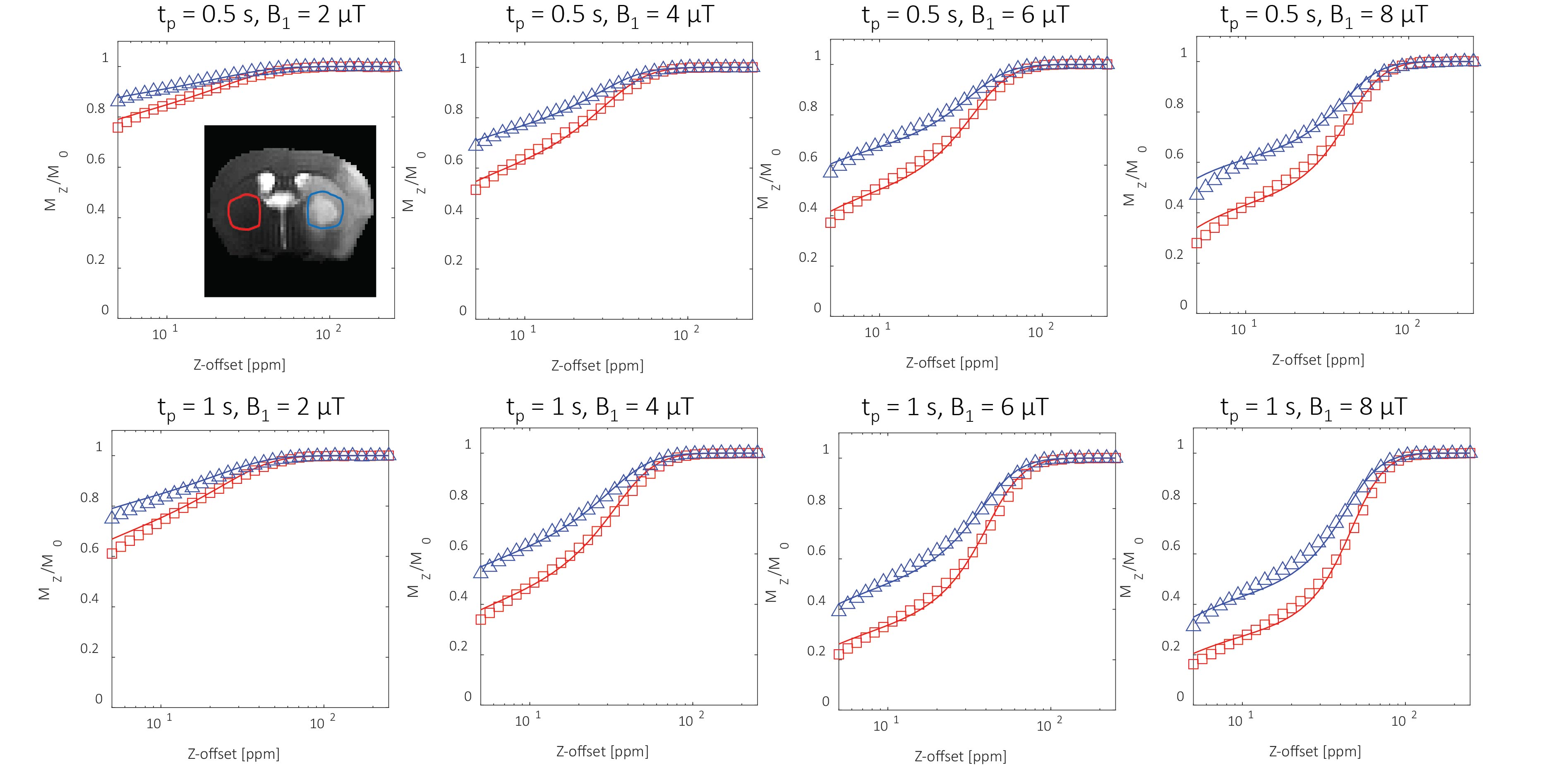
Figure
1: Example of magnetization transfer
saturation curves measured in contralateral (red) and ipsilateral ischemic
hemisphere (blue) of a mouse brain 24h post tMCAO. The curves were recorded
following saturation module applied at different off-resonance frequency, with
varying B1 power (2,4,6 and 8 μT) and saturation duration, tp (0.5 and 1 s).
Significant differences were observed for curves recorded at different brain
sides, reflecting changes in the magnetization transfer between free solute and
bound water protons of the underlying tissue.