Lucy Liu1,2, Andre Bongers2, Lynne Bilston1,2, and Lauriane Jugé1,2
1Neuroscience Research Australia, Sydney, Australia, 2University of New South Wales, Sydney, Australia
1Neuroscience Research Australia, Sydney, Australia, 2University of New South Wales, Sydney, Australia
Diffusion tensor imaging (DTI) and magnetic resonance
elastography can track neurodevelopmental microstructural changes in rat
brains, but only DTI detected subtle white matter injury in offspring of Poly(I:C)-induced maternal immune activated rats.

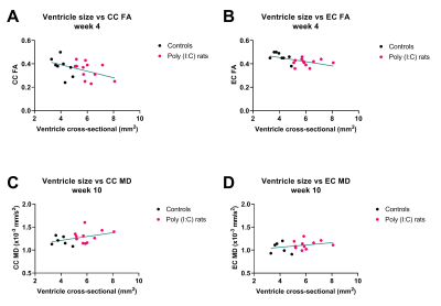
Figure 3: In the
corpus callosum (CC) and the external capsule (EC), an increase in ventricle size was associated with a decrease in
fractional anisotropy (FA) at 4 weeks (A and B, respectively), and an increase
in mean diffusivity (MD) at 10 weeks (C and D, respectively) after birth. These
results infer a temporal variation in the white matter injury in poly (I:C)
rats.

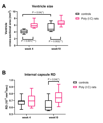
Figure 2: Ventricle
cross-sectional area (A) and radial diffusivity (RD) measured in the internal
capsule (B) of Poly (I:C) offspring rats and controls at weeks 4 and 10 after
birth. Significant Sidak’s comparisons are reported. Poly (I:C) rats had larger
ventricles than controls at both time-points and larger RD at week 10.