Zhenxiong Wang1,2, Wenzhen Zhu1, Shun Zhang1, Guiling Zhang1, Mehran Shaghaghi2, and Kejia Cai2
1Tongji Hospital, Tongji Medical College, Huazhong University of Science and Technology, Wuhan, China, 2Departments of Radiology, Department of Bioengineering, and the Center for MR Research, Chicago, IL, United States
1Tongji Hospital, Tongji Medical College, Huazhong University of Science and Technology, Wuhan, China, 2Departments of Radiology, Department of Bioengineering, and the Center for MR Research, Chicago, IL, United States
NODDI can characterize the microstructural alterations in rat brain tissues due to middle cerebral artery occlusion at a 3T MRI and its parameters were validated by the histology.
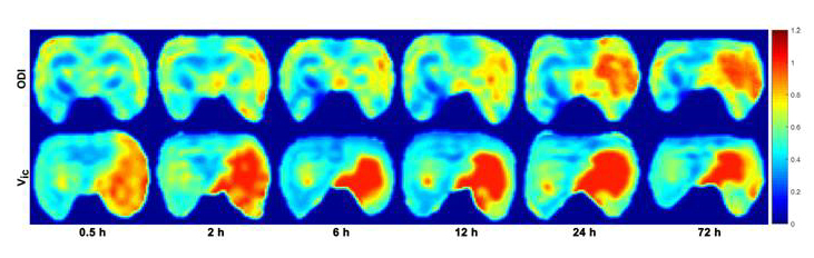
Figure 1. Representative NODDI metric maps at different
time points from a MCAO rat. The signal intensity of ODI and Vic maps in
the infarction areas is increased compared with contralateral brain tissues.
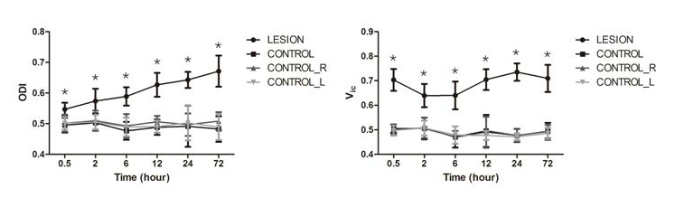
Figure 2. The temporal evolution of NODDI metrics from
different groups. * represents significant differences between LESION
and CONTROL.