Junhong Liu1 and Jingliang Cheng1
1The First Affiliated Hospital of Zhengzhou University, Zhengzhou, China
1The First Affiliated Hospital of Zhengzhou University, Zhengzhou, China
It’s considered that changes of abnormal resting-state networks might reveal the possible neural mechanism of OCD.
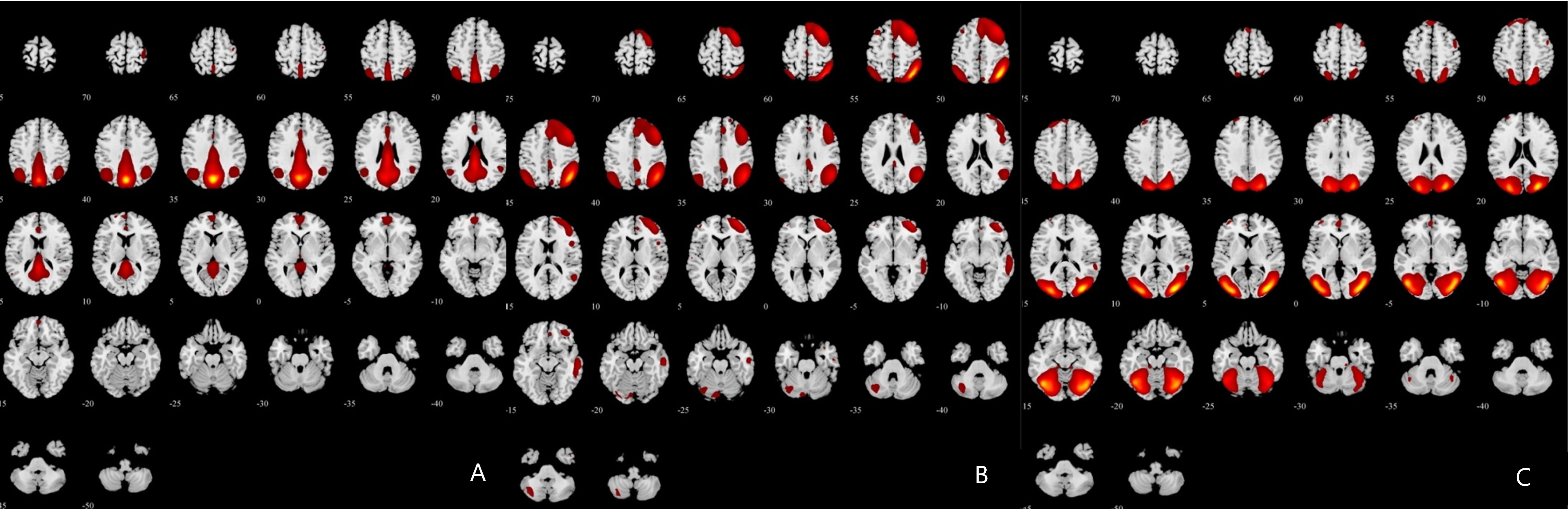
Figure
1. Three
significant different masks of resting-state networks between obsessive-compulsive
disorder patients and healthy controls. A) Posterior default-mode network
(pDMN); B) Right frontoparietal network (RFP); C) Lateral visual network (lVN)
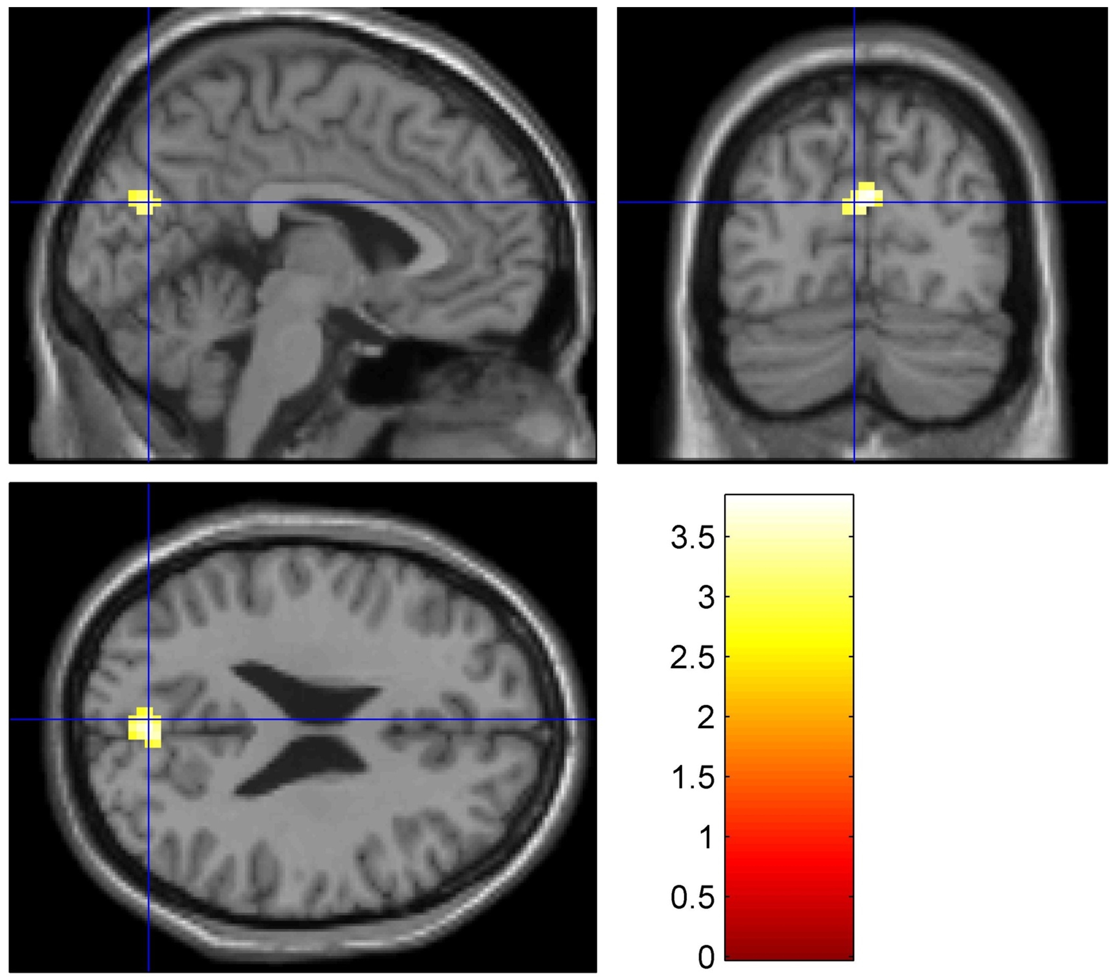
Figure
2. Compared
with healthy controls, OCD patients exhibited increased functional connectivity (FC) in pDMN in the bilateral cuneus. The pDMN FC of OCD
patients were higher than that of healthy controls in bilateral cuneus (T=3.822,P=0.005) in OCD patients. MNI
coordinate (3, -75, 27), Brodmann's area 31, voxel 50.